Lecture 04. Skeleton & Joints Flashcards
(59 cards)
Two Major Regions

Two major regions of the skeleton
-
Axial Skeleton:
Frontal Bone, Parietal Bone, Occipital Bone, Temporal Bone, Sphnoid Bone, Ethmoid Bone
Malleus, Incus, Stapes
Cervical Vertebrae (7), Thoracic Vertebrae (12), Lumbar Vertebrae (5), Saccrum, Coccyx
Ribs, Sternum -
Appendicular Skeleton:
Scapula, Clavicle
Humerus, Radius, Ulna, Carpals, Metacarpals, Phalanges
Hip Bones
Femur, Patella, Tibia, Fibula, Tarsals, Metatarsals, Phalanges
Typical number of bones
206 in typical adult skeleton (can range 204-207)
Skull
- 22 bones joined together by sutures
- Cranial bones surround cranial cavity
- *Frontal Bone** (1)
- *Parietal Bones** (2)
- *Temporal Bone** (1)
- *Occipital Bone **(1)
- *Sphenoid Bone** (1)
- *Ethmoid Bone** (1)
- **Facial bones support teeth and form nasal cavity and orbit **(make up for the other 14 bones)
- *Maxilla** (2)
- *Palatine Bone** (2)
- *Zygomatic Bone** (2)
- *Lacrimal Bone** (2)
- *Nasal Bone** (2)
- *Vomer** (1)
- *Inferior Nasal Concha** (2)
- *Mandible** (1)
Frontal Bone

- Forms forehead
- Forms roof of the orbit (Eye-socket)
- Contains frontal sinuses (openings in the bone which help lighten the bone)
Parietal Bone

- Cranial roof and part of its lateral walls
- Temporal lines of temporalis muscle
- One on each side, seperated by the Saggital Suture
Temporal Bone

Lateral wall and part of floor of cranial cavity
- Squamous part (flat part of bone)
- Tympanic part (external auditory canal/external auditory meatus)
- Mastoid part (mastoid process)
- Petrous portion (rocky, stlyoid process and surface)
Occipital Bone

Rear and base of skull
- Foramen magnum: Hole located in the mid-section of the occipital bone
- Skull rests on atlas
Sphenoid Bone

This bone helps form the base of the cranium, the sides of the skull, and the floors and sides of the orbits (eye sockets).
- Sella Turcica (Turk’s saddle): A saddle-shaped mass along the middle, within the cranial cavity. The depression of this saddle is occupied by the pituitary gland, which hangs from the base of the brain by a stalk.
- Sphenoidal Sinuses: Two sinesus which lie side by side and are separated by a bony septum that projects downward into the nasal cavity.
Ethmoid Bone

An anterior cranial bone located between the eyes. It is a very porous and delicate bone.
THREE major portions:
- Vertical Perpendicular Plate: A thin median plate that forms the superior two-thirds of the nasal septum (The lower part is formed by cartilages and the vomer) The septum divides the nasal cavity into right and left air spaces called the nasal fossae. The septum is often curved, or deviated, toward one nasal fossa or the other.
- Cribriform Plate: Horizontal plate, forms the roof of the nasal cavity. This plate has a median crest called the crista galli, an attachment point for the dura mater. On each side of the crista is an elongated depressed area perforated with numerous holes, the cribriform (olfactory) foramina. A pair of olfactory bulbs of the brain, concerned with the sense of smell, rests in these depressions, and the foramina allow passage for olfactory nerves from the nasal cavity to the bulbs.
- Labyrinth: A large mass on each side of the perpendicular plate. The labyrinth is named for the fact that internally, it has a maze of air spaces called the ethmoidal cells.
Malleus, Incus, Stapes

Maxillae

Largest facial bones.
They form the upper jaw and meet each other at a median intermaxillary suture.
- Alveolar Processes: Grow into the spaces between the bases of the teeth.
- Infraorbital Foramen: Just below the orbit, provides passage for a blood vessel to the face and a nerve that receives sensations from the nasal region and cheek.
- Inferior Orbital Fissure: A gash in the maxilla, angles downward and medially. The inferior and superior orbital fissures form a sideways V whose apex lies near the optic foramen. The inferior orbital fissure is a passage for blood vessels and sen- sory nerves from the face.
Palatine Bone
Located in the posterior nasal cavity.
- Horizontal Plate: (L-shape) form the posterior one-third of the bony palate.
- Perpendicular Plate: (L-shape) A thin, delicate, irregularly shaped plate that forms part of the wall between the nasal cavity and the orbit.
- Palatine Foramen: A nerve passage to the palate.
Zygomatic Bone
The zygomatic bones (cheekbones) form the angles of the cheeks inferolateral to the eyes and part of the lateral wall of each orbit; they extend about halfway to the ear.
- Zygomaticofacial Foramen: Inverted T shape on each zygomatic bone near the intersection of the stem and crossbar of the T.
- Zygomatic Arch: Flares from each side of the skull, formed by the union of the zygomatic bone, temporal bone, and maxilla.
Lacrimal Bone
The lacrimal bones form part of the medial wall of each orbit. These are the smallest bones of the skull, about the size of a small fingernail.
Lacrimal Fossa: Depression that houses a membranous lacrimal sac. Tears from the eye collect in this sac and drain into the nasal cavity.
Nasal Bones

Two small rectangular nasal bones form the bridge of the nose and support cartilages that shape the lower portion of the nose.
Vomer
The vomer forms the inferior portion of the nasal septum. Its name literally means “plowshare,” which refers to its resemblance to the blade of a plow. The superior half of the nasal septum is formed by the perpendicular plate of the ethmoid bone. The vomer and perpendicular plate support a wall of septal cartilage that forms most of the anterior part of the septum.
Inferior Nasal Concha

There are three conchae in the nasal cavity.
- Superior Conchae
- Middle Conchae
- Inferior Nasal Concha: Largest of the three.
Mandible

Only movable bone
- Holds the lower teeth
- Attachment of muscles for mastication
- Mandibular foramen: Two small holes located on both inner-sides of mandible (for alveolar nerve and vessel)
- Mental foramen: Two small holes located on both outter-sides of mandible (mental nerve and other veins pass through to supply the tissues of the mouth)
THREE parts:
- Ramus: The portion of the mandible from the condoyle to the angle.
- Angle: Small corner of the mandible where the bone begins to slop upward toward.
- Body: The portion of the mandible from the angle to the chin.
The Vertebral Column

- 33 vertebrae (Though variations in number of lumbar and sacral vertebrae)
Five vertebral groups
- Cervicle Vertebrae: (7) C1-C7
- Thoracic Vertebrae: (12) T1-T12 (Giraffes)
- Lumbar Vertebrae: (5) L1-L5 (Moose)
- Sacrum: (5) S1-S5
- Coccyx
Thoracic (Rib) Cage

Attachment site
- Protection
- Involved in respiration
Pectoral Girdle

Attaches upper extremity to the body
- Scapula and clavicle
- Clavicle attaches medially to the sternum and laterally to the scapula
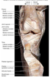
Brachium and Antebrachium

- Brachium: The arm, specifically the upper arm from shoulder to elbow.
- Antebrachium: The forearm.
Pelvic Girdle
Supports trunk on the legs and protects viscera.
Carpals, Metacarpals, Phalanges

Carpal Bones: Bones of the wrist.
- Some - Scaphoid
- Lovers - Lunate
- Try - Triquetrum
- Positions - Pisiform
- That - Trapezium
- They - Trapezoid
- Can’t - Capitate
- Handle - Hamate (has prominent hook)
Metacarpal Bones: Bones of the palm.
- Metacarpal I is located proximal to the thumb
- Metacarpal V is proximal to the little finger
- The proximal end of a metacarpal bone is called the base, the shaft is called the body, and the distal end is called the head. The heads of the metacarpals form the knuckles when you clench your fist.
Phalanges: Bones of the fingers.
- Phalanges are identified by roman numerals preceded by proximal, middle, and distal.
- The three parts of a phalanx are the same as in a metacarpal: base, body, and head.
- The ventral surface of a phalanx is slightly concave from end to end and flattened from side to side; the dorsal surface is rounder and slightly convex from end to end.
Comparison of Male and Female

- Female: Lighter, shallower pubic arch, pubic inlet round or oval
- Male: Heavier, upper pelvis nearly vertical, coccyx more vertical, and pelvic inlet heart-shaped