Lab Practical III Flashcards
Identify the Image
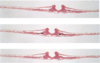
24 hr whole mount
Down the left, down the right, the two on the outside

Head fold
Notochord
Intersomitic furrow
Cranial intestintal portal
Somites
Segmental plate
Primitive knot
Blood islands
Area Vasculosa
Identify the image
24 hr cross section
Middle arrows point to the same thing

Foregut
Cardiac Primordia
Notochord
Identify the Image
24 hr chick cross section

Cardiac Primordia
Pericardial Cavity
Cranial Intestinal Portal
Somatapleure - ectoderm and mesoderm
Splanchnopleure - mesoderm and endoderm
Identify the Image
24 hr cross section
Somite
Identify the Image
24 hr cross section

Inner somitic furrow
Identify the Image
24 hr sagittal section
Left to right

Cardiac primordia
Pericardial cavity
Neural tube
Identify the Image
33hr sagital section
Top panel left to right
Bottom panel left to right

Somite
Intersomitic furrow
Ventricle (heart)
Pericardial cavity
Neural tube
Cranial intestinal portal
Sinoatrial region (heart)
Pericardial cavity
Subcephalic pocket
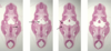
Identify the Image
33hr whole mount
down the left, down the right

Bulbus cordis
Cranial Intestinal Portal
Blood islands
Area vasculosa
Ventricle
Sinoatrial region
vitelline veins
intersomitic furrow
somites
segmental plates
Identify the Image
33hr cross-section

Foregut
Ventral Aorta
Dorsal Aorta

What is the path of the heart from top to bottom
Ventral aorta
Bulbus cortus
Ventrical
Sinoatrial Region
Vitelline Veins
Identify the Image
33hr cross section

Ventral Aorta
Bulbus Cortus
Identify the Image
33 hr cross section

Transitioning Bulbus Cortus
Ventricle
Identify the Image
33hr cross section

Ventricle
Endocardium
Myocardium
Identify the Image
33hr cross-section

Ventricle
Sino-atrial region
Identify the Image
33hr cross-section

Sinoatrial Region
One vitelline vein
Identify the Image
33hr cross section

Vitelline Veins
Cranial Intestinal Portal
Identify the Image
33hr cross section

Somatopleure - ectoderm, mesoderm
Splanchnopleure - mesoderm, endoderm
Identify the Image
33hr cross-section

Somites
Inner somitic furrow
Identify the Image
48hr injected whole mount
down the left
down the right

left vitelline vein
descending aorta
left dorsal aorta
left vitelline artery
right vitelline vein
ventricle
right dorsal aorta
right vitelline artery
Identify the Image
48hr uninjected whole mount
down the left, down the right

Aortic arch I
Aortic arch II
Aortic arch III
Atrium
Vitelline Artery
Bulbus cordis
Ventricle
Sinus Venosus
Vitelline Vein
Cranial Intestinal portal
Somites
Neural tube
Identify the Image
48 hr whole mount
top to bottom

Aortic arches 1-3
Atrium
Ventricle
Sinus venosus
Cranial intestinal portal
Dorsal aorta
Notochord
Identify the Image
48hr whole mount
top to the bottom

Notochord
Bulbus cordis
Ventricle
Atrium
Cranial Intestinal portal
Somites
Identify the Image
48hr heart
left side
down the right side

1st aortic arch
2nd aortic arch
3rd aortic arch
right dorsal aorta (paired)
left vitelline vein
right vitelline vien
bulbus cordis
ventricle
atrium
sinus venosus
descending aorta
Identify the Image
48hr whole mount
left side, right side

Pharyngeal pouches 1-3
Stomadeum
Bulbus cordis
ventricle
atrium
sinus venosus
cranial intestinal portal
Identify the Image
48hr whole mount
down the right, two on the left

Pharyngeal pouch 1
Aortic arch 1
Aortic arch 3
Pharyngeal pouch 2
Dorsal aorta
Cranial intestinal portal
Atrium
Ventricle
Identify the Image
48hr cross-section

Phayrnx
Dorsal aorta
Identify the Image
48hr cross-section

Closing Plate
1st Pharyngeal Arch
1st Aortic Arch
Identify the Image
48hr cross-section

1st pharyngeal arch & 1st aortic arch
Top: Mandibular
Bottom: Maxillar
Stomodeum
Identify the Image
48hr cross-section

2nd pharyngeal arch
2nd aortic arch
2nd pharyngeal pouch
Dorsal aorta
Identify the Image
48hr cross-section

Dorsal aorta
Single descending aorta
Identify the Image
48 hr cross-section

Bulbus Cortis
Atrium
Ventricle
Bulbus Cortis and Ventricle connect - developing heart
Identify the Image
48hr cross-section

Endocardium
Myocardium
Cranial Intestinal Portal
Identify the Image
48hr Cross-section

Mesonephric Duct
Mesonephric Tubular
Identify the Image
48 hr cross-section

Raske’s Pouch
Infundibulum Below
Identify the Image
Pig
1st pharyngeal pouch
1st branchial groove
1st closing plate
Identify the Image
Pig cross-section
1st pharyngeal pouch
Eustachian Tube
Tympanic Cavity
Pharynx
Identify the Image
Pig cross-section

Tuberculum Impar
1st pharyngeal arch
1st pharyngeal groove
Identify the Image
Pig cross-section

1st pharyngeal arch
1st pharyngeal groove
2nd pharyngeal arch
2nd pharyngeal groove
Pouches above groove
Identify the Image
Pig Cross-Section

Cervical Sinus (3rd & 4th arches connected)
Identify the Image
Pig cross-section

Epiglottis
Glottis
Identify the Image
Pig cross-section

Epiglottis
Arytenoid Swelling
Esophagus
Trachea
Identify the Image
Pig cross-section

Eparterial bronchus
Dorsal aorta
Esophagus
Trachea
Identify the Image
Pig Cross-section

Trachea
Separating Lung Buds
Identify the Image
Pig cross-section

Stomach
Identify the Image
Pig cross-section

Stomach
Omental Bursa (space around stomach > become peritoneal space)
Hepatogastric Ligament (connects stomach and liver)
Identify the Image
Pig cross-section

Pancreus
Dorsal pancreus (top)
Ventral pancreus
Duodenum
Identify the Image
Pig cross-section

Mesenephric Kidneys
Descending Aorta
Mesenephric tubules
Mesenephric ducts
Mesenephric Glomerulus
Identify the Image
Pig cross-section

Liver
Hepatic sinuses (spaced)
Hepatic chords (tissue)
Identify the Image
48hr chick cross-section

Rathke’s pouch
Infindibulum
What is the fate of the 1st pharyngeal pouch? (2)
Eustachian tubule
Tympanic cavity
What is the fate of the 2nd pharyngeal pouch? (2)
- Tonsils
- Thymus
What is the fate of the 3rd pharyngeal pouch?
- Thymus
- Parathyroids (1 pair)
What is the fate of the 4th pharyngeal pouch?
- Parathyroids (2nd pair)
- Respiratory diverticulum
