Lab Practical 1 Flashcards
Identify the Image
Top, right side, left side
- Ocular
- Stage
- Focus
- Objective
- Condenser diaphragm
- Field diaphragm
Identify the Image
Left, Right
- Manufacturer
- Magnification
- Tube length
- Magnification Color Code
- Lens Correction
- Numerical Aperature
- Indicates Oil Objective
- Coverglass Thickness
Identify the Type of Light Microscopy
- Bright field microscopy
- Darkfield microscopy
- Phase-contrast microscopy
- Transmission electron microscopy (TEM)
Identify the Type of Light Microscopy
- Nomarski (differential interference contrast)
- Fluorescence microscopy
- Confocal laser scanning microscopy
- Scanning Electron microscopy (SEM)
What is the purpose of the objective? What is the range in magnification?
Provides the main source of magnification
Range in magnifications of 2.5-100x
What is the purpose of an eyepiece?
Used to view the specimen image magnified by the objective
What is the purpose of a stage?
Holds the specimen and allows for movement within the optical path of the micrscope
What is the purpose of focus knobs (fine and coarse)?
Raise and lower the height of the objective, thereby changing the plane of focus
What is the purpose of the condenser?
Focuses the light passing form the lamp to the specimen
Its height relative to the specimen is essential in establishing Köhler Illumination
What is the purpose of the condenser knob?
Raises and lowers the condenser
What is the purpose of a condenser diaphragm?
Regulates the amount of light passing through the condenser
Adjusting its diameter regulates how much light illuminates the specimens - closing condenser diaphragm can reduce scattered light traveling through the microscope, thus improving resolution
What is the purpose of the field diaphragm?
Located above the lamp housing/light source
It regulates the amount of light traveling form the lamp to the condenser
What is the light source?
Consist of a bulb which provides illumination
What is Köhler illumination?
A lighting technique so the specimen can be uniformly lit with minimal interference from the internal components of the microscope
What are the two focal planes established in Köhler illumination?
- The Conjugate Field Planes; back of the retina, field stop of injective, the specimen, and the field diaphragm are all in focus at once
- The Conjugate Aperature Plane; iris of the eye, rear focal plane of the objective, the front focal plane of the condenser, and the lamp filaments are all in focus at once
Light Microscopy
Light microscopy utilized light to illuminate the speciment
Bright-field microscopy
The specimen is illuminated with the full wavelengths of visible light and contrast in the image is provided by the specimen itself
Both live and preserved specimens can be viewed
How do you improve contrast with bright field microscopy?
If the specimens are preserved you can use a Hematoxylin and Eosin (H&E) stain
Dark Field Microscopy
The exclusion of unscattered light, thus regions that don’t scatter light (no specimen) appear black
If you need increased contrast of a live specimen, what microscopy technique should you use?
Dark Field Microscopy
Phase Contrast Microscopy
Utilizes the differences in refractive indices within a specimen to improve contrast
What microscopy is especially useful for viewing unstained specimens and viewing edges of structures where refractive index becomes more evident?
Phase Contrast
Differential Interference Contrast (DIC)
Aka Nomarski
Similar to Phase Contrast microscopy
It utilizes polarized light to create two separate coherent image components which are then recombined
The interference generated during the recombination allows for difference in refractive index to be detected, thereby creating increased contrast
What microscopy technique is especially useful for viewing intracellular components such as vesicles
Differential Interference Contrast (DIC)
aka Nomarski
What are the two types of fluorescence microscopy?
- Epifluorescence Microscopy
- Confocal Microscopy
What are the three types of microscopy discussed in class?
- Light Microscopy
- Fluorescence Microscopy
- Electron Microscopy
What are the 4 types of light microscopy?
- Bright field
- Dark field
- Phase contrast
- Differential Interference contrast (DIC)
What are the two types of electron microscopy?
- Scanning Electron Microscopy (SEM)
- Transmission Electron Microscopy (TEM)
Fluorescence Microscopy
Fluorescent dyes (fluorochromes) are able to absorb wavelength at a particular light and re-emit light at a slightly longer wavelength of light
These dyes are then typically linked to antibodies or other compounds which bind to specific cellular structures
Epifluorescence Microscopy
A specimen is stained with a fluorescent dye and then illuminated with a fluorescent light source of a particular wavelength
Regions of the cell with the bound dye will emit light at a specific, slightly longer wavelength
What are some examples of frequently used dyes for epifluorescence microscopy?
- DAPI (illuminated by UV light and emits a blue light)
- FITC or GFP (excited by blue light and emits green light)
- Rhodamine/Red (excited by green light and emits a red light)
What is the pro and con of fluorescent microscopy?
- Pro = allows for visualization of specific subcellular components
- Con = Reliant on specific fluorescent dyes
Confocal Microscopy
utilizes lasers to illuminate the fluorescent dye
only collected data from a specific defined focal plane
Provides a more detailed fluorescent image
Electron Microscopy
Bombards the specimen with electrons
What is the biggest advantage of electron microscopy?
Allows for greater magnification and visualizing much smaller objects
In order to generate contrast in electron microscopy what needs to happen?
The specimens must be coated or stained with material with will prevent the passage of electrons
Scanning Electron Microscopy enables one to visualize what?
The outer surface of the speicimen
How does Scanning Electron Microscopy work?
The specimen is preserved and coated in a fine layer of gold
When electrons are projected at the specimen, they reflect off the gold and are detected by sensors which generates the image
Since the specimen is gold-coated only external details are evident
Transmission Electron Microscopy (TEM) enables one to view what?
Internal Structures
How does Transmission Electron Microscopy (TEM) work?
The specimen must be embedded in a hard resin and then cut into then secions
The sections are stained with electron-opaque stains which differentially bind to various intracellular components
Electrons are projected onto the specimens and will either pass through the specimens or are blocked
Detectors collect the transmitted electron to generate an image
Preparation of specimens for _____ requires significantly more work and skill relative to ____ (electron microscopy)
TEM
SEM
Label the Stages Of Cell Divison
M-phase Mitosis
G1
S Phase (DNA synthesis)
G2
Identify the Image
Diakinesis
Identify the Image
Top, Left, Right
Pachytene
Diakinesis
Late Diplotene
Identify the Image
X chromosomes
Leptotene
Identify the Image
Leptotone
Zygotene
Packytne
Diplotene
Diakinesis
Identify the Image
Top from left to right
Leptotene
Zygotene
Pachytene
Diplotene
Diakinesis
Identify the Image
Prophase I, Metaphase I, Anaphase I, Telophase I
Prophase II, Metaphase II, Anaphase II, Telophase II
Identify the Image
Metaphase
Identify the Image
Interphase
Identify the Image
Prophase
Centrioles
Identify the Image
Prometaphase
Microtubules
Nuclear Membrane Breakdown
Identify the Image
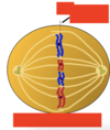
Metaphase
Metaphase Plate
Identify the Image
Anaphase
Identify the Image
Telophase
Cleavage Furrow
Identify the Image
Pachytene
Identify the Image
Prometaphase
Identify the Image
Prophase
Identify the Image
Telophase
Identify the Image
x-chromosomes
Zygotene
G1 Phase/ Gap 1
What is it? What is produced (4)? Length? Metabolically Active?
period of preparation for division
production of:
- Nucleotides
- Amino acids
- Enzyme systems
- Reserve Engergies
Long Phase
Metabolically Active
G2 Phase / Gap II Phase
What is it? What is being produced? Length?
Production of molecules necessary for mitotic development
- Spindle fiber protiens
- Microtubules
Fairly short
M Phase
what is it?
Separation of chromosomes followed by cell divison
Karyokinesis
Separation of chromosomes
The first stage of mitosis or meiosis
Cytokinesis
separation of the cell cytoplasm
resulting cells may be equal or unequal in size
Which phases comprise interphase?
G1-G2
During G1-G2 phases, can you see chromosomes?
No
G0
A special resting phase that terminally differentiated cells enter
Divisions are halted
Prophase (4 primary events)
Chromosomes condense,
nucleolus,
disappears,
centrioles replicate
Prometaphase (3 major events)
Nuclear envelop fragments
Microtubules bind to kinetochores
Non-kinetochore microtubules overlap
Metaphase (1 major event)
Mitotic spindle organizes and directs chromosomes to equatorial plate
Anaphase (2 events)
Chromatids separate
Chromatids are pulled to opposite ends of cell
Telophase (4 events)
Nuclear envelope reforms
Chromosomes uncoil and become indistinct
Nucleoli reappear
Cytoplasm divides
What happens during S period?
Chromosomes replicate again and prepare for the next division
The two daughter cells are mitosis are diploid or haploid?
Diploid
What are the stages of mitosis?
- Prophase
- Prometaphase
- Metaphase
- Anaphase
- Telophase
What are the stages of Meiosis?
Prophase I
Metaphase I
Anaphase I
Telophase I
Prophase II
Metaphase II
Anaphase II
Telophase II
Names have been assigned to distinguish the changing chromosomal states during prophase I stage. What are they?
- Leptotene
- Zygotene
- Packytene
- Diplotene
- Diakinesis
Leptotene - distinguishing features
Chromosomes have replicated forming sister chromatids
Cell in enlarged
The X chromosome is condensed and seen as a dark oval on one side of the chromosomal mass
Zygotene (synaptene)
Begins as sister chromatids start forming synapses with their homologous pair (tetrad)
Tetrad
Homologous pair of sister chromatids
Pachytene
starts at the conclusion of synapses formation
Some crossing over may happen at this stage
When does crossing over occur?
In the Pachytene and Diplotene stages of Prophase I
Diplotene
Homologous pairs start to separate (de-synapses) but remain attached at chiasmata
RNA synthesis is occurring