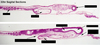
Lab Practical II Flashcards
Identify the Image
down the right and to the left

Early Head fold
Early Neural fold
Primitive Knot
Area Opaca
Area Pellucida
Primitive Streak
Identify the Image
Down the left, down the right, two on the far right

Head fold
Head mesenchyme
Neural fold
Area pellucida
Primitive streak
Epidermal ectoderm
Cranial intestinal portal
Notochord
Intersomitic furrow
somites
segmental plate
blood islands
area opaca
Identify the image
down the right, bottom left

Cranial neuropore
Lateral margin of the foregut
Cranial Intestinal Portal
Notochord
Intersomitic furrow
Somites
Identify the image
down the left, down the right

telencephalon
diencephalon
mesencephalon
metencephalon
myelencephalon
cranial intestinal portal
neural tube
blood islands
area opaca vasculosa
area pellucida
cranial neuropore
optic cup
head mesenchyme
notochord
isthmus
ventricle
sinoatrial region
vitelline veins
intersomitic furrow
somites
primitive knot
primitive streak
Identify the Image
down the left, down the right

head fold of amnion
forebrain
mesencephalon
cranial intestinal portal
somite
telencephalon
optic vesicle
isthmus
ventricle
vitelline vein
neural tube
primitive knot
primitive streak
Identify the image
Down the left, down the right

Isthmus
metencephalon
mylencephalon
auditory vesicle
amniotic fold
body wall
mesencephalon
diencephalon
infundibulum
optic cup
lens placode
optic fissure
telencephalon
bulbus cordis
sinus venosus
mesonephros
spinal cord
intraembryonic coelom
primitive knot
Identify the image
down the left, down the right

down the left, down the right

Cranial neuropore
Notochord
primitive streak
head fold
subcephalic pocket underlying head fold
neural folds closing
primitive pit
Identify the image
top, down the left, right

Proamnion
Head process
Primitive knot
Primitive ridge
Primitive groove
Primitive pit
Identify the image
Down the left, down the right

Foregut
Notochord
Neural plate
Somite 1
Primitive ridge
Identify the image
down left, down the right

cranial intertial oportal
notochord
primitive streak
forebrain
optic vesicle
mesencephalon
somite
notochord
neural fold closing
primitive streak
Identify the image
left, middle

Notochord
isthmus
ventricle
sinoatrial region
cranial intertinal portal
telencephalon
optic cup
dencephalon
mesencephalon
metencephalon
mylencephalon
neural tube
somites
intersomitic furrow
primitive knot
primitive streak
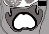
Identify the image
across the top, across the bottom

Blastodisc
Thin albumin
Thick albumin
Blastodisc
Thin albumin
Thick albumin
air space
chalaza
air space
Identify the image
top right, bottom left, bottom right

neural fold
cranial neruopore
endoderm
neural fold
neural groove
head mesechyme
subcephalic pocket
ectoderm
foregut
endoderm
Identify the image
top left, top right, bottom left, bottom right

neural tube
head mesenchyme
coelom
oral membrane
subcephalic pocket
future neural crest cells
foregut
proamnion
notochord
somatopleure
pericardial coelom
neural fold
splanchnopleure
anterior intestinal portal
Identify the image
across the top, across the bottom, across the top, across the bottom

ectoderm
lateral plate mesoerm
neural groove
neural fold
endoderm
notochord
somite mesoderm
ectoderm
open neural plate
mesoderm
endoderm
henson’s node
Identify the image
left, right

neural fold
region of neural plate
primitive knot
Identify the image

- telencephalon
- somatopleure
- subcephalic pocket
- splanchnoplesure
- optic visicle
- diencephalon
- proamnion
- internal carotid artery
- late diencephalon
- infundibulum
Identify the image

- late diencephalon
- oral membrane
- foregut
- mesenchephalon
- foregut
- notochord
- mesenchephalon
- proamnion
Identify the image

- pronephric cord
- floor plate
- spinal cord
- roof plate
- coelom
- notochord
- dorsal aorta
- neural groove
- neural folds
- notochord
- pronephric cord
- primitive groove
- primitive streak
Identify the image
left to right
Somite
Ventricle
Optic vesicle
cranial intestinal portal
sinoatrial region
neural tube
dienciphalon
subcephalic pocket
Identify the image

- amnion
- head mesenchyme
- mesencephalon
- yolk sac
- myelencephalon
- late diencephalon/early mesencephalon
- isthmus
- mesencephalon
- head mesenchyme
- myelencephalon
- per cardinal vein
- notochord
- infundibulum
- late dienchepalon / early mesencephalon
Identify the image

- roof plate of myelencephalon
- notochord
- dorsal aorta
- Rathke’s pouch
- infundibulum
- pigmented retina
- sensory tetina
- lens vesicle
- diencephalon
- amnion
- acousticofacialis ganglion
- pharynx
- myelencephalon
- auditory vesicle
Identify the image

- myelencephalon
- optic stalk
- diencephalon
- pharynx
- infundibulum
- nasal placode
- telencephalon
Identify the image

- bulbus cordis
- somite 1
- amniotic cavity
- spinal cord
- foregut
Identify the Image

- Sensory retina
- Pigmented retina
- lens vesicle
Identify the image
Leftmost, across the top and around

- neuromeres
- mesencephalon
- diencephalon
- telencephalon
- optic vessicle
- Subcephalon pocket
- metencephalon
- myelencephalon
Identify the image
top left, then down the right

notochord
infundibulum
optic stalk
oral plate
cranial intestinal portal
Identify the image

Sensory/Pigmented retina
lens vesicle
Identify the image
top then counter clockwise

lens vesicle
optic fissure
pigmented retina
sensory retina
opticoel (space)
Identify the Image

Neural plate
epidermal ectoderm, neural ectoderm
early neural fold
late neural fold
neural ridge, neural groove
neural tube
epidermal ectoderm, neural tube
Identify the Image
down middle, down right

Forebrain
Midbrain
Hindbrain
Telencephalon
Diencephalon
Mesencephalon
Metencephalon
Myelencephalon
Identify the image
Left top, right top, left bottom

Forebrain
Cerebral cortex
Midbrain
Hindbrain
Identify the image
a) top to bottom
b) top to bottom
c) top left then around clockwise

A) midbrain, hindbrain, forebrain
B) Mesencephalon
Metencephalon
Myelencephalon
Diencephalon
Telencephalon
C) Cerebrel hemisphere
Diencephalon
Midbrain
Pons
Medulla oblongata
Identify the image
top left, top right, bottom left to right

Lateral ventricles
Cerebral aqueduct
interventricular foramen
third ventricle
fourth ventricle
Identify the image

Identify the image
A-E

A. Telencephalon
B. Diencephalon
C. Mesencephalon
D. Metencephalon
E. Myelencephalon
What are the 12 cranial nerves?
- Olfactory
- Optic
- Oculomotor
- Trochlear
- Trigeminal
- Abducens
- Facial
- Vestibulocochlear
- Glassopharynx
- Vagus
- Accessory
- Hypoglossal
Oh oh oh to touch and feel a girls vagina, ah heaven
What are diencephalon derived structures? (5)
- thalamus
- optic nerve
- hypothalamus
- pituitary gland
- pineal gland
Identify the image
down the left, down the right

auditory vesicle
posterior of semi-circular canal
anterior of semi-circular canal
Identify the image
top, bottom

Auditory vesicle
Endolymphatic duct
Identify the image
left, right

gray matter
white matter
What does the blue indicate?

Myelencephalon

Identify the image
Nasal pits
Identify the image
down the left, down the right

amnion
myelencephalon
auditory vesicle
endolymphatic duct
metencephalon
isthmus
mesencephalon
fourth ventricle
cerebral aqueduct
Identify the image

Mylencephalon
superor ganglion
endolymphatic duct
cranial neves
cranial nerves
metencephalon
mesencephalon
cranial nerves
jugular ganglion
audiotory vesicle
semilunar ganglion
nueromeres
Identify the image
down left, down right

Cranial nerve
ganglion
mesencephalon
ganglion
myelencephalon
auditory vesicle
semilunar ganglion
cranial nerve
Identify the image
down left, down right

Roof plate
alar plate
basal plate
floor plate
cranial nerve
auditory vesicle
mesencephalon
Identify the image
down left, down right

Myelencephalon
brancial groove
mesencephalon
auditory vesicle
metencephalon
semilunar ganglion
Identify the image
down right, down left

Myelencephalon
branchial groove
infundibulum
diencephalon
Identify image
down left, down right

branchial groove
pharyngeal pouch
Rathke’s pouch
diencephalon
dorsal root ganglion
brancial groove
Identify the image
down left, down right

branchial groove
lens vesicle
optic cup
brancial groove
branchial groove
sensory retina
pigmented retina
Identify the image
down left, down right

Pharynx
optic stalk
dorsal root ganglion
branchial groove
stomodeum
optic fissure
Identify the image
down left, down right

pharynx
spinal chord
ventral root ganglion
pharyngeal pouch
stomoedeum
maxillary process
Identify the image
down left, down right

Pharynx
stomodeum
maxillar process
lateral lobes
spinal chord
telencephalon
Identify the image
down left, down right

telencephalon
pharynx
larynx
nasal pits
lateral lobes
Identify the image
top to bottom

sympathetic ganglia
nasal pits
Identify the image
top to bottom

internal organs
Identify the image
down the left, arcross the top (L to R), across bottom (R to L)

lens vesicle
sensory retina
pigmented retina
optic fissure
optic stalk
diencephalon
opticoel (space)
Identify the image
top, bottom

Metencephalon
Mesencephalon
Identify the image
bottom left, clockwise

Marginal zone
Intermediate zone
ventricular zone
sulcus limitans
roof plate
alar plate
central canal
basal plate
floot plate
Identify the image

gray matter
white matter
central canal
Identify the image
left most, top, right top, right lower, across bottom (L to R)

Dorsal root ganglia
roof plate
alar plate
basal plate
motor horn
floor plate
Identify the image
left to right

Marginal zone
intermediate zone
ventricular zone
