L11-12: Clinical reasoning in management of the shoulder Flashcards
What are 5 causes traumatic onset shoulder pain?

What are 3 categories of instability (& associated pathologies)?
- Traumatic unidirectional instability
- Typically anterior: Bankart lesion, bony Bankart lesion, Hill-Sach’s lesion
- Can be posterior: posterior labrum, posterior capsuloligamentous structures
- Most typical due to traumatic history
- Acquired instability due to overstress (sports-specific)
- Anterior capsular laxity
- Multidirectional instability
- Generalised ligament laxity
What are 5 managements for instability: acute traumatic injury for the shoulder?
- Hospital – reduction asap (easier)
- More muscle spasm the longer you leave it
- Ideally x-ray before reduction, otherwise after if not possible
- Usually stable unless in abd/ER
- Immobilisation:
- Traditional sling – IR position; Bankart lesion worsens by becoming detached from bone
- Can be quite practical –> able to protect joint, comfortable
- Would not allow healing of Bankart lesion (bring labrium away from glenoid)
- Position in 30o ER (pillow, brace) for 3/52 – reduces incidence of recurrent dislocation
- Not functional or feasible
- Systematic review: no difference in rate of recurrence between bracing in ER vs. IR
- Most important –> most comfortable and functional for patient
- Traditional sling – IR position; Bankart lesion worsens by becoming detached from bone
- Exercise therapy
- Pendular exercises –> passive gravity assisted (lean forward and swing arm around (use arm) –> prevent stiffness in joint
Why should physios not relocated the shoulder?
- Might fracture or dislocate fragment that has been fractured
- Neurovascular bundle (nerve praxia)
What is an athroscopy for repair of Bankart lesion for recurrent anterior dislocation?
- Repair of Bankart lesion (can reduce rate of redislocation to <5%)
- Bone graft
- Tighten capsule (e.g. stitches, heat shrink)
- Anterior capsule for more support and stability

What are 6 post-op protocol (example) for a Repair of Bankart lesion athroscopy for recurrent anterior dislocation?
- Sling 3-4 weeks
- Pendular movements after 24 hours
- Active ER once pain settles
- Scapulothoracic muscles & rotator cuff++
- Start active strengthening after 6 weeks
- More active and dynamic exercises
- Return to sports after 3-4 months
Timeframes are indicative only –> need also be adherent to exercise management
What are 6 post-op protocol (example) for a only Bankart lesion-Latarjet procedure athroscopy for recurrent anterior dislocation?
- Sling 3-4 weeks
- Pendular movements after 24 hours
- Active ER once pain settles
- Scapulothoracic muscles & rotator cuff++
- Start active strengthening after 6 weeks
- Return to sports after 3-4 months
What is an athroscopy for bony Bankart lesion- Latarjet procedure for recurrent anterior dislocation?
- If bony Bankart lesion - Latarjet procedure
- Move the horizontal part of the coracoid to the anterior inferior glenoid – bone graft
- Faster healing – bone vs. labrum- Due to blood supply of labrum
What are 3 characteristics of exercise therapy for posterior dislocation?
- Subscapularis is primary muscle preventing posterior translation
- BUT all rotator cuff muscles important (esp. transverse force couple)
- Less successful if posterior labral tear or traumatic event (16% success rate)

What are 4 characteristics of exercise therapy for posterior dislocation?
Surgery (anchor/suture repair of labral lesion)
- abduction brace (30o) for 4-6 weeks (Let the labrum heal) + passive pendulum & scaption exercises; avoid combined flex/IR (stretch capsule)
- IR and adduction restricted for 6 weeks
- Commence strengthening at 6 weeks
- Return to sport 4-6 months
What are 7 rehabilitation for traumatic unidirectional instability?
- scapular rehabilitation
- rotator cuff
- control/activation, then strengthening
- deficiencies in RC strength, accurate muscle activity and timing of activation
- force couple important for control
- closed chain → open chain
- can add stretching / manual therapy if needed (e.g. post-immobilisation)
- kinetic chain exercises

What are 3 categories of Long head of biceps (LHB) pathology?
- LHB inflammatory / degenerative conditions & partial tears
- Instability of LHB tendon in bicipital groove (injury to transverse humeral ligament)
- SLAP lesions
What are 3 physiotherapy treatments (which should be considered first) in biceps-related pathology?
- Similar return to sport as surgically-managed patients
- Phased progression of rotator cuff exercises, scapular exercises and stretching
- Take care with tensioning LHB – protect area first, gradual increase
What are 3 evidence for arthroscopy to repair SLAP lesions in biceps-related pathology?
- Good-excellent results in non-athletic patients
- 20-94% returned to previous level of sport
- RTS rates particularly low in specialist throwers (e.g. pitchers)
What are the 2 post-op rehab for arthroscopy to repair SLAP lesions in biceps-related pathology?
- No resisted biceps activity for first 8 weeks (protect healing of biceps anchor)
- No aggressive strengthening of biceps for 12 weeks
What ae the 16 Graded exercise program (increasing biceps EMG)?
- IR in 90o abduction
- Prone extension
- Knee push up plus
- Seated rowing
- IR in 20o abduction
- IR diagonal
- ER in 20o abduction
- Serratus punch
- Forward flexion in side lying
- ER in 90o abduction
- ER diagonal
- Forearm supination
- Uppercut
- Full can
- Elbow flexion in forearm supination
- Forward flexion in ER and forearm supination

What are the clinical features for Tendinopathy VS Tear?
- Pain when sleeping (more in tear than tendinopathy)
- More weakness (in tear)
- But is not enough alone –> only can do imaging (ultrasound than MRI –> narrow tuning and is quite expensive)
- Many people still have tears and have no symptoms
- Won’t change management
What are 2 managements for rotator cuff pathology?
- Reduce symptoms
- Exercise therapy
What are 6 features of reducing symptoms in a rotator cuff pathology?
- Avoid aggravating activity
- Ice
- Numbs and lessens inflammatory
- No evidence to support NSAIDs, ultrasound, IFT, laser, magnetic field therapy or local massage
- Do not massage tendon –> will aggravate tendon = cause more pain
- Corticosteroid injection into subacromial space may provide short-term pain relief, but impairs long-term recovery for tendinopathy
- Spike of improvement and then drop off
- Isometric exercise…?
- Could help but no evidence
- MWM (e.g. for impingement, painful arc)
- Increasing subacrominal space -> Repositioning of HOH or inferior glides
What are 4 features of rotator cuff strengthening (exercise therapy) in a rotator cuff pathology?
- Elevation in the scapular plane: supraspinatus, infraspinatus, subscapularis
- External rotation: supraspinatus, infraspinatus, teres minor
- Internal rotation: subscapularis
- Horizontal abduction with ER (e.g. prone): infraspinatus

What are 2 features of eccentric rotator cuff training (exercise therapy) in a rotator cuff pathology?
- Evidence for symptomatic effects
- Functional (especially for throwing athletes)

What is the most common tendon to get impinged in the subacromial space?
Supraspinatus tendon for impingement- Most common as it runs through the subacromial space
What are 3 features of physiotherapy management in an older patient (>50 years); small, partial thickness tears for rotator uff pathology?
- Consider avoiding excessive load on rotator cuff
- Semi-closed chain exercises with increasing gravity impact and increasing resistance
- Do not deload rather control the load
- Eg. weight loading on ball
- Shoulder balance exercises in increasing elevation
- Semi-closed chain exercises with increasing gravity impact and increasing resistance
- Focus on aiming to restore function – findings of TDT may help guide treatment selection
- Consider poor relationship between imaging findings and symptoms
What is the management in a young patient full thickness tears for rotator cuff pathology?
Full thickness tears in younger patients may need surgical repair – greater risk of progression
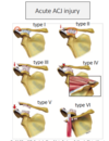
What are the 2 ACJ injuries?

What are 7 characteristics of an acute ACJ injury?
- Ligamentous injury management:
- Ice, immobilisation
- 2-3 days if type I
- 6 weeks if type II/III
- Isometric exercises when pain permits
- ACJ mobs as needed (pain, be careful if instability)
- For pain relieving and preventing stiffness
- Restore scapulohumeral rhythm, especially for higher grade injuries
- Return to function when full pain-free ROM and no local tenderness
- ACJ taping (comfort/confidence)
- Surgery:
- Type III failed physio; Type IV, V, VI

What are 6 characteristics of a chronic ACJ injury?
- Mobilisations either in neutral, add restricted movements (Can be more effective) (e.g. horizontal adduction, elevation)
- MWM
- Scapular retraining
- Strengthening
- Scapular and rotator cuff (if indicated)
- ACJ taping (deload)
- Corticosteroid + local anaesthetic injection – short-term pain relief
- Window to start rehab (if very painful)
- Do not do repeated injections

What are 10 causes of non-traumatic shoulder pain?

What is the rehabilitation for instability?

What is the rehabilitation for traumatic unidirectional instability?

What is the rehabilitation for acquired instability?

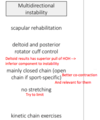
What is the rehabilitation for multidirectional instability?

What are the difference between the Rockwood and Watson program in instability rehab?

What is the Watson prgram compared to the Rockwood in instability rehab?

What is GIRD?
Pathological Glenohumeral IR Deficit
What are 3 characteristics of GIRD?
- Posterior shoulder stiffness – capsular tightness & muscular contraction
- Common adaptation in dominant limb of overhead athletes
- Can be associated with acquired instability
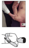
What are 3 ways to Increase IR ROM & ensure adequate scapular/humeral head control in GIRD?
- Sleeper stretch – 3x30secs, daily, 6 weeks: increases acromiohumeral distance
- Cross-body stretch
- Add hold-relax techniques to sleeper and cross-body stretch
- AP accessory glides in positions of increasing tension of posterior capsule (small advantage over sleeper stretch alone)
- MWM (e.g. posterior and inferior glides with movement)
- Taping to correct humeral head position
- Might not impart posterior glide yet -> need to check this can be done first
- Soft tissue techniques to posterior rotator cuff (e.g. dry needling, massage)
- Rotator cuff control/strengthening, esp if associated with acquired instability

What is Adhesive capsulitis?
Multiregional synovitis/inflammation; capsuloligamentous fibrosis & contracture
What are 4 phases of Adhesive capsulitis?
- Sharp pain at EOR, achy pain at rest, sleep disturbances
- Freezing – severe pain; early loss of ER is hallmark sign (DDx subacromial impingement)
- Frozen – pain & loss of ROM
- Thawing – resolving pain; significant persistent stiffness

What are 7 clinical practice guidelines: recommendations for adhesive capsulitis?
- Corticosteroid injections combined with shoulder mobility & stretching exercises are more effective than exercises alone (strong evidence)
- Condition of capsular inflammation –> that’s why injections can be effective compare to tears or tendinopathy
- Patient education should describe the natural course of the disease; promote activity modification to encourage functional pain-free ROM; and match the intensity of stretching to current level of irritability (moderate evidence)
- Stretching exercises (moderate evidence)
- Make sure to not irritate otherwise will cause muscle spasms which try to protect
- Shortwave diathermy, ultrasound or electrical stimulation combined with mobility and stretching exercises to improve pain and ROM (weak evidence)
- Heat deep into the joint (eg. possible benefit for OA compared to heat pack as can go deeper)
- GHJ mobilisations to reduce pain and increase ROM and function (weak evidence)
- Can try to improve symptoms (can work for some but not as a primary intervention)
- Manipulation under anaesthesia (weak evidence)
- Force joint into EOR while patient can’t feel (might possibly tear structures)
- hydrodilatation
- Inject saline into capsule to stretch it
- Dependent on stage of condition
- Conflicting evidence
What are 3 successful clinical practice guidelines: recommendations for adhesive capsulitis?
- Injections
- Patient education –> long term condition that will resolve but there is not quick fix
- Stretching
What is GHJ oesteoarthristis?
Degenerative arthritis of the GHJ; often secondary to trauma (acute or microtrauma) – damage to articular cartilage

What is a rotator cuff arthropathy?
- Degenerative arthritis of the GHJ that develops over time after RC damage
- Failure of RC force couple + unopposed pull of deltoid – upward translation HH
What are 5 techniques in the rotator cuff arthropathy?
- Manual therapy to increase subacromial space
- Esp. for rotator cuff arthropathy
- GHJ lateral glides
- MWM
- Open up joint space
- Rotator cuff control and strength (esp. if rotator cuff arthropathy)
- Scapular control
What are 4 typical deviations for scapular dyskinesis?
- lack of scapular upward rotation, posterior tilting & external rotation
- Scapula would be sitting in anterior, IR, protraction if they have dyskinesis
- increased clavicular elevation and retraction
- Scapular asymmetry at rest of during movement (abnormal scapulohumeral rhythm)
- Winging of the medial border (serratus anterior) or inferior angle (Lower trapezius)

What are 5 biomechanical mechanisms that can alter scapular kinematics in scapular dyskinesis?
- Pain
- Soft tissue tightness
- Muscle activation or strength imbalances
- Muscle fatigue
- Thoracic posture
What are 3 muscle impairments in scapular dyskinesis?
- Decreased strength of serratus anterior
- Results in winging of medial border of scapula
- Hyperactivity and early activation of upper trapezius
- Results in excessive elevation of the shoulder girdle during elevation
- Decreased activity and late activation of the middle and lower trapezius
- Results in winging of inferior border of scapula
What are 2 soft tissue findings in scapular dyskinesis?
- Tight pectoralis minor- Attachment to coracoid process
- Results in increased scapular internal rotation and increased anterior tilt
- Posterior GHJ capsular stiffness
What are the 3 managements for muscle impairments for scapular dyskinesis?

What are the 3 managements for soft tissue findings for scapular dyskinesis?

What is the Cools’ rehab algorithm for scapular duskinesis?

What are the origins and insertions for pect minor, levator scapulae, rhomboids, upper trapezius?
- Pectoralis minor… ribs & coracoid process
- Levator scapulae… Cx spine & supramedial scapula
- Rhomboids… Cx/Tx spine & medial border scapula
- Upper trapezius… occiput/lig nuchae/C7 & lateral clavicle
What are the 2 types of stretches for pec minor for scapular dyskinesis?

What is the pec minor release for scapular dyskinesis?

c
- AP (Humeral head will pull anteriorly) or PA (Can stretch it (anterior portion) when pushed) accessory glides… what grade would you choose?
- Combine with increasing abduction, ER, horizontal abduction
Capsule is one whole structure

What are 4 posterior capsule techniques for scapular dyskinesis?
- GHJ AP at a point in range; with restricted movement (IR, horizontal adduction, flexion); in HBB; EOR elevation (target posteroinferior capsule)
- MWM (AP glide + active movement in restricted direction)
- Sleeper stretch
- Horizontal adduction stretch

What are the 3 features of Stage 1: Conscious muscle control for Cools’ rehab algorithm for scapular dyskinesis?
- Improve proprioception
- Normalise scapular resting position
- Important to facilitate correct timing and control of muscle recruitment before commencing high-intensity strength training – avoid reinforcing poor kinematics

What are 4 characteristics of scapular orinetation ex. for scapular dyskinesis?
- Patient palpates coracoid process with contralateral hand
- Asked to pull the coracoid away from their finger, moving the scapula backwards
- Can teach consistent reproduction of scapular posterior tilt and upward rotation
- Increase in scapular muscle activity

What are the 4 features of Stage 2: muscle control & strength for daily activities for Cools’ rehab algorithm for scapular dyskinesis?
- Train scapular co-contraction in basic positions, movements and exercises
- Activate key scapular stabilising muscles without putting high demands on the shoulder joint
- Use open & closed chain exercises – If closed chain proprioception; rotator cuff cocontraction
- Exercises with ER component tend to improve scapular muscle recruitment
- Address strength deficits
- Aim for selective activation of weaker muscles, with minimal activity of overactive muscles
- Exercises with low UT/LT, UT/MT ratios (early activation of LT)
- Side lying ER
- Side lying forward flexion
- Prone horizontal abduction with ER
- Prone extension
- SA exercises (low UT/SA ratio):
- Elbow push-up (rather than push up plus – reduce demand on shoulder)
- Dynamic hug- Theraband around back, shoulders like a towel
- Supine punch
- Wall slides- Avoid lumbar lordosis when shoulder overhead

What do exercises for scapular dyskinesis?

What are the 5 features of Stage 3: advanced control during sports/occupational movements for Cools’ rehab algorithm for scapular dyskinesis?
- Integrate kinetic chain
- Plyometrics
- Pushup on a ball (some protraction, retraction) –> maintain control of scaoula
- Eccentric exercises
- Scapular control should be automatic
- Kinetic chain integration

What is 1 feature of Stage 3: advanced control during sports/occupational movements for throwing athletes for Cools’ rehab algorithm for scapular dyskinesis?
Eccentric loading of ERs (weighted balls, theraband)
What are 3 features of Stage 3: advanced control during sports/occupational movements forswimmers for Cools’ rehab algorithm for scapular dyskinesis?
- Train in prone or supine
- Integrate core control
- High repetitions
Scapular dyskinesis often related to_____ or _____ postures
thoracic kyphosis; flexed thoracic
- Control into extension coupled with rotation
What are 4 features of scapular dyskinesis for management?
- Correction of posture into extension
- Mobilisation into extension and rotation
- Core control and muscle retraining of trunk muscles
- Taping – e.g. thoracic spine and scapula into extension, posterior tilt and retraction
____ occurs in many patients with shoulder pain, injury and pathology
Scapular dyskinesis
What are 3 specific impairments that should be identified in assessment?
- lack of soft tissue flexibility?
- lack of muscle performance?
- combination
What are the only 2 baseline prognostic factors had a consistent association with outcome in two or more studies?
- Longer duration fo shoulder pain
- Poorer baseline function
It is important to intervene ____ with interventions aimed at improving _____ to optimising _____ .
early ; function; prognosis
Don’t forget _____ factors, and how they may impact on your treatment …fear avoidance, kinesiophobia, self-efficacy, anxiety, depression and treat the _____ , not just the shoulder
psychosocial ; whole person


