Knee and Thigh Flashcards
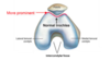
1
Q

label this femur

A
2
Q

describe the condyles of the distal femur and what they articualte with

A

the medial is larger than the lateral because it bears more weight in the standing position, since the centre of mass passes medial to the knee joint

lateral condyle is more prominent

the inferior and posterior condyles articualte with the menisci of the knee and tibia

3
Q

whats this groove called adn what does it articulate with

A

patellofemoral groove or trochlear goove

articualtes with the patella

4
Q

whats the function and feature of the lateral condyle

A

it is more prominent and prevent slateral displacement of the patella during patellar tracking.

5
Q

people with a more (less prominent) lateral condyle are more likely to experience what

A

patellar instability

lateral displacement

6
Q

describe the epiconyles

A

they orginate arebone bony elevations above the non-articular ares of the condyles

medial epicondyle is larger than the lateral one

they are attatchment centres for collateral ligaements

7
Q

what are the collateral ligaments ?

O and I ?

which is stronger

A

medial collateral ligament

  • O= medial epicondyle of the femur
  • I= medial condyle of the tibia

lateral collateral ligaement

  • O= lateral epicondyle of the femur
  • I= depression on the lateral surface of the fibular head

MCL isbroader butwaker than LCL, which in itself is weak when working in asolation but strong when working together with the arcuate ligament and popliteal tendon

8
Q

functionof the collateral ligaments

A

MCL= resists lateral (valgus) angulation of the tibia on the femur

LCL = resists varus angulation of the tibia on the femur

9
Q

which collateral ligament is stronger

A

LCL but it is itself weak when in isolation but strong when it works with the arcuate ligaemtn and popliteal tendon

10
Q

describe the patella

A

sits within the trochlear groove (patellofemoral groove) of femur

superiorly articulates with the quadriceps tendon

inferiorly articulates with the patellar ligament

  • the apex connects with the tibial tuberosity by patellar ligament
    • whilst the base forms superior aspect of the bone and provides the insertion area for the quadricpes tendon
  • 2 facets;
    • medial and lateral
11
Q

describe the facets of the patella

A

both the posteriorsurface of the patella articulate with the femur

  • medial ; articulates with the medial condyle
    • lateral ; articulate with lateral condyle
12
Q

functions of the patella

A
  • enable the quadriceps muscle to directly cross the anterior aspect of knee as it acts a fulcrum, the patella enhances the leverage that the quadricprs tendon an xert on the femur, increasing the mechanical efficacy of muscle by 30-55%
13
Q

whats the tibia adn describe it?

A

the shin bone

articulates with the knee and ankle joints, second largest bone in the body

proximal tibia is widened by medial and lateral condyles wc help with weith bearing

14
Q

label this

A
15
Q

whats special about the condyles of tibia

A

they forma flat surface known as tibial plateau wc articualtes with femoral condyles to form the major articulation of the knee joint

between the condyles is the intercondylar area and in the centre of that is teh intercondylar eminence

16
Q

describe the fibula

A

head of fibula articualtes withthe proximal tibia in the tibiofibular joint and doesnt form the knee joint

distal end widens to assits with weigth bearing

medial mallelous is bony projection infeirorly on medial aspect of tibia and articualtes ith tarsal bones to form part of the ankle joint,

lateraly theres a notch called the finular notch, where the fibular is bound to the tibia forming the inferior tibiofibular joint

17
Q

whats the intercondylar space

A

space between the two tibular condyles

at the centre is the intercondylar eminence and on either side of this emence is the lateral and medial intercondylar tubercules

these lateral and medial intercondylar tubercules of the tibia articulate with the intercondylar fossa of the femur

18
Q

whats special about the intercndylar eminence

A

attacthment for the cruiate ligaments nd menisci of the knee joint

19
Q

describe the attatchment for the cruiate ligaments

A

the posterior cruiate ligaments attatches from the posterior edge of the intercondylar area

20
Q

describe the tibia shaft

A

3 borders; anterior posterior and lateral

anterior

  • palpable subcutaneously down anterior aspect of leg
  • marked by tibial tiberosity - wc is the site of insertion of the patellar ligament

posterior

  • marked by ridge known as soleal line, wc is origin of soleus muscle. this line extends inferomedial and blends in with rdge of medial edge of tibia

lateral

  • aka interosseous boader and gives attatchemnt to the interosseous membrane that binds the tibia and the fibula together
21
Q

describe the distal fibular

A

tibia widesn to assist with weigth bearing

medial condyle extends inferiorly to form the medial malleous wc articulats with the tarsal bones to form the ankle joint

laterally theres a fibular nothc wc articulates where fibula is bound to tibia and forms the inferior tibiofibular joint

22
Q

describe the fibula

A

localated on the lateral aspect of the lef adn acts as a mucle attatchment

3 main articualtion :

distal tibiofibular jont (arituclates with teh tibia at the fibular notch

proxial tibiofibular joint (articulates witht eh alteral condyle of the tibia

ankle joint (articltes with the talus bones of the foot

23
Q

whats special about nerves and the fibula

A

the common peroneal (common fibular) nerve winds around the posterior and lateralneck of the fibular and so is vulnerable to damage in proximal fibular fracture

24
Q

descrube the boarders of the fibula

A

laeral

anteiror and postierior

25
describe the knee joint, what type of joint is it and wht movements does it permit? what are the articulation surfaces?
hinge joint allowing for flexionad extension with a small degree of medial and lateral rotation patealla, femur and tibia (fibula isnt)
26
what are the names for the articulation surfaces of the knee
tibiofemoral (*lateral and medial condyles of the femur artiualte with the medial and lateral condyles of the tibia)* and patellofemoral (patella articulates with the femur at the *patellofemoral/trochlear groove)*
27
describe the joint surfaces
lined with hyaline and enclosed in a single joint cavity
28
whats the function of the tibiofemoral joint
weightbearing joint of the knee
29
what tendon from the thighcompartment is associated with the knee joint and its funtion
* quadriceps femoris tendon (main exnsor of the muscle groupof knee) inserts directly over the knee increasing the efficiency of the muscle * the patella acts as a fulcrum to increase the power of the knee extesnion * it also reuces the frictional fores laces on the femoral condyle by the quardicpe tendon contracting * also provides protection from physical trauma ver anteiror knee
30
whats teh b supply to knee joint
genicular anastomes aroudn the knee wc are supplied the genciular branchs of the popliteal adn femoral arteries
31
what is the clinical relevance of the genicular branches
if popliteal artery is occluded by an atheroma, the geinuclar anastomes can dilate to maintain the b suply around the knee
32
33
what are he main b vessels that supply the muscles around the knee joint
common peroneal (common fibular) femoral popliteal tibial
34
how stable is the knee joint
inherently unstable toimporve stability the tibial articualr surface is deepened by the menisci and the joint is supported by the hoint capsule, ligaments and surrounding musclature
35
menisci describe it and its function
* fibrocartilaginous structures in the knee * lateral and medial * they are crescentric (cshaped) thicker peripherally than centrally and attach at both ends to the intercondylar area of the tibia f**ucntion:** * is to act as a shock absorbant by increasing surface area to further dissipate forces * deepen the articualr surface of the tibia, increasing the stability of the joint
36
whats different in the attatchements of the lateral and medial mensici and which is larger
the medial mensic is connected to the intercondylar medially, and peripherally the medial (tibial) collateral ligament and thejoint casule the medial mensci is larger the lateral meniscus is smaller and isnt attatched to the lateral collateral ligaemnt , rendering it fairly mobile
37
are the menisci connected to each other
yes anteriorly via a transvere ligament of knee
38
describe the attachments of the menisci
anteirorly they are connected to each other via transverse ligament of the knee posterioly they are attachemed bia a posterior meniscodemoralligament wc helps stabilise the posterior horn of the lateral meniscus by attatching ti to the medial femoral condyle peripheral rims are loosely attatched to the joint capsules and the tibia bia coronary ligametns
39
what are the main ligaments of the knee joint
ligaments that strengthenthe capsule = oblique poplitral liagament intracapsular = cruiate ligaments extracapsular = collaeral and patellar
40
describe the intracapsulare ligaemnts of the knee
cruiate ligaments connectfemur to the tibia and cross each other when doing so ACL = attatches proximally to the posterolateral aspect of the femoral intercondyle fossa and passes diagnoally adn attatches to the anteromedial aspect of the intercondylar eminence of the tibia where it also adheres to the medial meniscus PCL attatches proximally from the medial border and roof of the intecondylar fsaa abd passes diagnaooly to attatch to the posterior intercondylar area of tibia posterolaterally
41
ACL vs PCL
ACL = posterolateral interco.fossa of the femur to the medial anteromedial aspect of the int.emmience do the tibia + also adheres to the medial meniscus . F = resist translation and medial rotaton of the tibia in relation the femur PCL = medial border of the roof of the int. fossa od the femur posterolaterally to the posteror interco. area of the tibia F = stabaliser of the weightbearing knee and prevents the femur from sliding off anterior edge of the tibia when walking down the stairs, adn prevents posterior dislocation of the tibia and fmemur
42
PCL or ACL inserts medially? laterally?
PCL medial ACL lateral
43
44
45
what aspect of the joint capusel is strong?
posteiror and sides are strong but anteriorly its weak this deficency allows thesynovail membreane to extend up beneath the patella forming the suprapatellar burse larally and medially it is strengthened by the inferior fibres of the vastus lateralis adn vastus medialuis respectively posteriorly supported by the oblique popliteal ligament this is a combination of semitendinous tendon in a superolateral direction from its main insterion at teh medial tibial condyle, posterly actoss to the back of knee to te lateral femoral condyle
46
bursaa of the knee
suprapatella wc is located anteriorly and moves superiorly due tot he weak anterior aspect of the joint capsule it is locaed ewteen the quadrcipes femoris muscle and femur prepatellar burda - found bewteen the anteior surfae of the pateela and skin superfiial (subcutanoues) infrapatella bursa = between patellar ligaemnt adn skin deep infrapateallar bursa = between the tibia adn aptalla ligament semimembranous bursa = posterir to the knee joint bw the semimembranous meusce adn medial head of the gastronemius subsartorial bursa = betwen insertion of the pes ansernius tendon adn medial tibial condyle
47
bursitis what is it and the most common ones
* inflammatio of the bursa * most common are the : * prepatella (patellar and skin) * infrapateallae (deep is pat. tendon and medial tibia/ superficial is pat. tendon and skin) * subartorial (pes anerinus medial tibia) * suprapatealla (quadricep femoris tendon and femur) *
48
pre patellar bursitis what is it? fluid? aka?
between the skin and patellar minimmal fluid involved patient presents with knee paiin and swelling, finds it difficult to walk and cant kneel on the affected side this is due ot a history fo repetitive trauma of the knee like scrubbing floors and so it terms the housemaid's knee
49
infrapatellar bursitis
consits of two burst the deep adn superfical wc is skin adn patealla tendon or tibia and the patellar tendon mostly its the suprefical one thats affects microtrauma of kneeling
50
suprapatellar burtisis
bewteen the quadricep femoris and the patelalr is its a sign of knee pathology like: * gout and **pseudogout** * **osteoarthitis** * **RA** * **repetitivte trauma to the** joint
51
semimembranous bursitsi
posterior askect bw deep fascia of teh popliteal fossa in the interval bw the sem. musceladn medial head of the gastronimus when inflamed fluidforces itself inot the sem. bursa dn rsults in swelling of the poplital fossa aka its semimembranous bursitis and also know as popliteal cyst or baker's cyst
52
OSD
osgod-schlatter's disease inflammation of the apophysis (sit of inserition of th epateallar ligament) into the tibial tuberosity teens playign sport get it , bilateral in 20-30% of cases paieitns present with pain when exercisn , trartment is ice and seling and pain goes down with age adn sketela maturity as teh epiphyseal plates fuse, however the bony prominence remains froever
53
OA if the knee
knee stiffnes pai and swelling and crepitus (bone churning sound) pateint can have deforimtis like varus deformit (agulated inwards towards medial line) or valgus deformity 152% po affected 35% \>75 years treatment is exercsiifn t build the muscle and widhliss and alagesa
54
septic arthiris of the knee
invasion of the joint capsule by microorhanislu s like bacetria mostly , ut usually birsys or myocobacerim or fungi. different from reactive arthitis wc is sterle infalmmation that resits from extrarticualr infection like gastroenteritis 50% pf joint cases are septic arhtitis followed by hip20% most common pathogen is stap aerusys, risk factors age, diabetes, RA, immunospuression IV drugs, prosthetic legs, dental suegwry polymethycrylate cement is used to imphibit WBC and comonnet functio wc incraes risk of ifection maindamage these micororganisms cause is damag to catilage due to neutophils being strimulates and causes hydrolysis of proteilygancs and collagen when they are releaseign cytokines and inflammtoryproduct if suspected joint aspoitionshouldbe done asap
55
fractures of femoral shaft
due to high velocity trauma in kids and yougn afults like cara ccisan elderl due to osetoeporosis or bone metasis or boen elsion the sucualte acts as a deforming force afer the fmoral shaft fract, proximal femur adbcuted due to the oull of the gluteus medius and minimus on the greaer trochanter adn flexed due to the actions of the iliopsoas on thelesser trochanter distal is adducted due t the adductor muscles (ad. magus and gracilus) wc c varus deformity and extedne d due to the pull of the gastrocnemiuson posterior femur pateins present with swollen thifh,b lossin closedfemoral shfat fraionis 1-1.5L wc pts them in hypovolemic shock d
56
distal femoral fractures
ypung pateints dye to high enrgy spors and eldery d to osteip. or elerly falling from standing popliteal aertrt may becoe inlved f ehres eruocascular status of the limb before ad after reduction of the fracture is essetial
57
tbial plateua fratures
hgih enrgy injiry adn usualu axial pressyre with vaus or varus angles they affcet the articualting surafcae of the surface fo the tibian within the knee joint can be unicondylar or i, lateral tibila condyle most common articualr cartilage is alwasy famaed adn so most patioents develop post fractuer OA
58
what are the movements of nee
exte = quadricep femoris (rectus dfemorus, vastus med, avastus late abd vastus intermedius) felex = hamstrings assisted by gra, sartouis, popli, plantaris and gastrn later ro = bicep femoris medial ro = semi m sem t , fracilus ,, sartoius and popli
59
when does roationof the knee happen
when flexed
60
why does the patelala displace laterally dueing etenrion
anfe of line of pull of the quad muscle and pateallat ligament is the q angle things that resit the ateral displacemt = deep trochlar groove fibres of the vmo wc instert into pateall distally abd more hostilaly than thse of the vasus later
61
what stabilsies the posteiror aspect of knee joint
politeus iliotibia tract hamstring gastroneium
62
ant compartmetn of knee neerve? b? msucels?
femoral nerve femoral artery the alteral adn medial femroal circumflex arteirs and profundus brchi muscles are ; pectinus , sartorius and quadricep femoris (iliopsoas)
63
iliopsoas
madeup of psoas major and iliacus, 2 muscle with differne in but same fucntion adinsetion P O= T12-L5 laeral margins I= lesser trhocanter F = flex lowerllimb adn hip joint adn aleteral rotaton fo the femur at hipjoit IN =aneiror rami of L£ I O= iliac fossa of pelvis I = lesser troch F = lateral rotatio fo the femur at hip joint adn flex of lower limb a the hip joint IN= femoral nerve I = lesser trochanter F =
64
quadricep femoris
onsits fo 4 muscles wc have same tendonof insetion * rectus femrois * vastus intermedius * vastus lateralis * vastus medialis *
65
vastus alteralis
O = greater trochanter I = tendon q base of patealla F =ectend knee at knee joint ands tabilis the aptalla IN= femoral nerve
66
vastus medialis
O= anterior and lateral surface of femoral haft I= base of patealla into q. tendon F = entend knee adn knee joint IN= femoral
67
vastus intermedius
O]= itnterotrochanteric ine of femur and medial lip od linea spera I = bas eof patealla into q tendon inferior fibres are oreintented hsriszonatlyabd are knoen as vatus mdial obliquus VMO F = contractio of the VMO prevents lateral displacemtne of the aptealla and its F is to exten the knee at the knee joint IN = femoral nerve
68
rectus femoris
O = 2 tendons anterior from the AIIP and posterior from the groove above rimof the acetabulum , theu unite I = base of patealla via q tendon F = ened leg at knee join and flex thigh at the hip joint only muscle that crosses bth the hip and knee jint IN = femoral
69
satoorid
longest muscle of th ebonyd O = ASIS I =medial aspectasepc tof proximal tibia as part ofthe pers aneurinus (alongside gracilis adn semitendo) F= flex ad, abducts and externally roates thethigha t hip joint and can fle interally roate the tibia at knee jointlike asailor IN = femoral nerve
70
pectinus
flat muscleforms base of femroal triangle wc can have dual innervation , transition muscle bewttwht the anterior thigh ad medial thigh comaprtment O =oetial line on anterior surfce of the superior pubic ramus I =pectineal line of the psoterio aspec tofhe femur inferior tot the lesser trochanter F = adducts and flxes the thigh at the hip joint IN= femoral nerve but it can recieve a branch from the obturator nerve
71
medial compartmetn of the thigh nerve ? b ? msucel
hip adductios gracilus, obturator externus, adductor lingus and addutor magnus in = obtruator berve wc comes from L2 3 4 arterty obturator artery
72
aductor magns
largeest muscle in medial cpartet 2 parts to it adductor comaprotnet and hasmtring compaortemtn