Iris Flashcards

1
Q

What is the iris?

A

a thin, heavily pigmented, contractile circular disc analogous to the diaphragm of a camera

2
Q

Where is the anatomical location of the iris?

A

suspended in the frontal or coronal plane anterior to the lens and ciliary body

3
Q

What substance surrounds the iris?

A

aqueous humour

4
Q

What structures are separated by the iris?

A

anterior and posterior chambers

5
Q

What communication connects the anterior and posterior chambers of the eye?

A

the pupil - opening in the iris

6
Q

What is the anatomical location of the pupil?

A

lies slightly inferonasal to the centre of the iris

7
Q

What are the boundaries of the iris?

A
  • it is attached by its root at the angle (iridocorneal) of the anterior chamber where it merges with the ciliary body and trabecular meshwork
  • the free edge is known as the pupillary margin
8
Q

What is the diameter of the iris?

A

12mm

9
Q

What is the circumference of the iris?

A

37mm

10
Q

What is the general shape of the iris?

A

cone shaped with the pupil margin positioned more anteriorly than the root

11
Q

Where does the pupil margin rest?

A

on the lens

12
Q

What would happen if the pupil margin did not rest on the lens e.g. in aphakic patients?

A

the iris would become tremulous (iridonesis)

13
Q

What is meant by aphakia?

A

without a lens

14
Q

What is the function of the pupil and what is this function dependent on?

A

size of the pupil regulates the amount of light entering the eye

this is dependent on the state of contraction of the intrinsic pupillary muscles, the dilator and sphincter pupillae

15
Q

What are the 2 key types of muscles involved in regulating the size of the pupil and therefore the amount of light entering the eye?

A

the intrinsic pupillary muscles: dilator and sphincter pupillae

16
Q

By how much may the pupil diameter vary?

A

1 to 8 mm

17
Q

Is a slight degree of asymmetry between pupil size normal between individuals?

A

yes - may be slight degree of asymmetry between right and left

18
Q

What 2 parts of the iris are thin?

A

pupil margin

iris root

19
Q

What is a consequence of the pupil margin and iris root being thin parts of the iris?

A

they are more susceptible to tearing in contusion injuries (iridodialysis)

20
Q

What is the definition of iridodialysis?

A

separation of the iris from its attachment to the ciliary body, commonly occurs in penetrating or blunt trauma

21
Q

What are the two zones into which the anterior surface of the iris is divided?

A
  • pupil zone
  • ciliary zone
22
Q

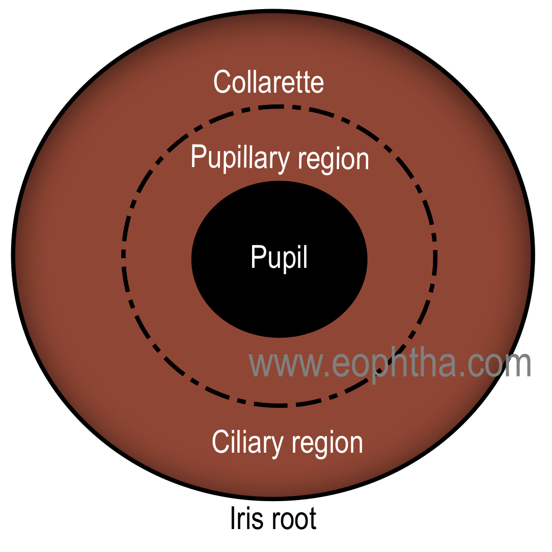
What structure divided the anterior iris into pupillary and ciliary zones?

A

the collarette, a thickened region

23
Q

What features characterise the anterior surface of the iris? 2 things

A
  1. radial streaks
  2. contraction furrows
24
Q

How do radial streaks on the anterior surface of the iris differ when the pupil is contracted vs dilated?

A

radial streaks are straight when pupil is contracted

wavy when pupil is dilated

25
When are contraction furrows on the anterior surface of the iris more noticeable?
in dilated irides
26
How does the visible appeares of dark irides vs blue irides differ? What causes this?
the surface of the iris appears smooth in dark irides in which the intrastromal melanocytes are heavily pigmented and more irregular in blue irides which have a less heavily pigmented stroma
27
What causes the blue appearances of some irides?
abosprtion of long wavelengths and reflectance of shorter wavelenghts, especially by the collagenous stroma
28
What are the 4 microscopic layers of the iris?
1. anterior border layer 2. stroma 3. dilator pupillae muscle 4. posterior pigment epithelium
29
What is the anterior border layer made up of?
modified stroma consisting of a dense collection of fibroblasts, melanocytes and a few interspersed collagen fibres (not epithelial cells)
30
What is the consequence of the anterior border layer of the iris being deficient in some areas?
the iris stroma is in free communication with the aqueous humour in the anterior chamber
31
What are large deficiencies in the anterior border layer of the iris macroscopically evident as?
crypts
32
What forms macroscopic naevi in the iris?
aggregates of heavily pigmented melanocytes in the anterior border layer of the iris
33
Why are melanocytes in the uveal tract unlike skin melanocytes?
in the uveal tract, melanocytes reside in connective tissue and **do** ***not*** **constantly shed melanin into adjacent cells** (the basis of skin pigmentation) and iris colour is a reflection of both the density of melanocytes, their location in the iris and their melanin content
34
How does iris colour relate to melanocytes in the anterior border layer?
iris colour is a reflection of both the **density** of melanocytes, their **location** in the iris and their **melanin** content
35
Label the following on the iris image: limbus, root, pupil, pigmented pupil margin, pupillary zone, collarete, collagenous trabecula, crypts.
36
Label the following on the diagram: radial folds, collarette, crypts, pupillary margins, melanocyte, blood vessel, stroma, anterior iris epithelium, dilator pupillae muscle, posterior iris pigment epithelium
37
Identify the following iris layers on the histological section of primate iris: * anterior border layer * stroma containing melanocytes * blood vessels * collagenous matrixp * posterior portion characterised by dilator pupillae muscle * posterior pigmented epithelium
38
What is shown in the electronic micrograph image?
iris stromal vessel surrounded by fibroblasts and melanocytes
39
What is shown in the image?
confocal microscopic image of iris and ciliary body (top of image) of mouse. shows nerves of iris as white and extensive network of tissue resident macrophages (red). Cx3cr1+ cells of myeloid origin are green
40
What is shown in the image?
ultrastructure of contractile myoepithelial portion of anterior iris epithelium, namely dilator pupillae muscle. shows cytoplasmic microfilaments and membranous densifications, both characteristic features of smooth msicle cells
41
What is shown in the image?
electronic micrograph of posterior iris epithelium - shows size, columnar shape and heavily pigmented nature of cells
42
What does the iris stroma layer consist of? 3 things within
Loose connective tissue containing 4 things: 1. fibroblasts 2. melanocytes 3. collagen fibres (types I and III)
43
What are the 2 types of ocllagen within the loose connective tissue of the iris stroma?
I and III
44
What allows fluid to move in and out of the iris stroma quickly during dilation and contraction?
loose nature of this tissue and its frree communication via openings in the anterior border layer
45
When does the iris stroma fluid move in and out quickly?
during dilation and contraction of the pupil
46
What are 2 types of immune cells in the human iris stroma and where are they found?
mast cells and macrophages many of which are perivascular
47
What is the nature of the macrophages in the iris stroma?
many are heavily pigmented and a subgroup may form large ovoid 'clump cells' of Koganei, which tend to accumulate near the iris root and sphincter pupillae muscle
48
What are clump cells (of Koganei)?
subgroup of **heavily pigmented macrophages** in iris stroma that accumulate near iris **root** and **sphincter pupillae** muscle
49
Where do clump cells tend to accumulate?
near iris root and sphincter pupillae muscle
50
What is the origin of the phagocytosed melanin in the iris stroma? 2 places
both from the ciliary / iris epithelium and stromal melanocytes
51
Where is the sphincter pupillae muscle located?
lying free within the stroma close to the pupil margin (see image)
52
What is the sphincter pupillae muscle?
circumferential ring of smooth muscle fibres about 1mm in width consists of muscle bundles eaching comprising 6-8 smooth muscle cells, which are continuous via gap junctions and surrounded by a basal lamina
53
What is the structure of the sphincter pupillae muscle?
consists of **muscle bundles** each comprising **6-8 smooth muscle cells**, which are continuous via gap junctions and surrounded by a basal lamina
54
What is the innervation of the sphincter pupillae muscle?
innervated by p**arasympathetic nerve fibres** derived from the **oculomotor** **nerve** (Edinger-Westphal nucleus houses preganglionic cell bodies whose fibres synapse in the ciliary ganglion from where postganglionic fibres travel via the short ciliary nerves) although **sympathetics** also terminate in this muscle
55
What is the course of parasympathetic nerve fibres of the oculomotor nerve which innervate the sphincter pupillae muscle?
Edinger-Westphal nucleus houses preganglionic cell bodies whose fibres synapse in ciliary ganglion from where postganglionic fibres travel via the short ciliary nerves
56
What is the unusual embryological origin of the sphincter pupillae muscle?
neuroectoderm
57
What is the dilator pupillae muscle?
layer of myoepithelial cells derived from the anterior iris epithelium
58
What are the myoepithelial cells of the dilator pupillae muscle derived from?
anterior iris epithelium
59
What is the structure of the iris epithelia?
two layers apposed to each other apex to apex with the gap between being theoretically continuous with the subretinal space
60
How thick are the basal processes of the anterior iris epithelium?
4 micrometres
61
How far do the processes of the anterior iris epithelium extend in the radial direction?
up to 50-60 micrometres
62
What are the characteristics of the apices of the myoepithelial cells of the anterior iris epithelium?
lightly pigmented, closely apposed to the apical aspect of the posterior pigment epithelium
63
What is the innervation of the dilator pupillae muscle?
non-myelinated sympathetic fibres whose cell bodies are situated in the superior cervical sympathetic ganglion parasympathetic innervation seems less significant
64
How far does the dilator pupillae muscle extend?
only as far centrally as the outer margin of the sphincter pupillae
65
What cells does the posterior pigment epithelium of the iris consist of?
large cuboidal epithelial cells (appear black macroscopically)
66
What is the embryological origin of the posterior layer of the posterior pigment epithelium?
derived from the inner neuroectoderm layer of the optic cup
67
How far does the posterior epithelium extend at the pupillary margin?
extends for a short distance on to the anterior iris surface
68
What is the effect of the posterior pigment epithelium of the iris extending onto the anterior iris surface at the pupillary margin?
forms the black ruff seen on the pupil margin during slit lamp examination
69
What are the 2 key macroscopic features created by the posterior pigmented epithelial layer of the iris?
1. radially arranged furrows (most evident near pupil margin) 2. circumferential contraction folds (most evident in the periphery)
70
What are the 2 key movements of the pupil?
1. mydriasis: dilation 2. miosis: contraction
71
In what conditions does mydriasis occur?
low light intensity and fear or excitement states
72
What muscle action brings about mydriasis?
action of the dilator pupillae muscle
73
What conditions cause miosis?
illuminated conditions, during convergence, and while sleeping
74
What muscle action brings about miosis?
sphincter pupillae muscle
75
Generally speaking, what is the blood supply of the iris like?
rich blood supply with extensive anastomoses
76
What is the overall arrangement of the arterial blood supply to the iris?
at iris **root** there is an incomplete **major ‘circle’** of the iris that is derived from anterior rami of the **anterior ciliary arteries** branches from here pass centripetally and form an incomplete **minor arterial 'circle**' at the level of the collarette
77
Where is the major arterial ring of the iris at the iris root derived from?
the anterior rami of the anterior ciliary arteries
78
What is the form of the arteries within the iris like and why?
unusual coiled form to accommodate the variable states of contraction of hte iris
79
Where are veins in relation to the arteries of the iris?
lies close to the arteries with larger veins primarily in the anterior stroma and smaller veins in the deeper layers
80
Where are large vs smaller veins found in the iris?
larger veins primarily in the anterior stroma smaller veins in deeper layers of the iris
81
Where do the veins of the iris drain to?
drain posteriorly/centrifugally into the ciliary body and eventually the vortex veins
82
What are 2 features of the iris capillaries?
1. tight junctions 2. non-fenestrated endothelial cells that have a high density of endocytotic vesicles and tight junctions
83
What are 2 key features of the non-fenestrated endothelial cells in iris capillaries?
* high density of endocytotic vesicles * high density of tight juntions
84
What is the relevance of the high density of tight junctions in the non-fenestrated endothelial cells of the iris capillaries?
less permeable to variety of solutes than normal somatic vessels
85
Why don't iris capillaries leak in fluorescein angiography?
due to being impermeable to variety of solutes due to high density of tight junctions in iris capillary endothelial cells
86
What overall phenomenon are the iris capillaries part of due to their properties and relative impermeability?
blood-ocular barrier
87
What are 2 features of the basal lamina of the endothelial cells in iris capillaries?
1. thickened (0.5 - 3 µm) 2. further strengthened by tube-like arrangements of perivascular collagenous/hyalinised layers
88
What is the function of the iris capillary endothelial basal lamina being thickened and strengthened?
helps preent collapse when the iris is fully dilated
89
Are there periarteriolar smooth muscle cells and elastic fibres in the iris?
periarteriolar smooth muscle cells - rare elastic fibres - absent
90
What is the arrangement of innervatino in the iris?
rich 3D nerve plexus of myelinated and non-myelinated nerves
91
What provides the **sensory** nerves to the iris?
branches of long and short ciliary nerves, which are branches of the nasociliary nerve (ophthalmic division of the trigeminal)
92
What provides the autonomic innervation of the iris muscles?
sphincter pupillae (pupil contraction) = **parasympathetic** nerve fibres dervied from **oculomotor** **nerve** (Edinger Westphal nucleus contains preganglionic cells bodies → synapse in ciliary ganglion → postganglionic fibres via short ciliary nerves) dilator pupillae (dilation) = non-myelinated **sympathetic** fibres, cell bodies in **superior cervical sympathetic ganglion**