Images2 Flashcards
Spinothalamic tract
(a) Synapses
(b) Decussations
Spinothalamic tract for pain/temp
(a) Synapses twices
- in dorsal horn
- at VPL of the thalamus
(b) Decussates across the anterior gray commisure 1-2 levels above where it enters the SC

Where in the brainstem do each of the cranial nerves sit?
CN I and II don’t go thru the brainstem
CN III, IV = midbrain
-midbrain stroke can cause 3rd nerve palsy
CN V, VI, VII, VIII = pons
=> brainstem glioma around the pons can present w/ 6th nerve palsy
CN XI, X, XI, XII = medulla

Describe the vascular territories of the following cross section

- note little sliver of ACA on top, then large chunk MCA b/c we’re deep here
- then inferiorly PCA

Pt w/ visual field deficit in the RUQ, locate the lesion (be specific)

Lesion = Optic radiations/Meyer’s loops = axonal connections btwn the lateral geniculate nucleus of the thalamus and the primary visual cortex of the occipital lobe
RUQ visual field deficit = optic radiations in the temporal lobe on the left
- superior quadrant affected 2/2 lesion of Meyer’s loops (temporal lobe)
- inferior quadrant 2/2 lesion to superior optic radiation (parietal lobe)

Locate the lesion

Left eye: mild ptosis w/ constricted pupil, also probably left sided anhidrosis = Horner’s syndrome 2/2 loss of sympathetic innervation to left side
Lesion of the superior cervical ganglion (houses the sympathetics)
-also can be 2/2 carotid artery dissection (or vertebral artery dissection = Wallenberg syndrome)
Expected MRA finding in a pt w/ 3rd nerve palsy
B/c of where the CN III exits the brainstem, right under the PCOMM => CNIII gets compressed by PCOMM aneurysm

Pt w/ the following MRI is likely to present w/ what visual complaint?

Pituitary pathology, possible etiologies adenoma, craniopharyngioma, etc
No matter what the etiology: getting compression of the optic nerve, causing bitemporal hemianopsia (loss of lateral fields of view)

Corticospinal tract
(a) Synapses
(b) Decussates
(b) Decussates at the medullary pryamids
(a) Synapses in the anteiror horn of the spinal gray matter before learing

Cuneate vs. gravile fasciculus
Both are dorsal column fibers
- cuneate fasciculus carries fibers from the arms (lateral): lateral b/c added on as the tract ascends
- gracile fasciculus carries fibers from the legs (medial)

Occlusion of the anterior spinal artery
Effect on each of the three spinal cord pathways
- weakness bilaterally (b/c hitting both lateral corticospinal tracts)
- loss of pain and temp bilaterally (b/c hitting both spinothalamic tracts)
- preservation of proprioception and vibration b/l (b/c dorsal columns not invovled

When pt looks to the right he cannot adduct his left eye, and there is abducting nysagmus of the right eye
-convergence preserved
Locate the lesion
Lesion of the medial longitudinal fasciculus (MLF) which connects CNIII and CNVI to allow for conjugate gaze
Conjugate gaze = simultaneous activation of medial and lateral rectus muscles to allow eyes to look in one direct simultaneously

Cavernous sinus
(a) Name 2 things in it besides cranial nerves
(b) What sits on top?
(b) Underneath?
Cavernous sinus
(a) Internal carotid artery and the pitutiary galnd
(b) Right on top sits the optic chiams
(c) Sinuses are right underneath

Differentiate the side effect by peripheral vs. central seventh nerve palsy
Central 7th = lesion of the motor pathway above the facial nucleus (supranuclear lesion)
- spares upper facial muscles, so you can move your forehead
- causes weakness of lower part of the phase CONTRALATERAL to the lesion
Peripheral 7th = lesion of the facial nucleus in the pons or the facial nerve itself
-weakness of the entire face IPSILATERAL to the lesion

Dorsal Column/Medial Lemniscus pathway
(a) Synapses
(b) Decussates
Dorsal columns for proprioception and fine touch
(a) Synapses twices
- in lower medulla just before decussating
- in VPL of thalamus
(b) Decussates in lower medulla forming the medial lemniscus throughout the brainstem

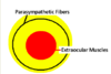
Initial sign of posterior communicating artery anurysm
Ipsilateral dilated pupil
-Compressive lesion (parasym fibers are on the outside) so parasymp fibers are lost before the EOM are affected
Pt p/w bitemporal hemianopsia and CT showing pitutiary mass w/ calcifications
(a) Dx
(b) Mechanism of visual complaints

(a) Dx = craniopharyngioma
- calcified on CT
(b) Compression on optic nerve

Describe Brown-Sequard syndrome
- weakness ipsilateral to the lesion
- loss of proprioception/vibration on same side of the lesion
- loss of pain/temp opposite the lesion

Describe finding’s of Bell’s palsy
Unilateral facial paralysis
- can’t close eye on affected side
- can’t smile (mouth drooped)
- smoothing of forehead (can’t raise eyebrown muscles)
- loss of nasolabial fold
Also may find: dry eyes, facial twitching, dry mouth, impaired taste

Pt p/w hearing difficulties in the left ear
- Weber’s: hears tuning fork louder in left ear
- when holding tuning fork next to each ear: hears better in the right ear
Dx
Dx = conductive hearing loss on the left
- air conduction normal on right => sounds louder when tuning fork just held next to ear
- Weber: if louder in affected ear = conductive hearing loss
What is the thalamic nuclei responsible for vision?
Lateral geniculate nucleus of the thalamus
= relay center for the visual pathway
-connects optic nerve to the occipital lobe by sending axons thru the optic radiations

What is the tract of Lissauer?
Gray matter tract of entering spinothalamic fibers
-right where the spinothalamic fibers enter

Pineal gland
(a) Function
(b) appearance on CT
(c) Most common pineal-region tumor
Pineal gland
(a) Secretes melatonin to regulate sleep/wake cycle
- stimulated by darkness, inhibited by light
(b) Physiologically calcified on CT
(c) Germinomas = most common pineal-region tumors
Pinealomas = primary pineal tumors, often clinically silent until large enough to compress vertical gaze center of the midbrain (=> vertical gaze palsy)

Diagnostic test for BPPV
Dix-Hallpike maneuver: basically move the pt (w/ head tilted at 45 degrees) from seated to supine and see if the nystagmus is in the same direction as gravity


