Images Flashcards
Pt can’t recognize coin or paper clip placed in her right hands if her eyes are closed, but has no trouble w/ left hand
Called asteroagnosia
(a) Locate the lesion
(a) Parietal lobe/primary somatosensory cortex

Differentiate the 3 types of MRI


Dx

FInding = adenoma sebaceum = facial angiofibromas in characteristic butterfly pattern = pathognomonic for tuberous sclerosis
MRI findings of hepatic encephalopathy
Hepatic encephalopathy (like alcoholic cirrhosis) => b/l, symmetric hyperintense lesions of the basal ganglia, most comonly in the globus pallidus

Dx

Hummingbird sign and Mickey Mouse sign both refer to midbrain atrophy seen in PSP (progressive supranuclear palsy)


Explain the concept of crossed findings seen in brainstem strokes
Crossed findings = ipsilateral CN findings (b/c CN don’t decussate) and contralateral sensory/motor findigns (corticospinal tract decussates caudally at medullary pyramids)
ex: lesion shown would give ipsilateral 3rd nerve palsy w/ contralateral motor/sensory deficits

MRI findings of pseudotumor cerebri
- slit like ventricles 2/2 compression by elevated ICP
- empty sella 2/2 pitutary flattening by elevated ICP
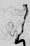
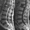


Name the vessels


Name ‘em


33 yo HIV+ F w/ low grade fever and HA x3 mo
- exam: cognitively slow, stiff neck
- LP shows 200 WBC
- CSF stain attached
(a) Dx
(b) Preferred Tx

(a) Cryptococcus neoformans = yeast that causes meningitis in immunocompromised, typically presents as lung infxn
(b) IV Amphotericin, then fluconazole

Name ‘em


Abduction vs. extension of the thumb
Adduction vs. flexion of the thumb


1 = anterior commisure
2 = lamina terminalis
3 = optic chiasm
4 = hypothalamic sulcus
5 = tuber cinereum
9 = infundibulum

Explain the vascular territories of the brain from the major arteries

What area of the brain is responsible for maintaining consciousness?

Reticular activating system (most influential component is the reticular formation): regulates wakefullness and sleep-wake transitions

P/w sudden inability to speak
After several days determine he can speak but only chooses to do so under extreme duress
(a) Dx
(b) Locate the lesion
(a) Akinetic mutism
(b) B/l cingulate gyrus = fold in brain superior to corpus callosum involved in emotiosn and regulation of aggressive behavior

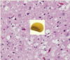
16 yo M p/w progressive muscle weakness, muscle biopsy below
Dx

Dx = mitochondrial myopathy
-biopsy: ‘ragged red’ muscle fibers = abnormal accumulations of mitochondria
Describe location of Wernicke’s area
Left superior temporal gyrus

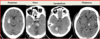
Describe the reversal sign on NCHCT and its indications
Reversal sign is when the cerebellum is brighter than the cortex parenchyma
-indicates no distinction btwn gray and white matter in the rest of the brain which is e/o global cerebral edema

45 yo F lost ability to recognize the face of her good friends
Locate the lesion
Prosopagnosia = inability to recognize known faces
Localizes to the fusiform gyrus of the temporal lobe- area for recognition
-color recognition, face recognition

Most common target for deep brain stimulation in Parkinson’s pts
Subthalamic nucleus (main part of the substantia nigra)
-can also target thalamus and globus pallidus

What do fibrillations and sharp waves indicate on EMG?

Fibrillations/positive sharp waves = spontaneous depolarization of individual fibers, not seen in normally innervated muscle which is resonsive as an entire motor unit
=> indicates acute (weeks to 12 mo) motor axon injury
Dx

Dx = Rabies
Negri bodies = pathognomonic inclusion bodies in the cytoplasm on Purkinjee cells containing the rabies virus
- especially in the Ammon’s horn of the hippocampus
- consist of ribonuclear proteins produced by the virus
Pt presents w/ ICH and this angiogram
(a) Dx
(b) Describe findings

ICH 2/2 AVM (arteriovenous malformation) = congenital abnormality in connection btwn arteries and veins
(b) Mass of blood vessels comprising an AVM
Etiology of this ICH

“popcorn” mass on imaging indicative of bleed into cavernoma = cavernous malformation = masses of abnormal vessels w/o any recognizable intervening neural tissue
Cause of watershed strokes
Watershed areas = farthest from direct perfusion of major cerebral arteries, frequently in overlap of ACA/MCA distribution
Etiologies: hypoperfusion of the brain from systemic hypotension, CHF, carotid stenosis

Name ‘em


Stroke in which large named artery would cause face/arm over leg weakness?
Face/arm worse than leg weakness (or more commonly) seen in MCA stroke
-while legs more commonly affected than face/arm in ACA strokes
2/2 the homonculus overlap w/ vascular territories
(obv both w/ contralateral findings, not ipsilateral)

Gerstmann’s Syndrome = lesion of dominant angular gyrus
(a) Location of angular gyrus
(b) Characteristic findings
(a) Angular gyrus = temporoparietnal jxn = region of the parietal near the superior edge of the temporal lobe
(b) Agraphia (can’t write), acalculia (can’t learn or comprehend math), finger angosia (can’t distinguish fingers on the hand), left-right confusion

Common locations for lacunar strokes
A- subcortical white matter
B- Basal ganglia/posterior limb of internal capsule
C- thalamus
D- pons
E- cerebellum
-Mirrors location of hemorrhagic strokes, given HTN is a major RF for both

Differentiate the arteries occluded in each of these 3 cerebellar strokes

Left pic = superior cerebellar
Middle = anterior inferior cerebellar artery
Right pic = posterior cerebellar artery infarct

Describe imaging findings of restricted diffusion
Hyperintensity on diffusion MRI w/ corresponding hypointensity on ADC map
-characteristic of ischemia, certain tumors, or active MS lesion (really ischemia tho)

Dx?

Dx = meningioma
Give away = dural tail: thickening of adjacent dura
-usually benign, slow growing masses

Name ‘em


Name 3 main Parkinson’s drugs and where they act at the dopaminergic synapse
- Levadopa works on presynaptic neuron to increase substrate
- COMT inhibitors work to increase half life of levadopa by inhibiting the enzyme that breaks it down
- Dopamine agonists work to activate the postsynaptic dopaminergic receptors

Primarily location of atrophy seen in Alzheimer’s
Mesial temporal lobe (hippocampi), then get diffuse atrophy over time

What is the goal of tPA
Save the penumbra! Tissue that is ischemic but not yet infarcted (lacking O2 but not yet dead)

MRI brain findings of Wilson’s disease
Abnormalities of the basal ganglion
-Wilson’s also called hepatolenticular degeneration, with the lenticular nucleus being the caudate and putamen

Describe the nuclei involved in lateral medullary syndrome

3 main features
- Loss of pain/temp in contralateral body (spinothalamic tract descending fibers) and ipsilateral face (spinal trigeminal tract)
- Horner’s (descending sympathetic tract) => anhidrosis, miosis, ptosis
- Other
- vestibular nuclei => nausea, vertigo, nystagmus
- Nucleus ambiggus (CN IX, X, XI) => dysphagia, dysarthria
- inferior cerebellar peduncle => gait/balance disturbance

Stroke of which territory?

MCA: see area of lateral sulcus
Dx (focus on pons/midbrain area)

‘hot cross buns’ sign of multiple system atrophy

15 yo boy w/ EEG: normal background w/ frequent generalized polyspike and wave discahrges diffusly throughout at 4-6 Hz
Dx?
Dx = Juvenile myoclonic epilepsy
-often confused as clumsy child, drops utensils


Characteristic angiographic finding of CNS vasculitis
CNS vasculitis presents w/ ‘beads on a string’
-esp PAN (polyarteritis nodosa) where the small aneurysms are strung like beads of a rosary

Describe location of Broca’s area

Pathologic findings of CJD
Spongiform degeneration of the gray matter due to neuronal loss

Differentiate the two pics

Infarct in superior vs. inferior division of the MCA
Superior division infarct => Broca’s (expressive) aphasia and prominent weakness
Inferior division infarct => Wernicke’s (receptive) aphasia and mild to no weakness

Name ‘em


Stroke of which territory?

PCA territory
- seeing effect on occipital lobe and inferior temporal lobe
12 yo boy found to have retinal angiomas on eye exam, then similar lesions found in his kidneys and pancreas
What is he at risk for developing in his brain?

Dx = Von-Hippau-Lindau syndrome = autosomal dominant 2/2 mutation in VHL tumor suppressor gene => formation of multiple hemangioblastomas
So at risk for hemangioblastomas of the brain
-usually neurologic symptoms are minimal unless a CNS hemangioblastoma ruptures

Wide-eyed stare and trouble looking down
Dx
‘Reptilian stare’ of progressive supranuclear palsy
Location of lesion when pt exhibits pure word deafness
Pure word deafness (can’t comprehend meaningful speech but can still hear sounds, speak, read, write) 2/2 lesion of b/l primary auditory cortex or 2/2 disruption of connection btwn the areas
Primary auditory cortex = posterior superior temporal lobes

Pt w/ sudden onset language difficulties. Mild word-finding problems, trouble naming objects, marked inability repeating phrases. Can follow simply commands
(a) Type of aphasia
(b) Locate the lesion
(a) Conduction aphasia
(b) Lesion in the arcuate fasciculus = deep white matter tract that connects Broca’s and Wernicke’s

Scan in pt s/p cardiac arrest
(a) Dx
(b) Etiology

(a) Dx = brain death
- see absolutely no intracranial blood flow
(b) Brain death 2/2 global cerebral anoxia 2/2 cardiac arrest
Name ‘em


Dx?

Imaging showing multiple meningiomas and multiple ependymomas
= NF2
NF 2 = multiple schwannomas (usually vestibular schwannoma), mulitple meningiomas, multiple ependymomas (brain or spinal cord)
-recall ependyma = epithelial lining of the ventricles
Name ‘em

Areas involved in language

MRI findings of Huntington’s
Caudate atrophy causing enlargement of anterior horn of the lateral ventricles
-ventricles just expand into the atrophic space

CT findings of Huntington’s chorea
Characteritic caudate nucleus atrophy seen on neuroimaging as enlargement of the lateral ventricles
Key: Huntington’s = atrophy of caudate nucleus

What is the most sensitive scan for acute ischemic stroke?

Diffusion-weight MRI
-local characteristics of water diffusion in the brain
5 mo old w/ seizures and this MRI
(a) Dx
(b) Other expected features

(a) Sturge-Weber Syndrome = seizures, MR, glaucoma, port-wine stain
- MRI: ipsilateral (to port-wine stain) leptomeningeal angioma that causes gradual calcification and atrophy of the underlying brain
(b) Port-wine stain 2/2 capillary overgrowth, usually in the V1 distribution
Explain alexia w/o agraphia and locate the lesion
(a) Infarction of which artery?
Alexia w/o agraphia = can write but cant read 2/2
(a) left posterior cerebral artery
- lesion of left occiptal lobe and the splenium of the corpus callosum => visual info only reaches the right occipital lobe, but not accessed by language areas of the brain => pt cannot read
- but language areas are otherwise fine => pt can write

3 clinical categories of TB in the CNS
- meningitis
- intracranial tuberculomas
- spinal tuberculosis arachnoiditis

Differentiate the territories of the 3 large named cerebral arteries
ACA: frontal lobe, superior sections
MCA: superior temporal lobe, basal ganglia stuff, region around lateral sulcus
PCA: parietal and occiptal lobe, inferior temporal lobe

EEG finding of absence seizures
3-Hz spike waves

Name the location of each lacunar stroke

A- subcortical white matter
B- cerebellum
C- thalamus
D- pons
E- internal capsule
F- basal ganglia
Locate the ICHs


67 yo M from rural china p/w rural back pain, weakness of legs, and incontinence
-MRI: pathologic fracture of T12 w/ herniation of disc material into spinal canal
Dx
Dx = Tb, Pott’s disease = infection of the vertebral bodies
- usually lower thoracic or upper lumbar
- infection spreads from two adjacent vertebrae into the adjoining intervertebral space

10 yo girl p/w seizures and confusion a week after mycoplasma pneumonia infection. MRI below
Dx?

Dx = acute disseminated encephalomyelitis
- acute autoimmune demyelinating disorder following viral infection (or atypical vaccination), p/w encephalopathy and siezure
- MRI showing white matter inflammatory lesions
Differentiate Kernig’s and Bruudzinski’s sign for bacterial meningitis
Kernig’s = pain when thigh bent at hip and knee at 90 degrees
Brudzinski’s = involuntary lifting of leg when head is lifted off the table

Mechanism of Kluver-Bucy syndrome
Damage to b/l amygdalae
-hypersexuality, hyperorality, lack of fear

Name ‘em


Location of berry aneurysms: anterior vs. posterior circulation
(a) Most common
Berry aneurysms- most common location is at the fork btwn the anterior communicating artery and the anterior cerebral artery
80% anterior circulation
20% posterior circulation

Name the vessels

