Images Flashcards
Which photo is diverticulosis and which is diverticulitis?

- diverticulosis = left
- diverticulitis = right
Which of the following V/Q scan studies demonstrates a PE?

Right image *right main branch PE*
What type of contrast is used in this study?

oral contrast/IV contrast
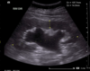
what study is this?

pelvic u/s
What type of malignancy is this?

liver cancer
What is the finding in this photo?

Volvulus *Coffee bean sign*
what is this finding on testicular u/s

hydrocele
What is the condition?

Cholelithiasis
What is the finding in this photo?

Appendicitis
What is this?

appendicitis
What is the finding in this photo?

small bowel obstruction
What liver condition is this?

acute hepatitis
What is the finding in this photo?

Large bowel obstruction
What is the finding in this photo?

Small bowel obstruction
What is this?

renal stones
what is this finding on testicular u/s

epididimytis
What is the finding in this photo?

Cholelithiasis
what is the finding on this pelvic u/s?

complex cyst
What is the condition?

Choledocholithiasis
What is the finding in this photo?

pancreatitis
What is the finding in this photo?

cholecystitis
what is this finding on testicular u/s

varicocele
What malignancy is this? What is this sign?

- Colon cancer
- “apple core” lesion
What is the finding in this photo?

normal KUB
What is the finding in this photo?

Small bowell obstruction
What is the finding in this photo?

foreign body
What is the problem?

polyps in gallbladder
what is this finding on testicular u/s

testicular torsion
what is this sign during the first trimester?

gestational sac
What is the finding in this photo?

colitis
What is this?

hydronephrosis d/t renal stone
What is the finding in this photo?

Hampton’s hump
What is the finding in this photo?

Saddle PE
What is the finding in this photo?

appendicitis *appendicolith*
What is the finding in this photo?

small bowel obstruction
What is the finding?

sludge
What is the finding in this photo?

AAA
What is this?
hydronephrosis
What is the condition?

cholecystitis
What is going on here?

trick question = normal transabdominal ultrasound
What type of malignancy is this?

right kidney cancer
What type of malignancy is this?

pancreatic cancer
What is the finding in this photo

right kidney stone + hydronephrosis
What is the finding in this photo?

Pneumoperitoneum
What is the finding in this photo?
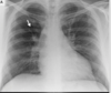
Westermark sign = PE
What is this sign?

fat stranding
What type of contrast is used in this study?

oral contrast
What is this sign?

contrast blush (hyperdensity representing active bleeding)
what is this

AAA
What is the finding in this photo?

Gall stones
What liver condition is this?

Hepatic Steatosis (Fatty liver)
what is this finding on testicular u/s

malignancy
What is the finding in this photo?

SBO *air fluid levels*
What is the finding in this photo?

appendicitis
What is this sign?

hyperemia (increased blood flow)
What is the finding in this photo?

Toxic megacolon
What type of contrast is used in this photo?

none!
Is this a cyst or free fluid?

cyst
Is this a cyst or free fluid?

free fluid
What is the recess where free fluid is accumulated called?

Morrison’s Pouch
This is an EFAST bladder u/s. Where is the fluid accumulated? Is this a male or female?

- accumulated in pouch of Douglas
- Female
This is a bladder u/s. Identify if this is male or female

- male
What is this sign called?

spine sign
