Histology Lecture 3b -- Salivary Glands Flashcards
From what are glands derived?
Ectoderm or endoderm layers
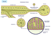
3 steps in the development of glands
- The epithelium invaginates into the underlying connective tissue, bringing the epithelial basal lamina with it
- The invagination develops a lumen that communicates to the original surface
- The invagination differentiates into a secretory unit as its furthest part and a duct that connects the secretory unit to the epithelium
Define exocrine glands
Product made by the secretory unit of the gland and is delivered by the duct to the surface from which the invagination occurred
Define endocrine glands
The duct loses its connection with the secretory unit and the unit secretes into the connective tissue around it and ultimately into the blood vessels (ductless glands)
3 classes of exocrine glands based on the shape of secretory unit
- Tubular
- Acinar
- Alveolar
Define tubular exocrine glands
Unifrom diameter for the secretory unit and the duct (i.e. sweat gland)
Define acinar exocrine glands
A grape-like secretory unit is attached to a duct (i.e. salivary glands)
Define alveolar exocrine glands
A flask-shaped secretory unit, but is not often used because it can be confused with an alveolus of the lung
2 classes of exocrine glands based on the relationship between secretory unit and duct
- Simple = one secretory unit to one duct (i.e. sweat gland)
- Compound = a branching tree-like system with secretory units at the ends of all the branches (i.e. salivary glands)
4 classes of glands based on the manner in which cells secrete
- Holocrine
- Merocrine
- Apocrine
- Cytocrine
Define holocrine glands
- Entire cell is the secretion product
- The gland cells become filled with the secretory substance and the cell dies and disintegrates as it forms the secretion product
- Example = sebaceous glands of the hair
Define merocrine glands
- Secretion based on production of membrane-bound secretory granules that are exocytosed or secreted to the outside of the cell
- No loss of cell material as membrane is derived from the Golgi apparatus and retrieved by fusion with cell membrane
- Secretory product is made de novo for export
- Example = all salivary glands
Define apocrine glands
- A small part of the cell cytoplasm is lost as part of the secretion
- Example = matrix vesicles from hypertrophic chondrocytes in the cartilaginous growth plate
Define cytocrine glands
- Part of one cell containing the secretory granule is phagocytosed by another cell
- Example = melanosomes produced by melanocytes phagocytosed by keratinocytes in the skin
3 classes of glands based on the type of secretion product
- Serous
- Mucous
- Mixed
Define serous glands
Producition of a watery secretion, usually glycoproteins that are enzymes
Define mucous glands
Production of a secretion that is high in carbohydrates (about 50% protein, 50% carbs)
Define mixed glands
Acinus is composed of both serous and mucous cells, or there is a mucous acinus with a serous demilune
Describe the arrangement of serous cells
Usually arranged as acinar secretory units. Their pyramidal shapes are arranged in a spherical unit with the apices of the cells meeting to form a small lumen
How may the luminal surface area of serous glands by increased?
Interncellular canalicules between serous cells
Describe the cell base of serous cells
- Basophilic
- Occupied by abundant parallel cisternae of rER
Describe the apex of serous cells
- Filled with eosinophilic zymogen granules
- Under certain conditions of poor fixation, the granules may be dissolved, leaving empty vacuoles
Describe the nuclei of serous cells
- Spherical and located towards the base of the cells, usually surrounded by rER
- Prominent nucleoli (high protein synthesis)
What drains the acini of serous glands?
Intercalated duct
How are mucous cells arranged?
- As acinar or tubular secretory units
- Cuboidal cells arranged around a large lumen
Describe the rim and base of mucous cells
- Rim of darkly stained cytoplasm rich in rER at the base
- Flattened dark-stained nuclei at base
Describe the apex of mucous cells
Filled with large mucinogen droplets or granules, whose proteins have a high sugar content
Why might it be hard to see the lumen of a mucous gland despite it being larger than a serous gland?
The secretory product of mucous glands, mucin, fills the large lumen when released and hydrated
How are mucous acini drained?
By intercalated or larger ducts
Describe the distribution of cells in mixed acini
Most often composed of a mucous acinus capped by a serous demilune (a cap of serous cells sitting on a mucous acinus)
What empties a mixed acinus?
The secretions enter a common duct, which may be intercalated
Describe what immediately surrounds all secretory units of glands and why
Since all secretory units are epithelially derived, they are surrounded by a basal lamina, which is continuous with the duct and ultimately the epithelium of origin
What lies between basal lamina and the base of secretory units in some acini?
Epithelial cells with contractile properties (myoepithelial cells) and therefore contain actin and myosin
3 divisions of duct systems (in sequence)
- Intercalated
- Striated
- Collecting
Smallest ducts
Intercalated ducts
Describe the histology of intercalated ducts
- Lined by simple squamous epithelium
- Very small lumen
- Difficut to see; only slightly bigger than capillaries
- Very regular structures with distinctive features
Diameter of striated ducts
Same as acini
Describe the histology of striated ducts (4)
- Regular and round in cross section
- Lateral borders of their columner or high cuboidal cells are indistinct due to cell interdigitation
- Round nuclei near the base
- Deeply eosinophilic cytoplasm that is striated due to extensive basal membrane infolding and elongated mitochondria
Function of striated ducts
Resorption of useful fluids and ions from the crude glandular secretion
Elements of the pumping mechanism in striated ducts
Extensive basal membrane infolding and elongated mitochondria
Largest ducts
Collecting ducts
Describe the histology of collecting ducts
- Lined by simple epithelium (cuboidal in small ducts; columnar in larger ducts)
- Largest duct joins with the epithelium (structure resembles epithelium here) –> secretions
What are glandular secretory units organized into?
Lobules
What are the lobules of glandular secretory units organized into?
Lobes
Define the capsule of a gland
A layer of dense connective tissue covering the outer aspect of the gland
How are glands divided into lobes and lobules?
Capsular connective tissue that continues into the gland
Function of the capsular connective tissue of the glands
Carries the blood supply to the glandular units, but also carries the duct system
Define intralobular structures of the glands and ducts
Structures located within the lobules:
- Acini
- Intercalated ducts
- Striated ducts
Define interlobular structures of the glands and ducts
Structures leaving the lobules to join to similar structures, such as striated ducts, which being intralobular non-striated ducts that leave the lobules immediately to join with similar ducts –> interlobular
Differentiate between intralobular and interlobular collecting ducts
Interlobular collecting ducts are larger
Differentiate between ducts outside and within a lobule
Outside = surrounded by much connective tissue
WIthin = little or no connective tissue around them
How are lobes of a gland drained?
Large interlobular ducts join to become large collecting ducts that drain the groups of lobules which make up a lobe of gland
Define minor salivary glands
Small compound glands located throughout the oral cavity beneath the epithelium
3 major salivary glands
- Parotid gland
- Submandibular gland
- Sublingual gland
Largest salivary gland
Parotid gland
Location of parotid gland
Well encapsulated gland located in the side of the face
What type of gland is the parotid gland?
Pure serous gland
What is located between the serous acini of the parotid gland?
Deposits of unilocular adipocytes
Describe the duct system of the parotid gland
Includes intercalated, striated and collecting ducts
Location of opening of the main duct of the parotid gland
Inside of the cheek, just oppostie to the upper 2nd molar tooth
Name of the main duct of the parotid gland
Stenson’s duct
Salivary gland with the greatest output
Submandibular gland
2 well-encapsulated major salivary glands
Parotid gland
Submandibular gland
Composition of the cells of the submandibular gland
90% serous acini
10% mucous acini
Describe the distribution of the acini in the submandibular gland
Mucous acini occur in small patches distributed throughout the gland. Some may be mixed or show serous demilunes
Location of the opening of the duct from the submandibular gland
Under the tongue
Name of the duct of the submandibular gland
Wharton’s duct
Location of the sublingual gland
Either side of the midline of the tongue in the floor of the mouth
Major salivary gland that is poorly encapsulated
Sublingual gland
Describe the composition of the sublingual gland
Mixed acini; mainly mucous with serous demilunes
Name of duct that empties the sublingual gland
Rivinus’ ducts
Where do Rivinus’ ducts open?
At numerous sites along the connection of the tongue to the floor of the mouth
Define xerostomia
Dry-mouth; ack of saliva
Consequences of xerostomia
- Difficulties in swallowing, tasting and speaking
- Can increase the chance of dental decay and other infections of the mouth
What can cause xerostomia
Certain medications (over 400) and medical treatments
Particular medications that may cause xerostomia
Those used for high blood pressure and depression treatments
Medical treatments that may cause xerostomia
Radiation therapy and chemotherapy
Diseases/pathologies that can affect salivary glands (5)
- Sjogren’s Syndrome
- HIV/AIDS
- Diabetes
- Parkinson’s disease
- Nerve damage
Symptoms related to xerostomia
Itchy, scratchy eyes from decreased tears
Define Sjogren’s disease
An autoimmune disease, where immune defenses destroy acinar salivary gland cells
Define sialoliths
Stone formation in the salivary glands and their duct system
Where can mineralized sialoliths occur most frequently
In the submandibular gland and its duct system (but also in other salivary glands and ducts)
Typical contents of sialoliths
Calcium-phostphate (hydroxyapatite) mineral and incorporated proteins
Consequence of sialoliths
If they do not pass through the duct system, blockage of ducts requires their surgical removal