Hand Flashcards

1
Q

What is the blood supply to the lunate?

A
  • Y-pattern
  • X-pattern
  • I-pattern -31% of patients
    • postulated to be at the highest risk for avascular necrosis
2
Q

Diagnosis? Etiology?

A

Keinbochs

  • Not well defined, multifactoria
  • biomechanical factors
    • ulnar negative variance
      • leads to increased radial-lunate contact stress
    • repetitive trauma
  • anatomic factors
    • geometry of lunate
    • vascular supply to lunate
      • “I” pattern
    • disruption of venous outflow
  • Patient factors - presumptive but not proven
    • Sickle cell
    • Steroids
    • Septic emboli
    • Gout
    • Carpal coalition
3
Q

Diagnosis? Classification

A

Keinboch’s - Litchman Classification

  • Stage I
    • No visible changes on xray, changes seen on MRI
      • Immobilization
      • NSAIDS
  • Stage II
    • Sclerosis of lunate
      • Ulnar negative - ulnar shortening
      • Ulnar positive - Radial wedge, STT fusion
      • Distal radius core decompression
      • Revascularization procedures
  • Stage IIIA
    • Lunate collapse, no scaphoid rotation
      • Same as Stage II above
  • Stage IIIB
    • Lunate collapse, fixed scaphoid rotation
      • Proximal row carpectomy
      • STT fusion
  • Stage IV
    • Degenerated adjacent intercarpal joints
      • Wrist fusion
      • proximal row carpectomy
      • limited intercarpal fusion
4
Q

Imaging to help define collapse of lunate

A
  • AP, lateral, oblique views of wrist
    • findings (see table above)
    • Can help define collapse and adjacent scelorsis
  • CT
    • most useful once lunate collapse has already occurred
    • best for showing
      • extent of necrosis
      • trabecular destruction
      • lunate geometry
  • MRI
    • best for diagnosing early disease
    • findings
      • decreased T1 signal intensity
      • reduced vascularity of lunate
5
Q

options in the treament

A
  • immobilization, NSAIDS
    • initial management for Stage 1 disease
    • Immobilize for up to 3 months
    • Outcomes
      • a majority of these patients will undergo further degeneration
      • BUT worth is to try because there are a small number of patient who will improve
  • temporary scaphotrapeziotrapezoidal pinning
    • adolescent with radiographic evidence of Kienbock’s and progressive wrist pain
  • Ulnar Negative Variance - Stage I, II, IIIa
    • joint leveling procedure
      • Stage I, II, IIIA disease with ulnar negative variance
      • technique
        • can be radial shortening osteotomy or ulnar lengthening
        • Goal is to have 1mm ulanr postive variance
        • Otherwise you risk ulnocarpal abutment
      • Outcomes
        • Changing ulnar variance by 4mm can offload lunate 45%
  • Ulnar positive variance - Stage I, II, IIIa
    • radial wedge osteotomy
    • Capitate shortening +/- hamate fusion
    • vascularized bone grafts
      • 2nd dorsal intermetacarpal artery is commonly used
      • early results promising, but long-term data lacking
      • greatest success in Stage II patients
    • distal radius core decompression
      • creates a local vascular healing response
  • Stage IIIb disease
    • Capitate lengthening with lunate excision
    • STT fusion
      • must address internal collapse pattern (DISI)
    • proximal row carpectomy (PRC)
      • some studies have shown superior results of STT fusion over PRC for stage IIIB disease
      • Others show no difference in outcome, grip strength, pain
  • Stage IV disease
    • Proximal row Carpectomy
      • Benefits
        • Preserved motion
        • Can do a wrist fusion later
      • Resect 1cm of the posterior interosseous nerve (which innervates the joint capsule in the floor of the 4th extensor compartment)
    • Contraindications
      • Severe arthritis of the head of the capitate
    • wrist fusion
      • must remove arthritic part of joint
    • total wrist arthroplasty
      • long-term results not available
6
Q

What are the components of the TFCC

A
  • dorsal and volar radioulnar ligaments
    • deep ligaments known as ligamentum subcruatum
  • central articular disc
  • meniscus homolog
  • ulno-lunate; ulno-triquetral (UCL)
  • ECU subsheath
  • origin of ulnolunate and ulnotriquetral ligaments
7
Q

What are the primary stabilizers of the DRUJ

A
  • Primary
    • volar radioulnar ligaments
      • volar translation
      • supination
    • dorsal radioulnar ligaments
      • dorsal translation
      • pronation
    • trigangular fibrocartilage
  • Secondary
    • ulnar head
    • sigmoid notch
    • interosseous membrane
    • pronator quadratus
8
Q

Presentation of TFCC tears

A
  • History
    • ulnar sided wrist pain
      • turning a door key often painful
    • Get a full and complete history of pain, may be associated with instability
    • Swelling
    • History of trauma
  • Physical exam
    • Examine for areas of fullness or swelling
    • ROM and instability
      • Assess stability in neutral, sup/pro
    • Hypersupnation will stress the DRUJ
    • Provacative test
      • Ulnocarpal stress test
        • Ulnar deviation, axial load, extension
      • positive “fovea” sign
        • tenderness in the soft spot between the ulnar styloid and flexor carpi ulnaris tendon, between the volar surface of the ulnar head and the pisiform
        • 95% sensitivity and 87% specificity for foveal disruptions of TFCC or ulnotriquetral ligament injuries
      • pain elicited with ulnar deviation (TFCC compression) or radial deviation (TFCC tension)
9
Q

What is the most sensitive test for TFCC tear

A

foveal sign

  • tenderness in the soft spot between the ulnar styloid and flexor carpi ulnaris tendon, between the volar surface of the ulnar head and the pisiform
  • 95% sensitivity and 87% specificity for foveal disruptions of TFCC or ulnotriquetral ligament injuries
10
Q

Palmar Classficiation of TFCC tears

A
  • Class 1 - Traumatic TFCC Injuries
    • 1A - Central perforation or tear
    • 1B - Ulnar avulsion (without ulnar styloid fx)
    • 1C - Volar avulsion (origin of UL, UT, UC ligaments)
      • usually results in dislocation
    • 1D - Radial avulsion
      • only time you might try to fix this is in a young patient
  • Class 2 - Degenerative TFCC Injuries
    • 2A - TFCC wear and thinning
    • 2B - Lunate and/or ulnar chondromalacia + 2A
    • 2C - TFCC perforation + 2B
    • 2D - Ligament disruption + 2C
    • 2E - Ulnocarpal and DRUJ arthritis + 2D
11
Q

How is the load distribution in the wrist affected by ulnar variance?

A
  • +2 mm ulnar variance approximately
    • 40% of the load goes to the ulna
    • 60% to the radius
  • normal neutral wrist approximately
    • 20%: ulna
    • 80%: radius
  • -2 ulnar variance
    • 5%: ulna
    • 95% radius
12
Q

What conditions are associated with the pathological finding of this XR? What would you expect to find on physical exam?

A

Ulnar Positive Variance

  • Load 60% radius, 40% ulna
  • Associated conditions
    • ulnar abutment syndrome
    • Scapho-lunate instability
    • TFCC tears
    • arthrosis
      • ulnar head
      • lunate
      • triquetrum
    • lunotriquetral ligament tears
  • Physical Exam
    • ulnar sided wrist pain from increased impact stress on the lunate and triquetrum

13
Q

What conditions are associated with the pathological finding of this XR?

A

Ulnar Negative Variance

  • Kienbock’s disease
  • ulnar impingement syndrome
    • ulna impinges on the radius proximal to the sigmoid notch
14
Q

How does position of the forearm affect ulnar variance?

A
  • Ulnar variance increases in pronation
  • decreases in supination
  • ulnar variane increases during grip

A pronated grip view is the best to determine your ulnar varience

15
Q

Pathophysiology of altered ulnar variance

A
  • congenital
    • Madelung deformity (positive UV)
    • reverse Madelung deformity (negative UV)
  • trauma/mechanical
    • distal radius/ulnar fracture with shortening
    • growth arrest (previous Salter-Harris fracture)
    • DRUJ injuries (Galeazzi and Essex-Lopresti)
  • iatrogenic
    • joint leveling procedures (radial or ulnar shortening/lengthening)
    • radial head resection (positive UV)
16
Q

Landmarks for wrist scope portals

A
  • Radiocarpar Portals
    • 3-4
      • Located just distal to Lister tubercle, between EPL and EDC
      • Established first, primary viewing portal
    • 4-5
      • Located in line with ring finger metacarpal, between EDC and EDM;
      • Portal for instrumentation, visualization of TFCC
    • 6R
      • Located just radial to ECU tendon;
      • Primary adjunct for visualization and instrumentation, ulnar-sided TFCC repairs
      • Dorsal sensory branch of ulnar nerve
    • 6U
      • Located just ulnar to ECU tendon;
      • Primary adjuct for visualization and instrumentation, ulnar-sided TFCC repairs
      • Dorsal sensory branch of ulnar nerve
    • 1-2
      • Located between APL and ECRB, along dorsal aspect of snuffbox;
      • Not often utilized, provides access to radial styloid and radial aspect of joint, sometimes used for inflow
      • Superficial branch of radial nerve; Radial artery
  • Midcarpal Portals
    • MCR
      • Located 1 cm distal to 3-4 portal along axis of radial border of middle finger metacarpal, between ECRB and EDC.
      • Allows visualization of scapholunate, scaphocapitate, and scaphotrapezoid joints.
    • MCU
      • Located 1 cm distal to 4-5 portal along axis of ring finger metacarpal, between
      • Allows visualization of lunocapitate, lunotriquetral, and triquetrohamate joints.
    • STT
      • Located along axis of index finger metacarpal just ulnar to EPL at level of STT joint
      • Allows visualization of scaphotrapezial and scaphotrapezoid joints.
  • First CMC Joint
    • 1U
      • Located on ulnar aspect of EPL at level of first CMC joint (basal joint)
      • Allows diagnosis of DJD of first CMC joint and arthroscopic debridement.
      • Superficial sensory branch of radial nerve
    • 1R
      • Located on radial aspect of EPL at level of thumb CMC joint, just volar to APL tendon
      • Allows diagnosis of DJD of first CMC joint and arthroscopic debridement.
      • Superficial sensory branch of radial nerve
17
Q

Indications for wrist arthroscopy

A
  • TFCC injuries
  • interosseous ligament injuries
  • anatomic reduction assistance (distal radius, scaphoid fxs)
  • ulnocarpal impaction
  • debridement of chondral lesions
  • removal of loose bodies
  • synovectomy
  • excision of dorsal wrist ganglia
  • assistance in treatment of SNAC and or SLAC wrist
  • septic wrist irrigation and debridement
  • diagnosis in unexplained mechanical wrist pain
18
Q

Complications associated with wrist arthroscopy

A

overall complication rate is 1-2%

  • Dorsal sensory branch of ulnar nerve
    • averages 8mm from 6R portal
    • at risk with establishment of 6U and 6R portals
      • to a lesser extent main ulnar nerve and artery also at risk
    • when performing a TFCC repair, small open incision is typically made prior to knot tying to prevent injury to this nerve.
  • Superficial sensory branch of radial nerve
    • averages 16mm from 3-4 portal
    • at risk during arthroscopy of basal joint, as 1U and 1R portals are on either side of the first branch of this nerve
    • at risk during placement of 1-2 portal
  • Radial artery Injury
    • associated with establishment 1-2 portal, used for arthroscopic radial styloidectomy.
  • Extensor tendon injury
    • most commonly EPL and EDM due to improper portal placement
  • Chondral injuries
    • iatrogenic from scope or instrument placement
  • Portal site infection
  • Stiffness
  • MCPJ pain
    • typically caused by over-distraction
19
Q

What are the common wrist extensor tendons that get injured during wrist athroscopy

A

most commonly EPL and EDM due to improper portal placement

20
Q

What portals do you need to be most concerned about neurovascular injury?

A
  • Dorsal sensory branch of ulnar nerve
    • averages 8mm from 6R portal
    • at risk with establishment of 6U and 6R portals
      • to a lesser extent main ulnar nerve and artery also at risk
    • when performing a TFCC repair, small open incision is typically made prior to knot tying to prevent injury to this nerve.
  • Superficial sensory branch of radial nerve
    • averages 16mm from 3-4 portal
    • at risk during arthroscopy of basal joint, as 1U and 1R portals are on either side of the first branch of this nerve
    • at risk during placement of 1-2 portal
  • Radial artery Injury
    • associated with establishment 1-2 portal, used for arthroscopic radial styloidectomy.
21
Q

Differential for DRUJ pain

A

DRUJ instability or arthritis
TFCC tear
LT ligament tear
pisotriquetral arthritis
ECU tendonitis or instability
Ulnocarpal impaction

22
Q

History and physical for a patient with ulnar sided wrist pain?

A
  • Differential for DRUJ Pain
    • DRUJ instability or arthritis
    • TFCC tear
    • LT ligament tear
    • pisotriquetral arthritis
    • ECU tendonitis or instability
    • Ulnocarpal impaction
  • History
    • Associations with unlocarpal abutment
      • pain on dorsal side of DRUJ
      • increased pain with ulnar deviation of wrist
      • pain with axial loading
      • ulna sided wrist pain
    • Painful snapping or instability
    • History of trauma or instability; gymnastics or repetative loading as a child
      • madelungs, distal radius malunion, TFCC
    • PMHx - RA, JRA
  • Physical exam
    • Examine
      • Assess the wrist for ulnar prominence
      • Irritation over the DRUJ
    • Full ROM
      • Especially sup/pro
        • DRUJ strained in hypersupnation
      • Assess for ECU subluxation
      • Assess for crepitus (OA)
    • Ballottement test
      • dorsal and palmar displacement of ulna with wrist in ulnar deviation
      • positive test produces pain
    • Ulnar stress test
      • ulnar deviation of pronated wrist while axially loading, flexing and extending the wrist
      • positive test produces pain
    • Fovea test
      • used to evaluate for TFCC tear or ulnotriquetral ligament tear (most sensitive for TFCC pathology)
      • palpation of the ulnar wrist between the styloid and FCU tendon to
23
Q

Radiographic work-up for ulnar sided wrist pain

A
  • Radiographs
    • AP radiograph with wrist in neutral supination/pronation and zero rotation
      • required to evaluate ulnar variance
      • Assess for congruence of the joint
      • Evidence of OA or ulnocarpal impaction
      • Assess for space between the proximal row carpi
    • pronated grip view
      • increases radiographic impaction
    • Findings assoicated with ulnocarpal abutment
      • ulna positive variance
      • sclerosis of lunate and ulnar head
  • Arthography
    • joint injection shows extravasation
    • can help determine
  • MRI
    • Important to determine presence of TFCC tear
      • Helps to determien procedure
    • has largely replaced arthrography
    • tear at ulnar part of lunate indicates ulnocarpal impaction
      • sensitivity = 74-100%
    • Not felt to be a good assessement tool for TFCC tear
      • Accuracy 55%
  • Arthroscopy
    • most accurate method of diagnosis
    • indicated in symptomatic patients after failing several months of splinting and activity modification
    • Benefit is that you can treat the pathology at the time of diagnosis
    • Components of the DRUJ you can see with a scope
      • Triangular fibrocartilage
      • Volar extrinsic ligaments
      • Components you can only see with a tear
        • Radial attachment of radial-ulnar ligaments
          • Can test indirectly with ballottement of the DRUJ
        • Ulnar attachement of the RU ligaments
24
Q

Patient with ulnar sided wrist pain. Young. Failed non-op. Options for treatment?

A

Ulnocarpal Abutment

  • Nonoperative
    • indications
      • may attempt supportive measures as first line of treatment
    • ECU strengthening, NSAID, Splint
  • Arthroscopy
    • any procedure can be combined with a diagnostic arthroscopy for further treatment determination, or to address isolated TFCC pathology
  • Ulnar shortening osteotomy
    • indications
      • most cases of ulnar positive variance
      • most cases of DRUJ incongruity
  • Wafer procedure
    • technique
      • 2 to 4mm of cartilage and bone removed from under TFCC arthroscopically
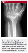
  • Darrach procedure (ulnar head resection)
    • indications
      • reserved for lower demand patients
    • technique
      • 40 deg oblique cut just proximal to the DRUJ
      • Try to preserve the R-U ligaments, TFCC and periosteium, ECU sheath
      • Some will leave the ulnar styloid to maintain staibilty
  • Sauvé-Kapandji procedure
    • indications
      • good option for manual laborers
      • Can be used in patients with a torn TFCC
    • technique
      • Dorsal-ulnar approach
      • Resect the distal ulna 10-15mm from the joint
      • Stabilize the DRUJ with k-wires and 3.5mm cannulated screws
      • creates a distal radioulnar fusion and a ulnar psuedoarthrosis proximal to the fusion site through which rotation can occur
      • ECU/FCU can be tenodesed in the ulnar stump for additional stability (to help reduce issues with post-op residual instability)
        • Proximal ECU slip
        • Distal FCU slip
        • Woven to create a stable stump
  • Ulnar hemiresection arthroplasty (HIT)
    • indications
      • usually requires an intact TFCC
      • appropriate treatment option in the presence of post-traumatic DRUJ with concomitant distal ulnar degenerative changes
  • ulnar head replacement
    • indications
      • Severe OA
      • salvage for failed Darrach
      • Isolated instability
      • Occasionally after trauma
    • Options
      • Partial ulnar head arthroplasty
        • There is also a hemiarthroplasty available, but can only be done after hit
        • Severe OA in an otherwise stable joint
      • Total ulnar head arthroplasty
        • Wider indications but can still have instability; often asymptomatic
    • Total DRUJ arthroplasty
      • Experimental, most often used in the setting of madelungs
      • This is commonly used by surgeons as a salvage technique
25
Q

Complications with DRUJ Resection

A
  • Pain
  • Residual ulnar instability
    • This is the number one problem with reported outcomes of up to 50% with any resection procedure
    • Short term outcomes are good, but there is difficulty to understand which patients will have persistent instability
  • Reossificaton (Sauve-kapanji)
  • Ulnar translation of the carpus
  • Painful abutment of stump on radius
  • Radioulnar convergence
    • Ulna abuts onto the radius from the loss of the bolster effect of the ulnar head
26
Q

What are the important ligaments associated with a DISI and VISI deformity

A

DISI

  • Scapholunate interosseous ligament
    • dorsal fiber thickened (2-3mm) compared to volar fibers
    • dorsal component provides the greatest constraint to translation between the scaphoid and lunate bones
    • proximal fibres have minimal mechanical strength

VISI

  • Lunotriquetral ligament
    • comprised of thick dorsal and volar regions and weak membranous portion
    • dorsal LT ligament most important as a rotational constraint
    • volar LT ligament thickest and strongest portion of the LT ligament
      • transmits extension moment of the triquetrum
  • Dorsal radiocarpal ligament (aka dorsal radiotriquetral ligament)
    • extrinsic ligament that serves as a secondary restraint to VISI deformity, and loss of integrity allows lunate to flex more easily
27
Q

Presentation and pertinant physical tests for this XR

A

DISI

  • Carpal instability disociative
    • instability with-in one row
  • Incidence
    • acute injury
      • occurs in approximately 10-30% of intra-articular distal radius fractures or carpal fractures
    • degenerative injury
      • degenerative tears in >50% of people over the age of 80 years old
  • History
    • acute FOOSH injury vs. degenerative rupture
    • age, nature of injury, duration since injury, degree of underlying arthritis, level of activity
    • usually dorsal and radial-sided wrist pain
    • Diminished grip strength
    • pain increased with loading across the wrist (e.g. push up position)
    • clicking or catching in the wrist
    • may be associated with wrist instability or weakness
  • Physical exam
    • inspection
      • may see swelling over the dorsal aspect of the wrist
    • palpation
      • tenderness in the anatomical snuffbox or over the dorsal scapholunate interval (just distal to Lister’s tubercle)
      • assess for weakend grip strength
    • motion
      • pain increased with extreme wrist extension and radial deviation
    • Watson test
      • when deviating from ulnar to radial, pressure over volar aspect of scaphoid produces a clunk secondary to dorsal subluxation of the scaphoid over the dorsal rim of the radius which induces pain
      • dorsal wrist pain or a clunk during this maneuver may indicate instability of scapholunate ligament
    • LT shuck test (aka ballottement test)
      • grasp the lunate between the thumb and index finger of one hand while applying alternative dorsal and palmar loads across the triquetrum with the thumb and index of the other hand
      • positive test elicits pain, crepitus or increased laxity, suggesting LT interosseous injury​
28
Q

What are the XR findings on this XR that give you your diagnosis? What other imaging might be helpful?

A

DISIS

  • Can use contralateral wrist for comparison
  • AP radiographs
    • SL gap > 3mm with clenched fist view (Terry Thomas sign)
    • cortical ring sign (caused by scaphoid malalignment)
    • humpback deformity with DISI associated with an unstable scaphoid fracture
    • scaphoid shortening
  • Lateral radiographs
    • SL angle > 70° on neutral rotation lateral
    • radiolunate angle > 15°
  • radial and ulnar deviation views
  • flexion and extension views
  • clenched fist (can attenuate the diastasis)
  • Arthrography
    • indications
      • may be used as screening tool for arthroscopy
    • views
      • radiocarpal and midcarpal views
      • always assess the contralateral wrist for comparison
    • findings
      • may demonstrate the presence of a tear but cannot determine the size of the tear
      • positive finding of a tear may indicate the need for wrist arthroscopy
  • MRI
    • often overused as a screening modality for SLIL tears
    • findings
      • requires careful inspection of the SLIL by a dedicated radiologist to confirm diagnosis
      • low sensitivity for tears
  • Arthroscopy
    • indications
      • considered the gold standard for diagnosis
    • Geissler Classification
      1. Attenuation or hemorrahage of ligament
      2. Hemorrhage with slight gap
      3. Incongruity of step-off or carpal alignment
      4. Incongruity from both radiocarpal and midcarpal space
29
Q

What are the radiographic findings associated with DISI

A
  • AP radiographs
    • SL gap > 3mm with clenched fist view (Terry Thomas sign)
    • cortical ring sign (caused by scaphoid flexion)
    • humpback deformity with DISI associated with an unstable scaphoid fracture
    • scaphoid shortening
  • Lateral radiographs
    • dorsal tilt of lunate leads to SL angle > 70° on neutral rotation lateral
    • radiolunate angle > 15°
30
Q

Treatment options

A

DISI

  • NSAIDS, rest +/- immobilization
    • indications
      • acute, undisplaced SLIL injuries
      • chronic, asymptomatic tears
    • technique
      • splinting and close follow-up with repeat imaging and clinical response with acute injuries
    • outcomes
      • most people feel casting alone is insufficient
      • may be effective with incomplete tears
  • scapholunate ligament repair or SL reconstruction (if repair not possible)
    • indications
      • acute scapholunate ligament injury without carpal malalignment
      • chronic but reducible scapholunate ligament injuries
      • primary repair can be performed up to 18 months from the time of injury
    • primary repair
      • SL joint pinning with k-wires
      • suture anchors with k-wires
      • Blatt dorsal capsulodesis
        • often added to a ligament repair and remains a viable alternative for a chronic instability when ligament repair is not feasible
    • tendon reconstruction
      • FCR tendon transfer (direct SL joint reduction)
      • ECRB tendonosis (indirect SL joint reduction)
      • weave not recommended due to high incidence of late failure
  • Stabilization with wrist fusion (STT or SLC)
    • indications
      • rigid and unreducible DISI deformity
      • DISI with severe DJD
    • technique
      • scaphotrapezialtrapezoidal (STT) fusion
      • scapholunocapitate (SLC) fusion
      • scapholunate fusion alone has highest nonunion rate
31
Q

Direct SL repair for acute injury

A
  • Approach
    • small incision is made just distal to the radial styloid
    • care to avoid cutting the radial sensory nerve branches
  • technique
    • place two k-wires in parallel into the scaphoid bone
    • reduce the SL joint by levering the scaphoid into extension, supination and ulnar deviation and lunate into flexion and radial deviation
    • pass the k wires into the lunate
    • confirm reduction of the SL joint under fluroscopy
  • place patient in short arm cast
  • post-operative care
    • remove k-wires in 8-10 weeks
    • no heavy labour for 4-6 months
32
Q

What is the staging of SLAC

A

Watson Staging

  • Stage I
    • Arthritis between scaphoid and radial styloid
  • Stage II
    • Arthritis between scaphoid and entire scaphoid facet of the radius
  • Stage III
    • Arthritis between capitate and lunate
  • The radiolunate joint is often preserved
  • some say there is pancarpal OA as IV; others will say a separate etiology needs to be present for this to occur
33
Q

Options for treatment of SLAC

A
  • NSAIDs, wrist splinting, and possible corticosteroid injections
    • Indications
      • mild disease
  • Radial styloidectomy and scaphoid stabilization
    • indications
      • Stage I
    • technique
      • prevents impingement between proximal scaphoid and radial styloid
      • may be performed open or arthroscopically via 1,2 portal for instrumentation
  • PIN and AIN denervation
    • indications
      • Stage I
    • technique
      • since posterior and anterior interosseous nerve only provide proprioception and sensation to wrist capsule at thier most distal branches, they can be safely dennervated to provide pain relief
      • can be used in combination with below procedures for Stage II or III
  • proximal row carpectomy
    • Indications
      • Stage II
      • contraindicated if there is an incompetant radioscaphocapitate ligament
      • contraindicated with caputolunate arthritis (Stage III) because capitate articulates with lunate fossa of the distal radius
    • technique
      • excising entire proximal row of carpal bones (scaphoid, lunate and triquetrum) while preserving radioscaphocapitate ligament (to prevent ulnar subluxation after proximal row carpectomy)
    • outcomes
      • provides relative preservation of strength and motion
  • scaphoid excision and four corner fusion
    • Indications
      • Stage II or III
    • technique
      • also provides relative preservation of strength and motion
      • wrist motion occurs through the preserved articulation between lunate and distal radius (lunate fossa)
    • outcomes
      • similar long term clinical results between
      • scaphoid excision/ four corner fusion and
      • proximal row carpectomy
  • wrist fusion
    • indications
      • Stage III
      • any form of pancarpal arthritis
    • outcomes
      • wrist fusion gives best pain relief and good grip strength at the cost of wrist motion
34
Q

Arthroscopic classfication of SL tear

A

1 - Attenuation or hemorrahage of ligament

2 - Hemorrhage with slight gap

3 - Incongruity of step-off or carpal alignment

4 - incongruity from both radiocarpal and midcarpal space

35
Q

Extrinsic wrist ligaments

A
  • Volar Ligaments
    • Radioscaphoid - ligament of testut
      • functions primarily as a neurovascular conduit.
    • Radioscaphocapitate
      • at risk for injury with excessively large radial styloid
      • Strongest ligament and needs to be repaired during volar appraoch to scaphoid (rousse)
    • Radioscapholunate
    • Short Radiolunate
    • Long Radiolunate
  • Dorsal Ligaments
    • Radiotriquetral
      • must also be disrupted for VISI deformity to form (in combination with rupture of lunotriquetral interosseous ligament rupture)
    • Radiolunate
    • Radioscaphoid
  • ​Ulnar Carpal Ligaments
    • ​Ulnocapitate
    • Ulnolunate
    • Ulnotriquetral

36
Q

Intrinsic Wrist Ligaments

A
  • Proximal row
    • lunotriquetral interosseous ligament
      • must also be disrupted for VISI deformity to form (in combination with rupture of dorsal radiotriquetral rupture)
    • scapholunate interosseous ligament
      • DISI deformity
  • Distal row
    • trapeziotrapezoid interosseous ligament
    • trapeziocapitate interosseous ligament
    • capitohamate interosseous ligament
37
Q

Mechanism and injured ligaments

A
  • Mechanism
    • LT ligament injury occurs with
      • wrist hyperextension or
      • extension and radial deviation
    • scaphoid induces the lunate into further flexion while triquetrum extends
  • Injured ligaments
    • lunotriquetral ligament
    • dorsal radiotriquetral ligament
      • allows the lunate to flex
    • volar radiolunate ligament

38
Q

Normal motion with-in the proximal row

A
  • Normal
    • scaphoid imparts a flexion moment
    • triquetrum imparts an extension moment
    • These opposing moments are balanced by the ligamentous attachment to the lunate.
  • DISI
    • With loss of the integrity of the scapholunate ligament, the scaphoid tends to flex, while the lunate and triquetrum tend to extend
  • VISI
    • Conversely, with loss of the integrity of the LT ligament, the triquetrum tends to extend, while the scaphoid and lunate attempt to flex
39
Q

History and physical exam for VISI

A
  • Symptoms
    • ulnar sides pain that is worse with pronation and ulnar deviation (power grip)
  • Physical exam
    • Will have ulnar sided wrist pain
    • Palpable clunk with ulnar deviation, pronation and axial loading
      • Maybe also get with non-dissociative
    • LT shuck test (aka ballottement test)
      • grasp the lunate between the thumb and index finger of one hand while applying alternative dorsal and palmar loads across the triquetrum with the thumb and index of the other hand
      • positive test elicits pain, crepitus or increased laxity, suggesting LT interosseous injury
    • Kleinman’s shear test
      • stabilize the radiolunate joint with the forearm in neutral rotation and with the contralateral hand load the triquetrum in the AP plane, producing shear across the LT joint
      • positive test produces pain or a clunk
    • Lunotriquetral compression test
      • displacement of triquetrum ulnarly during radioulnar deviation which is associated with pain
40
Q

Important radiological factors to make your diagnosis? What adjunt tests do you need?

A
  • Radiographs
    • lateral
      • volar flexion of lunate - SL angle < 30° (normal is 47°) and VISI deformity
      • capitolunate zigzag deformity seen with capitolunate angle > 15° (lunate and capitate normally co-linear)
    • AP
      • NO widening of LT interval
      • break in Gilula’s arc
      • may see proximal translation of triquetrum and/or LT overlap
  • Radial Deviation or Grip View
    • will accentuate the deformity and help with diagnosis on plain films
  • Arthroscopy
    • helpful in making diagnosis, as radiographs may be normal
  • MRI is not helpful
  • Intra-articular injections can help if you are unsure of the diagnosis
41
Q

Assessment of LT on arthroscopy and classification

A
  • helpful in making diagnosis, as radiographs may be normal
    • Volar and dorsal LT ligaments are assessed from 4/5
    • Step-off of LT joint is assessed from the mid-carpal joint
  • Geissler’s classification
    • I Attenuation or hemorrhage of the interosseous ligament as seen from the radiocarpal space. No incongruency of carpal alignment in the midcarpal space.
    • II Attenuation or hemorrhage of the interosseous ligament as seen from the radiocarpal space. Incongruency or step-off of carpal space. There may be slight gap (less than width of probe) between carpal bones.
    • III Incongruency or step-off of carpal alignment as seen from both radiocarpal and midcarpal space. Probe may be passed through gap between carpal bones.
    • IV Incongruency or step-off of carpal alignment as seen from both radiocarpal and midcarpal space. There is gross instability with manipulation. A 2.7-mm arthroscope may be passed through gap between carpal bones.
42
Q

Options for treatment

A

VISI

  • CRPP (multiple K-wire fixation) with acute ligament repair +/- dorsal capsulodesis
    • acute instability
    • technique
      • ligament reconstructions with bone-ligament-bone autograft and LT fusion have fallen out of favor in acute setting
      • Dorsal approach, ligament sparing
      • some will argue for second volar approach
      • technically demanding, nonabsorbable suture with k-wire augmentation
  • LT fusion
    • chronic instability
    • Technique
      • _​_dorsal ligament sparing approach
      • denude the articular surface, cortical bone graft with compression screw
    • complications
      • nonunion is a known complication
  • arthroscopic debridement of LT ligament with ulnar shortening
    • chronic instability secondary to ulnar positive variance
    • long ulna chronically impacts the triquetrum, resulting in LT tear with instability
    • often associated with degenerative tear of triangular fibrocartilage complex (TFCC)
43
Q

Mechanism and classification

A
  • Mechanism
    • Extension, ulnar deviation
    • intercarpal supnation
  • Description
    • Greater arc = fracture
    • Lesser arc = ligamentous
    • Named based on bone fracture (ie; trans-scaphoid perilunate dilsocation)
  • Herzberg
    • Stage 1 - perilunate dislocation
    • Stage 2 - lunate dislocation
      • ​A < 90%
      • B > 90%
  • Mayfeild Classification
    • Stage I
      • scapholunate dissociation
    • Stage II
        • lunocapitate disruption
      • space of porier
    • Stage III
      • lunotriquetral disruption, “perilunate”
    • Stage IV
      • dorsal radiolunate ligament torn
      • lunate dislocated from lunate fossa (usually volar)
        • volar ligaments remain intact
        • continues to receive blood supply from the ligament of testut
      • associated with median nerve compression

44
Q

Radiographic assessment of peri-lunate dislocation. Adjunt imaging?

A
  • AP Plain film
    • break in Gilula’s arc
    • lunate and capitate overlap
    • lunate appears triangular “piece-of-pie sign
  • lateral
    • loss of colinearity of radius, lunate, and capitate
    • SL angle >70 degrees
  • MRI
    • usually not required for diagnosis
  • ​CT
    • helpful to assess fractures for pre-op planning
45
Q

Injury? Treatment?

A

Trans-scaphoid perilunate dislocation

  • emergent closed reduction/splinting
    • Adequate local and concious sedation is key
    • Finger traps with 15lb weight, allow relaxation 10 min
    • Reduction is gentle!
    • Dorsal = traction with ext, then flex
    • IIA injury = flex wrist with volar support on the lunate, then extend and flex via capitate ​
  • open reduction, ligament repair, fixation, possible carpal tunnel release
    • 3-4 days following closed reduction when swelling goes down
    • indications
      • all acute injuries <8 weeks old
    • technique
      • _​_Dorsal - 3rd compartment with EPL transposition
        • can do scaphoid from here
        • SL and LT
      • Volar
        • carpal tunnel release
        • RSC ligament
        • tear in capsule threw space of poirier
        • scaphoid fracture
      • Fix the radius, don’t resect the radial syloid
      • 1 - fix associated fractures
      • 2- repair scapholunate ligament
        • suture anchor fixation to scaphoid (usually torn here)
        • The ligament is often in bad condition
        • Can use the radiocarpal ligament to agument your repair
      • 3 - protect scapholunate ligament repair
        • controversy of k-wire versus intraosseous cerclage wiring
      • 4 - repair of lunotriquetral interosseous ligament
        • decision to repair based on surgeon preference as no studies have shown improved results​
    • Postop
      • _​​_thumb spica with finger, elbow movement
      • 6-8 weeks, splint with gentle ROM
      • 8-10 weeks , pins out, full ROM
    • outcomes
      • emergent closed reduction leads to
        • decreased risk of median nerve damage
        • decreased risk of cartilage damage
      • return to full function unlikely
      • decreased grip strength and stiffness are common
  • proximal row carpectomy
    • indications
      • chronic injury (defined as >8 weeks after initial injury)
      • not uncommon, as initial diagnosis frequently missed
  • total wrist arthrodesis
    • indications
      • chronic injuries with degenerative changes

46
Q

Pros and cons of a combined dorsal/volar approach for perilunate dislocation

A
  • pros
    • added exposure
    • easier reduction
    • access to distal scaphoid fractures
    • ability to repair volar ligaments
    • carpal tunnel decompression
  • cons
    • some believe volar ligament repair not necessary
    • increased swelling
    • potential carpal devascularization
    • difficulty regaining digital flexion and grip
47
Q

Complications following perilunate dislocation

A
  • Surgical
    • pin tract infection
    • superficial or deep wound infection (including septic arthritis)
    • skin irritation
    • implant failure
    • flexor tendon adhesions
    • loss of reduction
    • scaphoid nonunion or malunion
    • painful implant
  • Post-traumatic
    • posttraumatic arthrosis
    • median nerve dysfunction
    • complex regional pain syndrome
    • hand or wrist weakness
    • tendon ruptures or dysfunction
    • residual carpal instability
    • wrist or hand stiffness
  • Transient ischemia is a known complication with no know permanent sequale
48
Q

Mechanism? Torn ligaments?

A

Radiocarpal dislocation

  • Fall from height, MVA, industrial
  • Short radiolunate - primary volar soft tissue restraint of carpus
  • RSC - volar retraint to ulnar translation of the carpus
  • Most commonly avulsed fractures
    • Radial styloid - RSC
    • Volar lunate facet - short RL
    • Ulnar styloid
49
Q

Necessary imaging and classification of radiocarpal dislocations

A
  • AP
    • guilula’s lines
    • Radial syloid fracture, ulnar styloid fracture
    • SL widening
  • Lateral
    • dislocation
    • Rim fracture
  • Tear drop - 10deg proximal
    • Assess for rim fractures
  • CT
  • Moneium classification​
    • Type I - without associated intercarpal dissociation
    • Type II - with an associated intercarpal dissociation
  • Doumontier
    • Group 1 - purely ligamentous or involves only a small cortical avulsion fracture off the radius
    • Group 2 - large radial styloid fracture fragment (involving at least one third of the scaphoid fossa)
50
Q

Management?

A
  • Emergent closed reduction with adequate sedation and traction
  • Principles
    • Concentric reduction of radiocarpal joint
    • Identify intercarpal ligament injuries
    • Stable repair of osseous-ligament avulsions
  • Steps
    • 1) provisional radiocarpal joint reduction
      • Volar-ulna approach
    • (2) decompression of neurovascular structures
      • Carpal tunnel
      • Guyon’s canal
    • (3) exposure and débridement of the joint
      • Provisional stay sutures
    • (4) treatment of intercarpal injuries
      • Use flouro to assess SL and LT joint
      • Separate dorsal incision via 3rd compartment
    • (5) fracture fixation and/or soft- tissue repair
      • Fix the radial styloid with compression screw or k-wires
      • Fix the lunate facet with screws if possible, or soft tissue repair
      • Short radiolunate and radioscaphocapitate are repaired
      • Address fractures or soft tissue deficiet of the ulna
      • Can pin the DRUJ if it’s very unstable
      • Ex-fix if still unstable or if open wound to monitors
51
Q

Factors associated with poor prognosis and complications of radiocarpal dislocation

A
  • Factors associated with poor outcome
    • open injury
    • complete radiocarpal ligamentous injury
    • associated nerve injury
    • intercarpal ligamentous injury
  • Complications
    • Stiffness
    • Post-traumatic OA
    • Chronic instability
    • Hardware irritation, tendon injury, infection
52
Q

What is the blood supply to the scaphoid?

A
  • major blood supply is dorsal carpal branch (branch of the radial artery)
    • enters scaphoid in a nonarticular ridge on the dorsal surface and supplies proximal 80% of scaphoid via retrograde blood flow
  • minor blood supply from superficial palmar arch (branch of volar radial artery)
    • enters distal tubercle and supplies distal 20% of scaphoid
53
Q

What measurements are you concerned about? What further imaging could be helpful?

A
  • AP, lateral, scaphoid view
    • AP view of the scaphoid with the hand in ulnar deviation
      • 20 deg ulnar deviation with 30 deg wrist ext, 45° pronation view
    • displacement > 1 mm
    • radiolunate angle > 15°
    • scapholunate angle > 60°
    • intrascaphoid angles > 35°
  • Bone scan
    • effective to diagnose occult fractures - have a good positive predicitve value
    • specificity of 98%, and sensitivity of 100%, PPV 85% to 93% when done at 72 hours
    • positive within 24 hours, perform at 72 hours
  • MRI
    • MRI has the highest positive and negative predictive values​
    • **Study in 2013 showing no cost benefit to immobilizing patients for 2 weeks and getting MRI at first appointment is just as effective with patient satisfaction
    • effective diagnose occult fractures
    • allows immediate identification of fractures and ligamentous injuries in addition to assessment of vascular status of bone (vascularity of proximal pole)
  • CT scan with 1mm cuts
    • less effective than bone scan and MRI to diagnose occult fracture
    • can be used to evaluate location of fracture, size of fragments, extent of collapse, and progression of nonunion
    • scapholunate angle > 60°
    • intrascaphoid angles > 35°
    • height ratio <0.6
54
Q

What is you approach to this patient with radial sided wrist pain following hyperextension injury

A

Clinical scaphoid fracture

  • Non-diagnostic in 30% of cases
  • if radiographs are negative and there is a high clinical suspicion options include
    • MRI within 24 hours
    • bone scan at 72 hours
    • repeat plain radiographs in 14 to 21 days
  • ***Recommendation is immobilization for 2 weeks, reimage with XR, and if continued pain get MRI
55
Q

Indications to fix an acute scaphoid fracture

A
  • These are poor prognostic factors that put patients at risk of nonunion and therefor should be treated surgically to prevent this
  • proximal pole fx
    • if presents with clear evidence of nonunion or AVN can treat initially with bone graft
  • displacement > 1 mm
  • radiolunate angle > 15°
  • scapholunate angle > 60°
  • intrascaphoid angles > 35°
  • scaphoid fx with perilunate dislocation
  • comminuted fx
  • vertical oblique fractures
  • following delay in treatment
56
Q

What is the benefit of percutaneous scaphoid fixation?

A
  • Faster union, faster return to activity than patients treated with a cast (this idea is because there are high rates of complications with open fixation and high rates of nonunion with casting)
  • Lower risk to damage blood supply and ligamentous support
  • There is increased risk of subchondral screw penetration with percutaenous
57
Q

23 yo male. FOOSH injury. diagnosed as sprain 2 weeks ago. This is his XR. Indications and surgical options.

A

Acute Scaphoid Fracture

  • Indications
    • proximal pole fx
    • displacement > 1 mm
    • radiolunate angle > 15°
    • scapholunate angle > 60°
    • intrascaphoid angles > 35°
    • comminution
    • vertical oblique fracture
    • associated dislocation
  • percutaneous approach
    • quicker healing and return to activity
    • less risk of vascular and ligamentous injury
  • Dorsal approach
    • indication
      • proximal pole fractures
    • care must be taken to preserve the blood supply when entering the dorsal ridge by limiting exposure to the proximal half of the scaphoid
    • Apporach
      • 3-4 cm straight up from listers tubercle
      • Release EPL sheath and tendon radially, and EDC ulnarly
      • small capsulotomy over the proxiaml scaphoid
      • care to preserve the SL ligament
  • volar approach
    • indications
      • waist and distal pole fractures
      • humpback flexion deformities
      • allows exposure of the entire scaphoid
    • Approach
      • uses the interval between the FCR and the radial artery​
      • Open the FCR sheath and ligate the superifical palmar branch of the radial artery
      • During this approach you will go threw the radioscaphocapetate ligament, this must be repaired at the end of the case
      • If you need to go proxiamally you can divide the long radiolunate ligament which also must be repair
58
Q

You discuss with a patient and decide to treat this nonoperativley. What is the appropriate technique?

A

Spica cast immobilization

  • indications
    • in stable nondisplaced fracture (majority of fractures)
    • if patient has normal xrays but there is a high level of suspicion immobilize in thumb spica and reevaluate in 12 to 21 days
  • ***controversial in the literature whether cast immobilizaiton or percutaneous is better
    • cast = high nonunion rates
    • Open = infection, stiffness, CRPS
  • Review of the literature showed no difference with various casting techniques
  • Several studies to suggest you don’t need to include the thumb in your cast
  • Don’t need to cast in flexion, they will just loose extension
  • Technique
    • start immobilization early (nonunion rates increase with delayed immobilization of > 4 wks)
    • duration of casting depends on location of fracture
      • distal-waist for 3 months
      • mid-waist for 4 months
      • proximal third for 5 months
    • athletes should not return to play until imaging shows a healed fracture
    • may opt to augment with pulsed electomagnetic field (studies show beneficial in delayed union)
59
Q

Complications associated with operative scaphoid fixation

A
  • Nonunion
  • AVN - especially to proxiaml pole
    • for proximal pole consider bone graft at intial treatment
  • Malunion
  • Infection
  • Subchondral screw protrusion
  • Injury to doral branch of radial sensory nerve
  • Extensor tendon injury
  • Comminution of proximal pole
  • Injury to scaphotriquetral joint
60
Q

Stages of SNAC

A
  1. Radial styloid OA
    • scaphoid facet OA
    • Captitate OA
  2. Pancarpal OA
61
Q

Treatment Options

A

Scaphoid Nonunion with no SNAC

  • Inlay (Russe) bone graft
    • indication
      • no adjacent carpal collapse
      • no excessive flexion deformity (humpback scaphoid)
    • Volar approach with cortical graft used as intramedullary fixation
    • If the fracture is stable you can put graft without fixation
    • More common to use cortical graft with k-wire or even better, cannulated screw (headless compression screw)
    • 92% union rate
  • Interposition (Fisk) bone graft
    • Modified by Fernandez (originally went radial and used radial bone graft)
    • an opening wedge graft that is designed to restore scaphoid length and angulation
    • Indication
      • adjacent carpal collapse (no OA)
      • excessive flexion deformity (humpback scaphoid)
      • Can be used for revision of a failed ORIF
    • results show 72-95% union rates
    • Contraindication
      • Avascular necrosis
      • SLAC
      • If patient doesn’t need extension of wrist
    • Technique
      • Volar approach
        • to preserve blood supply
        • Needs to be more extensile than incision used for acute
      • Pre-op - determine your angle
      • Curettage and debridement of nonunion site
      • Fill with tricortical graft (corticocancellous)
        • Radial cancellous graft
        • Iliac graft - best option
        • Synthetic
      • Take off the volar lip of the triquetrum to get a better angle on your screw
      • Stabilize with k-wire and derotational k-wire PRN
      • Cancellous screw
        • Better fixation with a cannulated screw
        • Use headless compression screw
  • Vascular bone graft from radius
    • gaining popularity and a good option for proximal pole fractures with osteonecrosis confirmed by MRI
    • Contraindications
      • SLAC - II/III
      • Damage to radial artery
      • Relative - smoker
    • Dorsal appraoch
    • vascularized graft is harvested from dorsal aspect of distal radius and is based on 1-2 intercompartmental supraretinacular artery (branch of radial artery)
      • 1.9mm from the styloid
      • 2cm graft with saw then osteotome
    • Secure with screw or k-wire
62
Q

Diagnosis? Treatment options

A

Stage 1 SNAC

  • Stage 1 (kissng osteophyte with styloid OA)
    • ​radial sylectomy
    • scaphoid ORIF with bone graft
  • ​Stage 2 (scaphoid facet OA)
    • ​Distal pole excision
    • 4 corner fusion (STT)
    • Proximal row carpectomy
      • ​ease of procedure
      • less immobilization
      • no risk nonunion
  • ​​Stage 3 (capitate OA)
    • ​STT
  • ​Stage 4 (pancarpal OA)
    • ​Wrist fusion
63
Q

Indications for 4-corner fusion

A

Capitate, hamate, triquetrum, lunate

  • Indications
    • Stage II/III SLAC
      • results similar to PRC
    • Stage II/III SNAC (scaphoid facet)
    • VISI
    • midcarpal instability
  • Dorsal approach between 3/4 compartments, need to open the dorsal ligaments
  • Excise the scaphoid preserving the volar ligaments
  • Decorticate the capitate/luntate/triquetrum/hamate
  • Use k-wires to line up appropriately
  • Can use a circular plate
    • the circular plates tend to lead to nonunion although there are new ones that are better
    • Better to use screws
      • lunate - capitate
      • triquetrum - hamate
  • Leave the EPL transposed, close the retinaculum
  • also provides relative preservation of strength and motion
  • wrist motion occurs through the preserved articulation between lunate and distal radius (lunate fossa)

64
Q

Proximal Row Carpectomy Indications

A
  • Indications
    • Stage II SNAC
    • Stage II SLAC
    • Stage IIIB, IV keinbochs
    • Chronic perilunate dislocation
    • Scaphoid AVN (Preser’s)
    • Wrist contracture
  • Contraindications
    • Inflammatory OA
    • Capitate OA
    • Ulnar carpal translocation
    • Controversial
      • Heavy labourer
  • Approach
    • Dorsal longitudinal incision in line with 4th extensor compartment
    • Incise retinaculum in line with incision, protect the dorsal radial artery
    • Excise the PIN in the capsule
    • Examine the capitate - if arthritis perform an arthrodesis
    • Protect the radioscapholunate ligament, avoid iatrogenic cartilage injury
  • Place osteomte parallel to FCR, excise the proximal scaphoid
  • Remove the lunate, scaphoid, triquetrum and pisiform whole,
  • Seat the capitate in the lunate fossa and assess for impingement on the trapesium
    • do a radial styloid excision if necessary
  • Maintenance of the volar ligaments is important to prevent subluxation
  • Close capsule and retinaculum seperately
65
Q

Anatomy of the first CMC. Which ligament is important for OA pathogenesis?

A
  • Beak ligament - anterior oblique
  • Biconcave or saddle joint
  • 9 muscles give the joint dynamic support
    • Volar - APB, FPB, opponens, FPL, adductor
      Dorsal - EPL, APL, EPB, 1st interosseous
66
Q

Causes of 1st CMC OA

A
  • female (hormones)
  • trauma (bennet’s)
  • hyperlaxity (>90deg 5th digit extension)
  • BMI
  • incompeant oblique ligament
67
Q

Classification

A

Eaton and Littler

  • Stage I
    • slight joint space widening (pre-arthritis)
  • Stage II
    • slight narrowing of CMC joint with sclerosis
    • osteophytes <2mm
  • Stage III
    • marked narrowing of CMC joint
    • osteophytes >2mm
  • Stage IV
    • pantrapezial arthritis (STT involved)
68
Q

How would this patient present? What clinical findings would they have? What view is this?

A

1st CMC OA

  • Symptoms
    • pain at base of thumb
    • difficulty pinching and grasping
    • concomitant carpal tunnel syndrome
      • up to 50% incidence
  • Physical exam
    • painful CMC grind test
      • combined axial compression and circumduction
    • swelling and crepitus
    • metacarpal adduction and web space contractures
      • are later findings
    • may have adjacent MCP fixed hyperextension
      • during pinch
    • Do a full NV exam to rule out carpal tunnel
  • Imaging
    • beam centered on trapezium with thumb hyperpronated
69
Q

Options for treatment?

A
  • NSAIDS, bracing, symptomatic treatment
    • indicated as first line of treatment for mild symptoms
    • technique
      • splints (thumb spica orthosis)
      • PT - first inerosseous ligament stretching
      • Strengthening
    • hyalgan injections
      • show no difference for the relief of pain and improvement in function when compared to placebo and corticosteroids
      • NOT FDA approved
    • Cortisone injections
      • Should always try cortisone injections first
      • The evidence behind this is controversial​
  • trapezial resection with LRTI (ligament reconstruction and tendon interposition)
    • indications - gold standard
      • Stage II-IV disease
      • most common procedure and favored in most patients
    • technique
      • there are many different surgical options available
      • trapezial excision appears to be the most important step to this procedure
      • FCR tendon most commonly used in reconstruction to suspend metacarpal
        • APL, palmaris
      • outcomes
        • can expect 25% subsidence postoperatively
        • results in improved grip and pinch strengths
  • Ligament reconstruction with FCR
    • indications
      • Failed nonoperative attempt at treatment
      • Stage I disease
      • joint is hypermobile
      • Stress view will shows radial subluxation
    • Technique
      • Volar approach
      • FCR or APL
      • Looped threw thumb metacarpal ulanr to radial
  • CMC arthroscopy and debridement
    • indications
      • early stages of disease
      • Especially good for post-traumatic OA for debridement of the joint
    • Technique
      • 1U and 1R portals with 2.7mm scope
  • extension osteotomy of the first metacarpal
    • Offloads the arthritis volar compartment
    • indications
      • early Stage disease
      • minimal arthritic degeneration of CMC joint
      • No evidence of joint subluxation (this is better for ligament reconstruction)
    • technique
      • Dorsal closing wedge osteotomy
      • Redirects the force to the dorsal, more uninvolved portion of the first carpometacarpal joint
      • Fix with plate or k-wires
    • outcomes
      • has gained in popularity and studies show that 93% are improved at seven years out
  • trapeziometacarpal arthrodesis and fusion
    • indications
      • Stage II and Stage III disease
      • young male
      • heavy laborers
    • technique
      • S-shape incision over APL and APB
      • Incise the capsule, ensure no ST invovlement
      • TM joint fused in “key pinch position”
        • 35° radial abduction
        • 30° palmar abduction
        • 15° pronation
    • outcomes
      • Advantage
        • good pain relief
        • Good stability
        • length preservation
        • improved strength
      • Disadvantage
        • decreased ROM (casting 3 months); difficult getting hands into pockets
        • Adjacent DJD
        • nonunion rate of 12%
  • volar capsulodesis, EPB tendon transfer, sesamoid fusion, or MCP fusion
    • indications
      • if thumb MCP hyperextension instability (joint can hyperextend > 30°)
      • otherwise a Swan neck deformity will arise
  • silicone replacements
    • indications
      • not recommended due to complications of prosthesis fracture, subluxation, or silicone synovitis
70
Q

Principles of tendon transfers

A
  • basic principles
    • donor must be expendable and of similar excursion and power
    • one tendon transfer performs one function
    • synergistic transfers rehabilitate more easily
    • it is optimal to have a straight line of pull
    • one grade of motor strength is lost following transfer
  • match muscle strength
    • force proportional to cross-sectional area
      • greatest force of contraction exerted when muscle is at resting length
      • amplitude proportional to length of muscle
    • work capacity = (force) x (amplitude)
      • motor strength will decrease one grade after transfer
      • should transfer motor grade 5
  • appropriate tensioning
  • joint surround tendon transfer should have full ROM
  • adequat soft tissue bed
  • appropriate excursion
    • can adjust with pulley or tenodesis effect
    • Smith 3-5-7 rule
      • 3 cm excursion - wrist flexors, wrist extensors
      • 5 cm excursion - EDC, FPL, EPL
      • 7 cm excursion - FDS, FDP
  • surgical priorities
    • elbow flexion (musculocutaneous n.)
    • shoulder stabilization (suprascapular n.)
    • brachiothoracic pinch (pectoral n.)
    • sensation C6-7 (lateral cord)
    • wrist extension and finger flexion (lateral and posterior cords)
  • selection
    • determine what function is missing
    • determine what muscle-tendon units are available
    • evaluate the options for transfer
71
Q

Radial nerve plasy findings and tendon transfers

A
  • classified according to location of lesion proximal or distal to the origin of PIN
    • low radial nerve palsy
      • PIN syndrome
      • Radial deviation of the wrist
      • Loss of thumb and finger extension
    • high radial nerve palsy
      • loss of radial nerve proper function (triceps, brachioradialis, ECRL plus muscles innervated by PIN)
      • Loss for wrist strength = loss of grip strength
      • Reduced 2 point discrimination
  • ​Elbow extension
    • ​Lat dorsi/deltoid/biceps to triceps
  • Ulnar wrist extension
    • ​Pronator to ECRB
  • Finger Extension
    • ​FDS/FCR/FCU to EDC
  • ​Thumb Extension
    • ​Palmaris/FDS to EPL
  • Post-op
    • Immobilize for 4 weeks
    • Start active ROM fingers
    • 6 weeks active ROM wrist with start to retrain
    • 8 weeks strengthening
72
Q

Median nerve palsy and tendon transfers

A
  • low median nerve palsy
    • loss of thumb opposition (ABP function)
  • high median nerve palsy
    • loss of thumb opposition
    • loss of thumb, index finger, and middle finger flexion
    • weak pronation, sensory palsy
  • Thumb opposition and abduction
    • _​_Ring FDS to APB (Bunnel opponenplasty)
      • Bunnel pulley uses the pisiform to redirect the line of pull
    • EIP to APB (pulley around the wrist)
      • routed around the ulnar side of the wrist and through the palm
  • Thump IP Flexion
    • _​_Brachioradialis to FPL
  • Index and Long Finger
    • _​_Side to side transfer of FDP
73
Q

Ulnar nerve palsy findings and transfers

A
  • Low ulnar nerve palsy
    • loss of power pinch
    • froment sign (loss adduction)
    • abduction of the small finger (Wartenberg sign)
    • clawing
      • results from imbalance between intrinsic and extrinsic muscles
    • Thumb Adduction
      • FDS/ECRB to adductor
    • Finger abduction
      • APL/EIP to 1st interosseous
    • ​Clawing
      • ​FDS/ECRL to lateral bands ulnar digit
        • ​must pass volar to transverse metacarpal ligament
        • goal is to prevent MCP hyperextension to allow IP extension
  • High ulnar nerve palsy
    • loss of ring and small finger FDP function
    • primary distinguishing deficit
    • clawing less pronounced because extrinsic flexors are not functioning
    • Thumb IP Flexion
      • BR to FPL
    • Finger Flexion
      • Side to side of FDP
74
Q

Management of upper extremity nerve palsy

A
  • Studies
    • Sensory and motor evoked potentials
    • better than standard EMG/NCS
  • Treatment
    • Nonoperative
      • physical therapy, splinting, and antispasticity medications
    • indications
      • decreased passive range of motion
      • spasticity
  • Operative
    • early surgical intervention (3 weeks to 3 months)
      • indications
        • total or near-total brachial plexus injury
        • high energy injury
    • late surgical intervention (3 to 6 months)
      • indications
        • partial upper-level brachial plexus palsy
        • low energy injury
  • postoperative care
    • protect for 3-4 weeks then begin ROM of fingers
    • continue with protective splint for 3-6 weeks, start ROM of wrist
    • strength and re-training to start at 8 weeks
    • synergistic transfers are easier to rehabilitate
75
Q

Tendon transfers for MC nerve

A
  • Elbow flexion
    • Pec major or lat dorsi to biceps
    • Stindler’s flexorplasty
76
Q

Risk factors for carpal tunnel

A

female sex
obesity
pregnancy
hypothyroidism
rheumatoid arthritis

advanced age
chronic renal failure
smoking
alcoholism
repetitive motion activities
mucopolysaccharidosis
mucolipidosis

77
Q

Associated conditions and risk factors of carpal tunnel

A
  • Associated conditions
    • diabetes mellitus
    • hypothyroidism
    • rheumatoid arthritis
    • pregnancy
    • amyloidosis
  • good prognostic indicators include
    • night symptoms
    • short incisions
    • relief of symptoms with steroid injections
    • not improved when incomplete release of transverse carpal ligament is discovered
78
Q

borders of the carpal tunnel

A
  • scaphoid tubercle and trapezium radially
  • hook of hamate and pisiform ulnarly
    • ​tunnel is narrowest at the hook of hamate
  • transverse carpal ligament palmarly (roof)
  • proximal carpal row dorsally (floor)
79
Q

Where is the palmar cutaneous branch of the medial nerve?

A
  • palmar cutaneous branch of median nerve
  • lies between PL and FCR at level of the wrist flexion crease
80
Q

anatomy of the recurrent branch of the median nerve

A
  • 50% are extraligamentous with recurrent innervation
  • 30% are subligamentous with recurrent innervation
  • 20% are transligamentous with recurrent innervation
  • cut transverse ligament far ulnar to avoid cutting if nerve is transligamentous
81
Q

Patient comes in with night pain in their wrist and decreased sensation of their first and second digit. Work-up.

A

CARPAL TUNNEL SYNDROME

  • Symptoms
    • numbness and tingling in radial 3-1/2 digits
    • clumsiness
    • pain and paresthesias that awaken patient at night
    • self administered hand diagram
      • the most specific test (76%) for carpal tunnel syndrome
    • ​rule out neck pain, radiulopathy
    • Vocation, Smoking, EtOH
    • PMHx - RA, diabetes, amyloidosis, thyroid, pregnancy
  • Physical exam
    • inspection may show thenar atrophy
    • Examine neck
    • carpal tunnel compression test (Durkan’s test)
      • is the most sensitive test to diagnose carpal tunnels syndrome
      • performed by pressing thumbs over the carpal tunnel and holding pressure for 30 seconds.
      • onset of pain or paresthesia in the median nerve distribution within 30 seconds is a positive result.
    • Phalen test
      • wrist flexed with elbow extended for ~60 sec produces symptoms
    • Tinel’s test
      • provocative tests performed by tapping the median nerve over the volar carpal tunnel
    • Provocative tests (will cause numbness)
      • ​hand above head 60 sec
      • grip test
      • tourniquette test
    • Semmes-Weinstein testing
      • most sensitive sensory test for detecting early carpal tunnel syndrome
      • measures a single nerve fiber innervating a receptor or group of receptors
    • innervation density test
      • static and moving two-point discrimination
      • measures multiple overlapping of different sensory units and complex cortical integration
      • the test is a good measure for assessing functional nerve regeneration after nerve repair
  • EMG and NCV
    • overview
      • often the only objective evidence of a compressive neuropathy (valuable in work comp patients with secondary gain issues)
      • not needed to establish diagnosis (diagnosis is clinical)
    • demyelination leads to
      • NCV - Nerve conduction velocity
        • increase latencies (slowing) of NCV
          • distal sensory latency > 3.2 ms
          • motor latencies > 4.3 ms
        • decreased conduction velocities less specific than latencies
          • velocity of < 52 m/sec is abnormal
      • EMG
        • test the electrical activity of individual muscle fibers and motor units
        • detail insertional and spontaneous activity
        • potential pathologic findings
          • decreased insertional activity
          • sharp waves
          • fibrillations
          • fasciculations
          • complex repetitive discharges
  • Histology
    • edema, fibrosis, and vascular sclerosis are most common findings
    • scattered lymphocytes
    • amyloid deposits shown with special stains in some cases
82
Q

Patient has pain in their wrist with sensory latency >3.2 and sharp waves with fibillations on EMG in the thenar emenance. What is your treatment?

A

CARPAL TUNNEL SYNDROME

  • NSAIDS, night splints, activity modifications
    • indications
      • first line of treatment
    • modalities
      • night splint in neutral (good for patients with nocturnal symptoms only)
      • activity modification (avoid aggravating activity) and ergonomic assessement
      • medication - NSAIDS, diuretics, cortisone
  • steroid injections
    • indications
      • second line of treatment
    • outcomes
      • 80% have transient improvement of symptoms (of these 22% remain symptoms free at one year)
      • failure to improve after injection is poor prognostic factor
  • ​Steroids more beneficial short term, surgery more benefical > 1 yr
  • Carpal tunnel release
    • indications
      • failure of nonoperative treatment (including steroid injections)
      • temporary improvement with steroid injections is a good prognostic factor that the patient will have a good result with surgery)
      • acute CTS following ORIF of a distal radius fx
    • outcomes
      • pinch strength return in 6 week
      • grip strength is expected to return to 100% preoperative levels by 12 weeks postop
    • Endoscopic vs open are similar, endoscopic has faster return to work with lower patient satisfaction
  • Revision CTR for incomplete release
    • Indications
      • failure to improve following primary surgery
      • incomplete release most common reason
    • outcomes
      • only 25% will have complete relief after revision CTR
      • 50% some relief
      • 25% will have no relief
83
Q

Expected outcomes of CTR

A
  • pinch strength return in 6 week
  • grip strength is expected to return to 100% preoperative levels by 12 weeks postop
84
Q

Complications associated with CTR

A
  • inadequate release (most common)
  • motor branch disection
  • hypertrophic scar
  • pillar pain - pain over trapeizium and hamate prominence
    • can result from division of the palmar cuteanous branch
    • some sources quote this as more common
  • adhesions
  • laceration superificial palmar arch
  • hematoma
  • recurrence
85
Q

Compression sites of the PIN

A
  • fibrous tissue anterior to the radiocapitellar joint
    • between the brachialis and brachioradialis
  • “leash of Henry”
    • are recurrent radial vessels that fan out across the PIN at the level of the radial neck
  • extensor carpi radialis brevis edge
    • medio-proximal edge of the extensor carpi radialis brevis
  • “arcade of Fröhse”
    • which is the proximal edge of the superficial portion of the supinator
    • most common
  • supinator muscle edge
    • distal edge of the supinator muscle
    • least common
86
Q

Course of the PIN and muscular innervations

A
  • course
    • passes between the two heads of origin of the supinator muscle
    • direct contact with the radial neck osteology
    • passes over abductor pollicis longus muscle origin to reach interosseous membrance
    • transverses along the posterior interosseous membrane
  • innervation
    • common extensors
      • ECRB (often from radial nerve proper, but can be from PIN)
      • Extensor digitorum communis (EDC)
      • Extensor digiti minimi (EDM)
      • Extensor carpi ulnaris (ECU)
    • deep extensors
      • Supinator
      • Abductor pollicis longus (APL)
      • Extensor pollicus brevis (EPB)
      • Extensor pollicus longus (EPL)
      • Extensor indicis proprius (EIP)
  • sensory fibers to dorsal wrist capusle
    • provided by terminal branch which is located on the floor of the 4th extensor compartment
87
Q

Common presentation of PIN palsy

A
  • Symptoms
    • insidious onset, often goes undiagnosed
    • defining symptoms
      • pain in the forearm and wrist
      • location depends on site of PIN compression
        • e.g., pain just distal to the lateral epicondyle of the elbow may be caused by compression at the arcade of Frohse
    • weakness with finger, wrist and thumb movements
    • reduced grip strength associated with lack of extension
  • Physical exam
    • inspection
      • chronic compression may cause forearm extensor compartment muscle atrophy
    • motion
      • finger metacarpal extension weakness
      • wrist extension weakness
      • inability to extend wrist in neutral or ulnar deviation
        • the wrist will extend with radial deviation due to intact ECRL (radial n.) and absent ECU (PIN).
    • provocative tests
      • resisted supination
        • will increase pain symptoms
      • normal tenodesis test
        • tenodesis test is used to differentiate from extensor tendon rupture from RA
  • ​​MRI
    • might be helpful to look for compression, edema in the nerve
    • space occupying lesion
88
Q

Treatment and complications associated with PIN compression

A
  • rest, activity modification, streching, splinting, NSAIDS
    • indications
      • recommended as first-line treatment for all cases
  • lidocaine/corticosteroid injection
    • a compressive mass, such as lipoma or ganglion, has been ruled out
    • isolated tenderness distal to lateral epicondyle
    • trial of rest, activity modification, anti-inflammatories were not effective
    • technique
      • single injection 3-4 cm distal to lateral epicondyle at site of compression
  • surgical decompression
    • indications
      • symptoms persist for greater than three months of nonoperative treatment
      • compressive mass detected on imaging
    • outcomes
      • results are variable
      • spontaneous recovery of motor function was seen in 75 - 97% of non-traumatic case series
      • may continue to improve for up to 18 months
  • Complications​​
    • Neglected PIN compression syndrome
      • muscle fibrosis of PIN innervated muscles
      • resulting in tendon transfer procedures to re-establish function
    • Chronic pain
89
Q

Flexor pulley system of the finger

A
  • Annular ligaments
    • A2 and A4 are critical to prevent bowstringing
    • A1, A3, and A5 overlie the MP, PIP and DIP joints respectively
    • A1 pulley most commonly involved in trigger finger
  • Cruciate pulleys
    • function to prevent sheath collapse and expansion during digital motion
    • 3 total at the level of the joints
90
Q

Flexor Pulley’s of the thumb

A
  • Oblique pulley
    • originates at proximal half of proximal phalanx
    • most important pulley in thumb
      • facilitates full excursion of flexor pollicis longus
      • prevents bowstringing of flexor pollicis longus
  • Annular pulleys
    • A1 pulley
      • at the level of the volar plate at the MCP joint
      • ~6mm in length
    • A2 pulley
      • contributes least to arc of motion of thumb
91
Q

Pathology of this hand and tests

A
  • intrinsic plus
  • causes
    • trauma
    • rheumatoid arthritis (secondary to distal joint malalignment)
  • pathology
    • spastic intrinsics
      • lead to flexion of the MCP and extension of the IP joints
    • weak EDC
      • fails to provide balancing extension force of MCP
    • weak FDS & FDP
      • fails to provide balancing flexion force on PIP and DIP
  • ​​Bunnell test - for intrinsic
    • ​diff between intrinsic and extrinsic
    • test is positive when there is less PIP flexion with MCP extension than when the MCP is flexed
92
Q

Pathology of this hand and tests

A
  • Intrinsic minus hand (or tight extrinsics)
  • causes
    • ​ulnar nerve palsy
      • cubital tunnel syndrome
      • ulnar tunnel syndrome
    • median nerve palsy
      • Volkmann’s ischemic contracture
      • leprosy (Hansen’s disease)
    • failure to splint the hand in an intrinsic-plus posture following a crush injury
    • Charcot-Marie-Tooth disease (hereditary motor-sensory neuropathy)
    • compartment syndrome of the hand
  • ​Pathoanatomy
    • ​​loss of intrinsics
      • leads to loss of baseline MCP flexion and loss of IP extension
    • strong extrinsic EDC
      • leads to unopposed extension of the MCP joint
      • remember the EDC is not a significant extensor of the PIP joint
      • most of the MCP extension forces on the terminal insertion of the central slip come from the interosseous muscles
    • strong FDP and FDS
      • leads to unopposed flexion of the PIP and DIP
  • ​​Tests
    • ​ulnar nerve worse in 4/5
    • median nerve worse in 2/3
    • Opposite to bunnels - get less IP flexion with MTP flexion, and more IP flexion with MTP extension
    • passive MTP flexion, the IP joints will extend
93
Q

Pathoanatomy of this deformity?

A
  • Boutinnere = button hole
    • A Zone III extensor tendon injury characterized by
      • PIP flexion
      • DIP extension
  • Mechanism
    • caused by rupture of the central slip over PIP
      • laceration
      • traumatic avulsion (jammed finger)
      • capsular distension in rheumatoid arthritis
  • Pathoanatomy
    • caused by
      • disruption of central slip
      • attenuation of triangular ligament
      • palmar migration of collateral bands (and lateral bands)
    • deforming forces
      • rupture of central slip leads to lack of extension force of PIP
      • because the lateral band has migrated volar, it now places a flexion force on the PIP joint and an extension force on the DIP joint.
  • Associated conditions
    • rheumatoid arthritis
    • pseudo-boutonniere
      • refers to PIP joint flexion contracture in the absence of DIP extension
94
Q

Test for subtle boutinnerre?

A

ELSON TEST

  • is the most reliable way to diagnose a central slip injury before the deformity is evident
  • bend PIP 90° over edge of a table and extend middle phalanx against resistance.
    • in presence of central slip injury there will be
      • weak PIP extension
      • the DIP will go rigid
    • in absence of central slip injury DIP remains floppy because the extension force is now placed entirely on maintaining extension of the PIP joint; the lateral bands are not activated
95
Q

Treatment?

A

Boutinnerre Deformity

  • Nonoperative
    • splint PIP joint in full extension for 6 weeks
    • indications
      • acute closed injuries (< 4 weeks)
    • technique
      • encourage active DIP extension and flexion in splint to avoid contraction of oblique retinacular ligament
      • complete part-time splinting for an additional 4-6 weeks
  • Primary central band repair
    • indications
      • acute displaced avulsion fx (proximal MP avulsion seen on x-ray)
      • open wound that needs I&D
  • lateral band relocation vs. terminal tendon tenotomy vs. tendon reconstruction
    • indications
      • in chronic injuries after FROM is obtained with therapy or surgical release
    • technique
      • Brayden - may have to plicate the lateral bands to the central slip to help move the tension dorsally
        • Still stiff, but more functional
      • terminal tendon tenotomy (Fowler)(never central slip tenotomy)
      • secondary tendon reconstruction (tendon graft, Littler, Matev)
      • triangular ligament reconstruction
  • PIP arthrodesis
    • indications
      • rheumatoid patients
      • painful, stiff and arthritic PIP joint
    • Technique
      • Decorticate the cartilage
      • Single antegrade screw
      • Position
        • 20-25 deg of flexion in the index finger
        • 30 deg of flexion in the long finger
        • 40 deg of flexion in the ring finger
        • 40-50 deg of flexion in the little finger​
96
Q

Etiology and Pathoanatomy

A

Swan Neck Deformity

  • Characterized by
    • hyperextension of PIP
    • flexion of DIP
  • Caused by
    • lax volar plate and collateral ligaments
    • imbalance of muscles forces on PIP (extension force > flexion force)
  • Injuries include
    • MCP joint volar subluxation (rheumatoid arthritis)
    • mallet finger
    • FDS laceration
    • intrinsic contracture
  • Primary lesion is lax volar plate that allows hyperextension of PIP
    • trauma
    • RA
    • Hyperlaxity
  • Secondary lesion is imbalance of forces on the PIP joint (PIP extension forces that is greater than the PIP flexion force)
    • mallet injury
      • leads to transfer of DIP extension force into PIP extension forces
    • FDS rupture
      • leads to unopposed PIP extension with an intact FDP
    • intrinsic contracture
      • tethering of the lateral (collateral) bands by the transverse retinacular ligament as a result of PIP hyperextension.
      • if the lateral (collateral) bands are tethered, excursion is restricted and the extension force is not transmitted to the terminal tendon, and is instead transmitted to the PIP joint
    • RA
      • MCP joint volar subluxation
        • instability contributes to swan neck
      • lateral bands sublux superiorly and become contracted with the triangular ligament - accentuates extension at PIP
      • Deviation at the wrist may also contribute
97
Q

Treatment

A
  • Nonoperative
    • double ring splint
    • indications
      • can prevent hyperextension of PIP
  • In RA you must address the wrist and MCP pathology as well as it can contribute to the deformity
  • Volar plate advancement and PIP balancing with central slip tenotomy
    • indications
      • progressive deformity
      • flexible joint
    • technique
      • address volar plate laxity with volar plate advancement
      • correct PIP joint muscles imbalances with either
        • FDS tenodesis indicated with FDS rupture
          • or lateral band tenodesis is an option in RA, both reconstitute the checkrein
        • spiral oblique retinacular ligament reconstruction
        • central slip tenotomy (Fowler)
  • DIP Arthrodesis
    • Dorsal approach
    • Extensor tenodesis with capsulotomy
98
Q

Diagnosis? Mechanism of Injury? Assessment?

A

Mallet Finger

  • A finger deformity caused by disruption of the terminal extensor tendon distal to DIP joint
    • the disruption may be bony or tendinous
  • Mechanism
    • traumatic impaction blow
      • usually caused by a traumatic impaction blow to the tip of the finger in the extended position.
    • dorsal laceration
      • a less common mechanism of injury is a sharp or crushing-type laceration to the dorsal DIP joint
  • Symptoms
    • painful and swollen DIP joint following impaction injury to finger
    • often in ball sports
    • ask about chronicity, associated injuries
    • Vocation, PMHx
  • Physical exam
    • fingertip rest at ~45° of flexion
    • lack of active DIP extension
      • <20 deg extensor lag is ok
    • assess for open injury
    • assess for swan neck defomity in chronic wounds
  • Imaging
    • AP/Lateral
    • assess for joint congruity
    • avulsion fracture > 1/3 of joint surface
  • Classification
    • I - Closed injury, with or without small dorsal avulsion fracture
    • II - Open injury (laceration)
    • III - Open injury (deep abrasion
      • involving skin and tendon
    • IV - Mallet fracture
      • A - Distal phalanx physeal injury (pediatric)
      • B - 20% to 50% of articular surface (adult)
      • C - >50% of articular surface (adult)
99
Q

Painful finger, 4 week old injury. Treatment?

A

Mallet Finger

  • Extension splinting of DIP joint for 6-8 weeks
    • Can be done even for an injury > 4 week out
      • Often times splinting will improve outcomes
      • Surgery can be done early or much later (>6months) to allow correction of extensor lag
    • Casts can be used in children and noncompliant patients
    • indications
      • acute soft tissue injury (< than 12 weeks)
      • nondisplaced bony mallet injury
        • < 1/3 articular surface, no subluxation
    • technique
      • maintain free movement of the PIP joint
      • worn for 6-8 weeks
      • volar splinting has less complications than dorsal splinting
      • avoid hyperextension (can comprimise skin)
      • begin progressive flexion exercises at 6 weeks
  • Measurements of good outcome
    • Extensory lag <20
    • DIP flexion arc > 50 deg
    • Minimal pain
100
Q

Acute injury (<12 weeks). Treatment?

A

Mallet Finger

  • CRPP vs ORIF
    • indications
      • absolute indications
        • volar subluxation of distal phalanx
    • relative indications
      • >50% of articular surface involved
      • >2mm articular gap
    • technique
      • simple pin fixation
      • dorsal blocking pin
    • Post-op
      • K-Wire is removed at 6 weeks
      • Followed by 2 weeks of nighttime splinting
  • CRPP with percutnaeous tendon repair
    • Indications
      • Open injury (type II/III)
    • Technique
      • Nonabsorbable suture fixation of skin and tendon together
      • Pin the DIP
    • Remove sutures in 12 days with pins out at 6 weeks
  • For severe soft tissue deficit
    • Stage procedure with soft tissue closure and a free tendon graft when soft tissues have healed
101
Q

Complications of chronic mallet finger and options for treatment

A
  • Complications
    • Extensor lag
      • a slight residual extensor lag of < 10° may be present at completion of closed treatment
      • <20 is considered a good outcome
    • Swan neck deformities
      • attenuation of volar plate and transverse retinacular ligament at PIP joint
      • dorsal subluxation of lateral bands
      • resulting PIP hyperextension
      • contracture of triangular ligament maintains deformity
  • surgical reconstruction of terminal tendon
    • indications
      • chronic injury (> 12 weeks) with healthy joint
    • technique
      • this may be done with direct repair/tendon advancement, tenodermodesis, or spiral oblique retinacular ligament reconstruction
      • Goal is to correct the flexor-extensor balance of the finger
    • outcomes
      • tendon reconstruction has a high complication rate (~ 50%)
  • DIP arthrodesis
    • indications
      • painful, stiff, arthritic DIP joint
    • Technique
      • Neutral-10 deg of flexion
  • Swan neck deformity correction
    • techniques to correct Swan neck deformity include
      • lateral band tenodesis
      • FDS tenodesis
      • Fowler central slip tenotomy
        • Be careful not to cut the triagnular ligament
      • minimal Swan Neck deformities may correct with treatment of the DIP pathology alone
  • Complications of surgery vs non-op
    • Associated with nonoperative - short term
      • Skin maceration/ulceration, tape allergy, splint related pain
    • Associated with operative - long term
      • Infection, nail plate deformity, hardware failure, joint incongruity, DIP prominence
102
Q

Classification of this injury

A

Jersey Finger

  • Refers to an avulsion injury of FDP from insertion at base of distal phalanx
    • a Zone I flexor tendon injury
    • FDP muscle belly in maximal contraction during forceful DIP extension
  • Ring finger involved in 75% of cases
    • during grip ring fingertip is 5 mm more prominent than other digits in ~90% of patients
    • Weaker insertion point
    • Least independent movement
  • _​_Proximal to A5 there is a dual vascualr supply, treatment will depend on whether one or both are disrupted
    • ​Vinculum Longus Profundus (VLP)
    • Vinculum Longus Superficialis (VSP)
  • Leddy Packer Classification
    • Type I
      • FDP tendon retracted to palm
      • Leads to disruption of the vascular supply
      • Prompt surgical treatment within 7 to 10 days
    • Type II
      • FDP retracts to level of PIP joint
      • VLP intact
      • Attempt to repair within several weeks for optimal outcome
    • Type III
      • Large avulsion fracture limits retraction to the level of the DIP joint (A4)
      • All blood supply intact
      • Attempt to repair within several weeks for optimal outcome
    • Type IV
      • Osseous fragment incarcerated in A4 and simultaneous avulsion of the tendon from the fracture fragment (“Double avulsion” with subsequent retraction of the tendon usually into palm)
      • If tendon separated from fracture fragment, first fix fracture via ORIF then reattach tendon as for Type I/II injuries
    • Type V
      • boney avulsion with comminution
103
Q

Acute injury. Treatment?

A

Jersey Finger

  • Physical exam
    • pain and tenderness over volar distal finger
    • finger lies in slight extension relative to other fingers in resting position
      • Disruption of the cascade
    • no active flexion of DIP
    • may be able to palpate flexor tendon retracted proximally along flexor sheath
      • Find the point of maximal tenderness
      • Look for tendon retraction of boney avulsion
  • Prognostic factors
    • Tendon retraction
    • Preservation of VLP/VBP or synovial sheath blood supply
    • Chroncity
    • Presence of osseous fragments
  • Radiograhs
    • may see avulsion fragment
  • MRI
    • May be useful if no identified fragment to identify location of tendon
  • Direct tendon repair or tendon reinsertion with dorsal button
    • indications
      • acute injury (< 3 months)
    • technique
      • Must be able to dilate pulley system and advance the tendon
        • Can incise the cruciates
        • Make sure to set the tendon under A2/A4 and in camper’s chiasm
        • advancement of > 1 cm carries risk of a DIP flexion contracture or quadrigia
        • Restore digit cascade
      • Once the tendon is advance the classic bunnel method is to repair with a button
      • Can also do an osseous tunnel or suture anchor to avoid nailbed comlications (combination is also an option)
      • Do not overtighten, buttress the button, use the concave side, trim the button
    • postoperative rehab should include either
      • Wrist at neutral, MCP at 90
        • Ensure nothing will cause resistance at the repair site
      • early patient assisted passive ROM (Duran) or
      • dynamic splint-assisted passive ROM (Kleinert)
    • Options if you can’t repair the tendon
      • Close and follow - fuse if necessary
      • Primary fusion
      • Immediate tendon graft - if you can dilate the pulleys, but can’t repair without >1cm tip-palm
      • Staged pulley dilation with later tendon repair (reserved for a certain population)
        • ​muscians, labourers, athletes
        • not good outcomes
  • ORIF fracture fragment with repair
    • indications
      • types III and IV (for type IV then repair as for Type I/II injuries)
    • techniques
      • with K-wire, mini frag screw or pull out wire
      • once osseous fragment fixed then you can repair the tendon
      • examine for symmetric cascade once fixation completed
104
Q

Chronic injury. Options for treatment?

A
  • Two stage flexor tendon grafting
    • indications
      • chronic injury (> 3 months) in patient with full PROM of the DIP joint
      • Can’t dilate the pulleys
      • Young patient
      • Labourer with requirements for dexterity (musician, athelte, skilled labourer)
    • Technique
      • Use a silastic spacer to create space in the pulleys
      • Can attempt to fix the tendon or use a free graft
    • Outcomes
      • Not as predictable, should only be used in special circumstances
  • DIP arthrodesis
    • indicated as salvage procedure in chronic injury (> 3 months) with chronic stiffness or OA
    • More predictable outcomes
105
Q

Complications associated with jersey finger

A
  • Stiffness/contracture
    • Importance of post-op PT
  • Quadragia
    • <1cm tip-palm is necessary to prevent this
  • Joint instability
  • Arthrosis
  • Re-rupture
106
Q

What is the quadragia effect? What are the common mechanisms?

A
  • The quadrigia effect is characterized by an active flexion lag in fingers adjacent to a digit with a previously injured or repaired flexor digitorum profundus tendon.
  • most commonly caused by a functional shortening of the FDP tendon due to
    • over-advancement of the FDP during tendon repair
    • >1 cm advancement associated with quadrigia
    • adhesions
    • retraction of the tendon
    • “over-the-top” FDP repair of the distal phalanx after amputation
  • Pathoanatomy
    • Usually following injury of zone 1
    • FDP tendons of long, ring, and little fingers share a common muscle belly
    • therefore excursion of the combined tendons is equal to the shortest tendon
    • improper shortening of a tendon during repair results in inability to fully flex adjacent fingers
  • FDP release only indicated in severe limitation of function
    • prevention is the best strategy
107
Q

What is a lumbrical plus finger?

A
  • Characterized by paradoxical extension of the IP joints while attempting to flex the fingers
  • Mechanism
    • most commonly caused by FDP laceration distal to the origin of the lumbicals (FDP avulsion)
    • DIP amputation
    • Conditions that lead to lumbrical plus
      • FDP transection
      • FDP avulsion
      • “too long” tendon graft
      • amputation through middle phalanx shaft
  • Location
    • commonest in middle finger (2nd lumbrical)
    • FDP 3, 4, 5 share a common muscle belly
    • cannot independently flex 2 digits without pulling on the third
    • index finger has independent FDP belly, and when making a fist following FDP2 transection, it is possible to only contract FDS2 (and not FDP2), thus avoiding paradoxical extension
  • Pathoanatomy
    • lumbricals originate from FDP
    • with FDP laceration, FDP contraction leads to pull on lumbricals
    • lumbricals pull on lateral bands leading to PIP and DIP extension of involved digit
    • with the middle finger, when the FDP is cut distally, the FDP shifts ulnarly (because of the pull of the 3rd lumbrical origin)(bipennate)
    • this leads to tightening of the middle finger lumbrical (2nd lumbrical, unipennate), and amplifies the “lumbrical plus” effect
108
Q

Treatment of lumbrical plus finger

A
  • tenodesis of FDP to terminal tendon or reinsertion to distal phalanx
    • indications
      • FDP lacerations
  • lumbrical release
    • indications
      • if FDP is retracted or segmental loss makes it impossible to fix
    • contraindications
      • do not transect lumbricals 1 & 2 if there is concomitant ulnar nerve palsy
      • with ulnar nerve paralysis, the interosseous muscles are also lost
        • (interosseus muscles extend the IP joints)
    • technique
      • transect at base of flexor sheath (in the palm)
109
Q

What is the appropriate pre-op work-up for a rheumatoid patient?

A
  • Flex-ex views C-spine
    • Treat appropriately
  • Medicine Consult
    • cardiovascular assessment
    • high risk of MI and cariac events
  • Consider stress dose steroid
    • Give stress dose if > 10mg/day
    • Follow for AI post-op if patient has taken steroids >1yr ago
    • Adrenal insufficiency
      • Weakness, fatigue
      • Hypovolemia and Na depletion
  • Medications to hold (topic of hot debate, some will just go at the end of their cycle so the drugs are lower in their system)
    • NSAIDS - 5-10 days
    • ASA - 7-10 days
    • Predisone - stress dose
    • Methotrexate - continue
    • Hydroxycholroquinilone (Plaquinel) - continue
    • TFF - alpha
      • hold enteracept - 1 week, restart 10-14 days post-op
      • others plan for the end of cycle
110
Q

Differential and assessment of RA patient unable to extend fingers

A
  • Differential for inability to extend fingers
    • Extensor tendon rupture
    • Saggital band rupture and subluxation
    • MCP dislocation
    • Consider PIN palsy if all invovled
  • Physical exam - Assess passive and active ROM
    • Assess ability to independantly extend EDQ
    • With intact tendons, tenodesis will cause MCP extension on wrist flexion
    • If the finger can be held in extension following passive positioning = saggital band
    • Arthritis/dislocation = inability to extend MCP
    • If all digits/thumb invovled = pin palsy
111
Q

Diagnosis? Differential? Treatment?

A

Vaughn-Jackson Syndrome

  • Differential for inability to extend fingers
    • Extensor tendon rupture
    • Saggital band rupture and subluxation
    • MCP dislocation
    • Consider PIN palsy if all invovled
  • Extensor Tendon Rupture
    • epidemiology
      • frequency EDM > EDC (ring) > EDC (small) > EPL
    • treatment
      • tendon transfer, interposition graft, or Darrach’s procedure
      • Tendon transfer is usually more reliable than interposition
      • Side to side is easiest for isolated EDM
      • Darrach’s/Sauve-kapanji procedure necessary to help prevent further tendon irriation
  • Vaughan-Jackson syndrome
    • describes the rupture of the hand digital extensor tendons which occur from the ulnar side of the wrist first then moves radially
    • pathoanatomy
      • results when DRUJ instability results in dorsal prominence of the ulnar head which results in an attritional rupture of the extensor tendons
      • EDM is the first extensor ruptured.
    • treatment
      • EIP to EDC transfer and distal ulna resection
  • Common extensor transfers
    • EPL - EIP to EPL
    • EDM - leave alone
    • EDM and EDC5
      • EIP to EDC5
    • EDM, EDC5, EDC4
      • EIP to EDM, EDC4 side to side to EDC3
112
Q

What is a mannerfelt lesion?

A

Mannerfelt syndrome

  • introduction
    • rupture of FPL (most common flexor rupture) in carpal tunnel due to scaphoid osteophytes
  • Presentation
    • Loss of thumb flexion
    • Physical - assess AIN, IP joint, passive and active ROM
  • DDX
    • AIN palsy
    • IP OA
    • Trigger thumb
  • XR - assess IP joint, if bad OA then FPL transfer won’t have much functional gain
  • FDS to FPL tendon transfer (or FPL pullthrough with spicule excision, or PIP fusion)
    • Need to assess for bone spicule on scaphoid or will damage your new tendon
    • Usually use index FDS; if FDP is rupture can use middle or ring FDS
113
Q

Diagnosis? Treatment?

A

Caput-ulna syndrome

  • pathoanatomy
    • synovitis in the DRUJ > ECU subsheath stretching > ECU subluxation > supination of the carpal bones away from the head of the ulna > volar subluxation of the carpus away from the ulna > increased pressure over the extensor compartments > tendon rupture
    • be sure to distinguish from extensor lag caused by PIN compression neuropathy (seen in RA due to elbow synovitis)
  • distal ulna resection (Darrach)
    • must also relocate ECU dorsally with a retinacular flap or perform ECU stabilization of ulna
    • More commonly used, but more unpredictible with more complications
  • Sauvé-Kapandji ulnar pseudoarthrosis
    • has advantage of preserving the TFCC
    • good option for younger patients, more active patients
    • Can still get instability and ECU subluxation, but it is not as common
114
Q

Treatment options for radiocarpal RA

A
  • synovectomy
    • indications
      • early disease
    • technique
      • Can be performed open or arthroscopically
      • Between 5th/6th compartments
      • Debride all prominences - listers, ulna
      • If ulna is unstable perfrom darrach or suave/kapanji
  • transfer of ECRL to ECU to diminish deforming forces (Clayton’s procedure)
    • Helps to prevent ulnar subluxation of the carpus and the zig-zag deformity they often get
      • Some think it comes from the wrist
  • radiolunate fusion (Chamay)
    • indications
      • intermediate disease
    • Technique
      • Bone graft
      • Radius is fused to the lunate preserving the mid-carpal joint
      • If scaphoid joint is invovled can inculde it as well
      • Wrist denervation can help, especially if there is mild midcarpal OA
    • Outcomes
      • Successful fusion
      • Predictable pain relief even with midcarpal OA
      • No evidence of rapid degeneration of midcarpal joint
  • wrist fusion
    • indications
      • advanced disease
      • remains gold standard
      • often combined with Darrach
    • Technique
      • Doral approach
      • Radius to 3rd MC
      • Plate is a reliable option with minimal need for hardware removal (15%)
    • Outcomes
      • Predictive pain control with less complications than arthroplasty
      • Can exacerbate issues with more proximal joints
  • total wrist arthroplasty
    • indications
      • is an option in some patients
    • advantages over fusion is motion and best in patients with reasonable motion preop
    • Patients often prefer this
115
Q

Diagnosis? Treatment?

A

Rheumatoid Nodules

  • epidemiology
    • most common extra-articular manifestation of RA
    • seen in 25% of patients with RA and associated with aggressive disease
    • an extraarticular process found over IP joints, over olecranon, and over ulnar border of the forearm
  • prognosis
    • erosion through skin may lead to formation of sinus tract
  • presentation
    • patients complain of pain and cosmetic concerns
  • non operative
    • steroid injection
  • surgical excision
    • indications
      • cosmetic concerns, pain relief, diagnostic biopsy
116
Q

Approach to ulnar drift at MTP joint

A
  • introduction
    • volar subluxation associated with ulnar drifting of digits
  • pathoanatomy
    • joint synovitits > radial hood sagittal fiber stretching > concomittant volar plate stretching extrinsic extensors subluxate ulnarly > lax collateral ligaments allow ulnar deviation deformity > ulnar intrinsics contract further worsening the deformity > wrist radial deviation further worsens > flexor tendon eventually drifts ulnar
  • presentation
    • extensor lag at level of MCP joint
  • synovectomy, extensor tendon centralization, and intrinsic release
    • indications
      • early disease
  • MCP arthroplasty
    • silicone MCP arthroplasty is most common
    • indications
      • late disease
    • techniques
      • important to correct wrist deformity at same time if it is radially deviated
      • synovectomy, volar capsular resection, ulnar collateral ligament release, radial collateral ligament repair/reconstruction, extensor tendon realignment, intrinsic tendon release
    • outcomes
      • ultimate function is less predictable
      • overall patient satisfaction of 70%
      • 1 year followup shows improved ulnar drift and extensor lag
  • complications
    • infection
    • implant failure
    • deformity recurrence
117
Q

Nalebuff classification of RA thumb deformity

A
  • Type 1
    • Boutonniere
      • Stage 1: Synovectomy with extensor hood reconstruction
      • Stage 2: MCP fusion or arthroplasty
      • Stage 3: IP and MCP fusion (if CMC is normal). IP fusion and MCP arthroplasty (if CMC is diseased)
  • Type 2
    • Boutonniere with CMC subluxation
    • Same as Type 1 and 3
  • Type 3
    • Swan neck deformity
      • Stage 1:splinting vs CMC arthroplasty
      • Stage 2: MCP fusion
      • Stage 3: MCP fusion with first web release
  • Type 4
    • Gamekeeper deformity
      • Stage 1 (passively correctable): synovectomy, UCL reconstruction, and adductor fascia release
      • Stage 2 (fixed deformity) MP arthroplasty or fusion
  • Type 5
    • Swan neck with MPJ disease
    • MP stabilized in flexion by volar capsulodesis
  • Type 6
    • Skeletal collapse (arthritis mutilans)
    • Combination of arthrodesis
118
Q

Sutherland classification of nerve injury

A
  • First-degree (Class I)
    • Seddon’s neuropraxia and first-degree are the same
    • nerve is stretched
    • will have a negative tinnel sign
  • Second-degree (Class II)
    • Seddon’s axonotmesis and second-degree are the same
    • Endoneurium, epineurium and perineurium remain intact
  • Third-degree (Class II)
    • Axonotemsis
    • lesion of the endoneurium, but the epineurium and perineurium remain intact
    • Recovery from a third-degree injury is possible, but surgical intervention may be required
  • Fourth-degree (Class II)
    • Axonotemesis
    • In fourth-degree injury, only the epineurium remain intact
    • In this case, surgical repair is required.
  • Fifth-degree (Class III)
    • complete transection of the nerve
    • Recovery is not possible without an appropriate surgical treatment.
119
Q

What are the most common sites of compression of the ulnar nerve

A

arcade of Struthers
medial intermuscular septumm
edial epicondyle
cubital tunnel
deep flexor pronator aponeurosis

120
Q

What are the zones of guyons canal

A
  • Zone 1
    • Proximal to bifurcation of the nerve
    • Ganglia and hook of hamate fractures
    • Mixed motor and sensory
  • Zone 2
    • Surrounds deep motor branch
    • Ganglia and hook of hamate fractures
    • Motor only
  • Zone 3
    • Surrounds superficial sensory branch
    • Ulnar artery thrombosis or aneurysm
    • Sensory only
  • Zone 4
    • compression of the motor branch with sparing of hypothenar
  • Zone 5
    • compression of only index/middle interosseous with adductor
121
Q

What are the boundaries of guyons canal

A
  • Floor
    • Transverse carpal ligament, hypothenar muscles
  • Roof
    • Volar carpal ligament
  • Ulnar border
    • Pisiform and pisohamate ligament, abductor digiti minimi muscle belly
  • Radial border
    • Hook of hamate
122
Q

Common etiologies of ulnar tunnel syndrome

A
  • gaglia are most common
    • if making a list you can list any benign tumor of the hand or several carpal articulations
  • vibratory compression is second most common
    • or prolong pressure, as in cycling
  • hook of hamate - most common traumatic finding
    • can also list pisiform, MT base, DR, ulnar styloid
  • arterial thrombosis/anerysm (hypothenar hammer syndrome)
  • Anomalous muscle, fibrous bands
  • OA/RA - synovitis, pannus, boney deformity
  • DM, EtOH, renal failure, scleroderma
  • Iatrogenic - CTR
123
Q

Conditions that can present with ulnar neuropathy as their main complaint

A

peripheral neuropathy (cubital tunnel more common location of compression)
infectious/polio neuropathy
brachial plexopathy
malignant nerve sheath tumors
CMT
ALS

124
Q

What tests help you distinguish elbow vs wrist ulnar nerve compression

A
  • palmaris brevis sign = excessive contraction of palmaris brevis with 5th digit abduction
    • loss brevis with compression at guyons canal, but not cubital tunnel…need to clarify; potentially martin-gruber???
    • PB is innervated by the superficial sensory branch of the ulnar nerve, so you will loose function with a zone 3 injury
  • Sensation of dorsal 4/5 digits
  • Tinels sign at elbow
  • numbness with 1 min elbow flexion
125
Q

Describe findings associated with zone 2 guyon canal compression

A
  • decreased grip/pinch - 1st interosseous/adductor
  • Froment sign - weak adductor
  • Wartenberg sign - inability to aDduct the 5th digit (loss 3rd interosseous)
  • Inability to cross index/middle finger - weak interosseous
126
Q

Indications for surgical release of guyons canal

A

NOT DUE TO VIBRATION
Symptoms worsening over 2-4 months
Intrinsic muscle weaknes, hypothenar atrophy
persistent sensory deficiet

127
Q

What are options for leg muscle flaps

A
  • Medial Gastroc flap
    • Used for medial and midline defects over proximal third of tibia
    • Pedicle supplied by medial sural artery
  • Lateral Gastroc flap
    • Used for lateral defects over proximal third of tibia
  • Soleus
    • Used for wounds over middle third of tibia
    • Supplied by branches of the popliteal artery trunk, the posterior tibial artery (medial), and the peroneal artery (proximal)
  • Gracilis
    • Most common donor for free muscle transfer
    • Nerve is anterior division of obturator nerve
    • Artery is branch of medial femoral circumflex artery
  • Free flaps
    • Used for wound coverage over distal third of tibia, or in the middle and proximal leg when soleus and gastrocnemius are damaged
  • Groin flap
    • Axial flap that has been a mainstay of providing soft-tissue coverage of the upper extremity
    • Based on the superficial circumflex iliac artery
128
Q

Options for flaps and soft tissue coverage in the hand

A
  • Finger Tip
    • Straight or Dorsal Oblique laceration
      • V-Y Advancement flap
      • Digital island artery
    • Volar Oblique laceration
      • Cross finger flap (if > 30 yrs)
      • Thenar flap (if< 30 yrs)
      • Digital island artery
  • Volar Proximal Finger
    • Cross finger (if > 30 yrs)
      • Axial flag flap from long finger
  • Dorsal Proximal Finger & MCP
    • Reverse cross finger
    • Axial flag flap from long finger
  • Volar Thumb
    • Moberg Advancement Volar Flap (if < 2 cm)
    • FDMA (if > 2 cm)
    • Neurovascular Island Flap (up to 4 cm)
  • Dorsal Thumb
    • FDMA
  • First Web Space
    • Z-plasty with 60 degree flaps
    • Posterior interosseous fasciocutaneous flap (if > 75%)
  • Dorsal Hand
    • Groin Flap
129
Q

Complications associated with fingertip flaps

A
  • Flap failure
    • inadequate arterial flow
      • vasospasm often leads to thombosis at anastamosis
      • need to take them back to OR
    • inadequate venous outflow
      • heparin, pie-crust nailbed
      • leeches
        • treat with cipro
  • Hook nail deformity
    • tight tip closure
    • insufficient bony support
    • treatment
      • variety of reconstructive procedures have been described
130
Q

Indications for treatment of soft tissue deficiet in the hand

A
  • healing by secondary intention
    • indications
      • adults and children with no bone or tendon exposed with < 2cm of skin loss
      • children with exposed bone
  • primary closure (revision amputation)
    • indications
      • finger amputation with exposed bone and the ability to rongeur bone proximally without compromising bony support to nail bed
  • full thickness skin grafting from hypothenar region
    • indications
      • fingertip amputation with no exposed bone and > 2cm of tissue loss
  • flap reconstruction
    • indications
      • exposed bone or tendon where rongeuring bone proximally is not an option
131
Q

What are the extensor tendon zones and their associated injuries

A

Zone I

  • Disruption of terminal extensor tendon distal to or at the DIP joint of the fingers and IP joint of the thumb (EPL)
  • Mallet Finger

Zone II

• Disruption of tendon over middle phalanx or proximal phalanx of thumb (EPL)

Zone III

• Disruption over the PIP joint of digit (central slip) or MCP joint of thumb (EPL and EPB

• Boutonneire deformity

Zone IV

• Disruption over the proximal phalanx of digit or metacarpal of thumb (EPL and EPB)

Zone V

• Disruption over MCP joint of digit or CMC joint of thumb (EPL and EPB)

•”Fight bite” common

Sagittal band rupture

Zone VI

  • Disruption over the metacarpal
  • Nerve and vessel injury likely

Zone VII

  • Disruption at the wrist joint
  • Must repair retinaculum to prevent bowstringing
  • Tendon repair followed by immobilization with wrist in 40° extension and MCP joint in 20° flexion for 3-4 weeks

Zone VIII

• Disruption at the distal forearm

Zone VIII

  • Extensor muscle belly
  • Usually from penetrating trauma
  • Often have associated neurologic injury
  • Tendon repair followed by immobilization with elbow in flexion and wrist in extension
132
Q

What are non-operative options for extensor tendon injureis

A
  • immobilization with early protected motion
    • lacerations < 50% of tendon in all zones if patient can extend digit against resistance
  • DIP extension splinting
    • indications
      • acute (<12 weeks) Zone 1 injury (mallet finger)
      • nondisplaced bony mallet
      • chronic mallet finger (>12 weeks) if joint supple, congruent
    • techniques
      • full-time splinting for six weeks
      • part-time splinting for four to six weeks
      • avoid hyperextension, which may cause skin necrosis
      • maintain PIP motion
    • outcomes
      • noncompliance is a common problem
  • PIP extension splinting
    • Indications
      • closed central slip injury (zone III)
    • techniques
      • full-time splinting for six weeks
      • part-time splinting for four to six weeks
      • maintain DIP flexion
  • MCP extension splinting
    • Indications
      • closed zone V sagittal band rupture
    • techniques
      • full-time splinting for four to six weeks
133
Q

Options for operative treatment of extensor tendon injuries

A
  • immediate I&D
    • fight bite to MCP joint (zone V)
    • techniques
      • close loosely or in delayed fashion
      • treat with culture-specific antibiotics, although Eikenella corrodens is a common mouth organism
  • tendon repair
    • laceration > 50% of tendon width in all zones
  • fixation of bony avulsion
    • boney mallet finger with P3 volar subluxation
    • techniques
      • closed reduction and percutaneous pinning through DIP joint
      • extension block pinning
      • ORIF if it involves >50% of the articular surface
  • tendon reconstruction
    • indications
      • chronic tendon injury or when repair not possible
  • central slip reconstruction
    • techniques
      • tendon graft
      • extensor turndown
      • lateral band mobilization
      • transverse retinacular ligament
      • FDS slip
  • EIP to EPL tendon transfer
    • indications
      • chronic EPL rupture
      • This tendon is not worth repairing
134
Q

Complications associated with extensor tendon rupture

A
  • Adhesion formation
    • leads to loss of finger flexion
    • common in zone IV and VII and older patients
    • prevented with early protected ROM and dynamic splinting (zone IV)
    • treatment
      • extensor tenolysis with early motion indicated after failure of nonoperative management, usually 3-6 months
      • tenolysis contraindicated if done in conjunction with other procedures that require joint immobilization
  • Tendon rupture
    • causes include poor surture material or surgical technique, aggressive therapy, and noncompliance
    • incidence = 5%
      • most frequently during first 7 to 10 days post-op
    • treatment
      • early recognition may allow revision repair
      • tendon reconstruction for late rupture or rupture with excessive scarring
  • Swan neck deformity
    • caused by prolonged DIP flexion with dorsal subluxation of lateral bands and PIP joint hyperextension
    • treatment
      • Fowler central slip tenotomy
      • spiral oblique ligament reconstruction
  • Boutonniere deformity (DIP hyperextension)
    • caused by central slip disruption and lateral band volar subluxation
    • treatment
      • dynamic splinting or serial casting for maximal passive motion
      • terminal extensor tenotomy, PIP volar plate release
135
Q

What are the phases of tendon healing

A
  • Inflammatory
    • 0-5
    • cellular proliferation
    • no strength
  • Fibroblastic
    • 5-28
    • fibroblastic proliferation with disorganized collagen
    • increasing strength
  • Remodeling
    • >28
    • linear collagen organization
    • will tolerate controlled active motion
136
Q

What are the flexor tendon zones and their associated injuries

A
  • Zone I
    • Distal to FDS insertion
    • Jersey finger
  • Zone II
    • DIP to distal palmar crease
    • Zone is unique in that FDP and FDS in same tendon sheath (both injured within the flexor retinaculum)
    • Direct repair of both tendons
      • early ROM (Duran, Kleinert)
      • preserve A2 and A4 pulley
    • Historically poor results
  • Zone III
    • Palm
    • Often associated with neurovascular injury which carries a worse prognosis
    • Direct tendon repair.
      • Good results from direct repair can be expected
  • Zone IV
    • Carpal tunnel
    • Often complicated by postoperative adhesions due to close quarters and synovial sheath of the carpal tunnel
    • Direct tendon repair.
      • Transverse carpal ligament should be repaired in a lengthened fashion
  • Zone V
    • Wrist to forearm
    • Often associated with neurovascular injury which carries a worse prognosis
    • Direct tendon repair
  • Thumb
    • TI, TII, TIII
    • Outcomes different than fingers
      • Early motion protocols do not improve long-term results
      • higher re-rupture rate
    • Direct end-to-end repair of FPL is advocated
      • Avoid Zone III - recurrent motor branch of the median nerve
      • Oblique pulley is more important than the A1 pulley; however both may be incised if necessary
      • Attempt to leave one pulley intact to prevent bowstringing
137
Q

Approach and controversy to flexor tendon repair

A
  • >50% of tendon laceration
    • outcome will depend on the zone
  • approach
    • incisions should always cross flexion creases transversely or obliquely to avoid contractures (never longitudinal)
  • timing of repair
    • within three weeks of injury (2 weeks ideal)
    • waiting longer leads to difficulty due to tendon retraction
  • # of suture strands that cross the repair site is more important than the number of grasping loops
    • linear relationship between strength of repair and # of sutures crossing repair
    • 4-6 strands provide adequate strength for early active motion
    • high-caliber suture material increases strength and stiffness and decreases gap formation
    • locking-loops decrease gap formation
    • ideal suture purchase is 10mm from cut edge
    • core sutures placed dorsally are stronger
    • meticulous atraumatic tendon handling minimizes adhesions
  • circumferential epitendinous suture
    • improves tendon gliding
    • improves strength of repair (adds 20% to tensile strength)
    • allows for less gap formation (first step in repair failure)
    • simple running suture is recommended
  • sheath repair is controversial
    • theoretically improves tendon nutrition through synovial pathway
    • clinical studies show no difference with or without sheath repair
    • most surgeons will repair if it is easy to do
  • pulley management
    • critical to preserve A2 and A4 pulleys in digits and oblique pulley in thumb
  • FDS repair
    • in zone 2 injuries, repair of one slip alone improves gliding when compared to repair of both slips
  • outcomes
    • repair failure
      • tendon repairs are weakest between postoperative day 6 and 12
      • repair usually fails at suture knots
138
Q

When and how to flexor tendon repair fail?

A
  • tendon repairs are weakest between postoperative day 6 and 12
  • repair usually fails at suture knots
139
Q

Approach to flexor tendon reconstruction

A
  • Indications
    • ​failed primary repair
    • chronic injury
  • requirements
    • supple skin
    • sensate digit
    • adequate vascularity
    • full passive range of motion of adjacent joints
  • usually performed as a two-stage procedure
    • first a silicone tendon implant is placed to create a favorable tendon bed
    • wait 3-4 months and then pass a biologic tendon graft through the sheath after the silicone rod is removed
    • only perform a single-stage reconstruction if the flexor sheath is pristine and the digit has full ROM
  • graft choices
    • palmaris longus (absent in 15% of population)
      • most common
    • plantaris (absent in 19%)
      • indicated if longer graft is needed
    • long toe extensor
  • pulley reconstruction
    • one pulley should be reconstructed proximal and distal to each joint
    • methods include belt loop method and FDS tail method
140
Q

Indications for tenolysis

A
  • indications
    • localized tendon adhesions with minimal to no joint contracture and full passive digital motion
    • may be required if a discrepancy between active and passive motion exists after therapy
  • timing of procedure
    • wait for soft tissue stabilization (> 3 months) and full passive motion of all joints
  • technique
    • careful technique to preserve A2 and A4 pulleys
  • postoperative care
    • follow with extensive therapy
141
Q

Options for rehab of flexor tendon injuries

A
  • Postoperative controlled mobilization has been the major reason for improved results with tendon repair
    • especially in zone II
    • improved tendon healing biology
    • limits restrictive adhesions
    • increased tendon excursion
  • Early active motion protocols
    • moderate force and potentially high excursion
    • dorsal blocking splint limiting wrist extension
    • perform “place and hold” exercises with digits
  • Early passive motion protocols
    • Duran protocol
      • low force and low excursion
      • active finger extension with patient-assisted passive finger flexion
    • Kleinert protocol
      • low force and low excursion
      • active finger extension, dynamic splint-assisted passive finger flexion
    • Mayo synergistic splint
      • low force and high tendon excursion
      • adds active wrist motion which increases flexor tendon excursion the most
  • Immobilize children and noncompliant patients
    • Children should be immobilized following repair
    • Casts or splints are applied with the wrist and MCP joints positioned in flexion and the IP joints in extension
142
Q

Nutrient supply to flexor tendons

A
  • diffuses through synovial fluid in sheath
  • comes from vinacular system
143
Q

Complications associated with flexor tendon repair

A
  • Tendon adhesions
    • most common complication following flexor tendon repair (causing stiffness, non complicance is an issue)
    • Need to wait until tissues reach equilibrium
    • Release tendons without releaseing pulley
    • If not possible need to do a staged procedure
  • Tendon re-rupture
    • 15-25% rerupture rate
    • treatment
      • if <1cm of scar is present, resect the scar and perform primary repair
      • if >1cm of scar is present, perform tendon graft
      • if the sheath is intact and allows passage of a pediatric urethral catheter or vascular dilator, perform primary tendon grafting
      • if the sheath is collapsed, place Hunter rod and perform staged grafting
  • Joint contracture
    • rates as high as 17%
  • Swan-neck deformity
  • Trigger finger
  • Lumbrical plus finger
  • Quadrigia or will be unble to fully extend the finger (which is what is more common than quadrigia)
144
Q

Indications and contraindications to replantation

A
  • Indications
    • thumb
    • multiple digits
    • almost all parts in children
    • wrist or forearm
    • elbow and above elbow
    • individual digits distal to insertion of FDS
  • Contraindications
    • single digit proximal to FDS insertion (Zone II)
    • mangled parts
    • multiple levels
    • other serious medical conditions
    • prolonged ischemia time
    • self-inflicted wound
    • mentally unstable patients
145
Q

Instruction for transport of the mangled digit

A
  • Transport of amputated tissue
    • keep wrapped in moist gauze in lactate ringers solution
    • place in sealed plastic bag and place on ice
  • Time to replantation
    • proximal to carpus
      • warm ischemia time < 6 hours
      • cold ischemia time < 12 hours
    • distal to carpus (digit)
      • warm ischemia time < 12 hours
      • cold ischemia time < 24 hours
146
Q

Order of repair for implantation

A
  • sequence of repair (befanvs)
    • bone
    • extensor tendons
    • flexor tendons
    • arteries
    • nerves
    • veins
    • skin
  • Finger order
    • thumb, long, ring, small, index
  • For multiple amputations structure-by-structure sequence is most efficient
    • digit-by-digit sequence takes the most time
147
Q

Post-op care for re-implantation

A
  • Environment
    • keep patient in warm room (80°F)
    • avoid caffeine, chocolate, and nicotine
  • Replant monitoring
    • skin temperture most reliable
      • concerning changes include a > 2° drop in skin temp in less than one hour or a temperture below 30° celcius
    • pulse oximetry
      • < 94% indicates potential vascular compromise
  • Anticoagulation
    • adequate hydration
    • medications (aspirin, dipyridamile, low-molecular weight dextram, heparin)
  • Arterial Insufficiency
    • release constricitng bandages
    • place extremity in dependent position
    • consider heparinization
    • consider stellate ganglion blockade
    • early surgical exploration if previous measures unsuccessful
    • thrombosis secondary to vasospasm is most common cause of early replant failure
  • Venous congestion
    • treatment
      • elevate extremity
      • leech appliation
        • releases Hirudin (powerful anticoagulant)
        • Aeromonos hydrophila infection can occur (prophylax with Bacrtim or ciprofloxacin)
      • heparin soaked pledgets if leeches not available
        • nail bed pie-crusting
148
Q

Complications of replanation

A
  • Infection
  • Cold intolerence
  • Stiffness
    • replanted digits have 50% of total motion
    • tenolysis is most common secondary surgery
  • Myoglobinuria
    • caused by muscle necrosis in larger replants (forearm and arm)
    • can lead to renal failure and be fatal
  • Reperfusion injury
    • mechanism thought to be related to ischemia-induced hypoxanthine conversion to xanthine
    • allopurinol is the best adjunctive therapy agent to decrease xanthine production
149
Q

How much pressure can disrupt nerve function

A

30mm Hg can cause paresthesias (increased latencies)

60 mm Hg can cause complete block of conduction

150
Q

Good prognostic factors associated with nerve repair

A
  • age
    • is single most important factor influencing success of nerve recovery
  • level of injury
    • is second most important (the more distal the injury the better the chance of recovery)
  • sharp transections
    • have better prognosis than crush injuries
  • repair delay
    • worsen prognosis of recovery (time limit for repair is 18 months)
151
Q

What are characteristic changes of an EMG

A
  • denervation of muscle
    • fibrillations
    • positive sharp waves (PSW)
    • fasiculations
  • neurogenic lesions
    • fasiculations
    • myokymic potentials
  • myopathies
    • complex repetitive discharges
    • myotonic discharges
152
Q

What findings would you see on NCS that would indicate demyelination

A
  • increase latencies (slowing) of NCV
    • distal sensory latency of > 3.2 ms are abnormal for CTS
    • motor latencies > 4.3 ms are abnormal for CTS
  • decreased conduction velocities less specific that latencies
    • velocity of < 52 m/sec is abnormal
  • motor action potential (MAP) decreases in amplitude
  • sensory nerve action potential (SNAP) decreases in amplitude
153
Q

What are options for surgical treatment of neuronotemesis

A
  • If delayed
    • EMG at 6 weeks
    • then repeat at 3 months to assess improvement
  • At time of injury
    • tag the ends if you are there
    • sooner the better (up to 12 months)
  • Direct muscular neurotization
    • insert proximal nerve stump into affected muscle belly
    • results in less than normal function but is indicated in certain cases
  • Epineural Repair
    • primary repair of the epineurium in a tension free fashion (<2.5cm)
    • first resect proximal neuroma and distal glioma
      • “breadloafing” fashion
    • it is critical to properly align nerve ends during repair to maximize potential of recovery
  • Grouped Fasicular repair
    • indications
      • median nerve in distal third of forearm
      • ulnar nerve in distal third of forearm
      • sciatic nerve in thigh
    • technique
      • similar to epineural repair, but in addition repair the perineural sheaths (individual fascicles are approximated under a microscope)
    • outcomes
      • no improved results have been demonstrated over epineural repair
  • Nerve cable grafting
    • autologous graft
      • remains the gold standard of repair for segmental defects > 5cm
      • sural, saphenous, lateral antebrachial, medial antebrachial
    • allograft
      • the only synthetic graft which shows equal results to autologous nerve grafting is a collagen conduit
      • collagen conduits allow for nutrient exchange and accessibility of neurotrophic factors to the axonal growth zone during regeneration
154
Q

What is the anatomy of the lumbrical muscles

A
  • 1st & 2nd lumbricals
    • originate from radial side of FDP tendons & insert onto dorsal expansion as radial lateral bands
  • 3rd & 4th lumbricals
    • bipennate, with origins from middle & ring FDP (3rd), ring & small (4th)
155
Q

What is the biomechanics of the interosseous to make you loose grip

A
  • extension → occurs by tension on sagittal bands (@MCP), central slip (@PIP), & terminal tendons (@DIP)
    • there must be a balance b/w intrinsics & extrinsics
  • intrinsics contribute 53% of grip strength & 85% of pinch
  • *w/o intrinsics, grasp of cylinder not possible → they must flex MCP when FDS flexes PIP & FDP flexes DIP
156
Q

Causes of intrinsic tightness

A
  • trauma/infection → edema & immobilization → adhesions & fibrosis
  • ischemia/compartment syndrome → fibrosis & dysfunction
  • lumbrical+ finger:
    • rupture of FDP
    • lax FDP graft
    • fingertip amputation
    • converts that tendon into an extensor via its associated lumbrical (paradoxical extension when trying to make fist)
  • spasticity: CP, stroke, TBI
  • congenital: arthrogrypotic syndromes
  • RA
157
Q

Options for operative treament of intrinsic tightness

A
  • Prevention is the best
    • can try splints for swan neck
    • progressive static splints
    • antispasmatic medications
    • hand therapy
  • Distal intrinsic release
    • resection of ulnar and/or radial lateral bands can improve PIP ROM
    • amount of fiber resection determined by degree of PIP flexion obtained during intra-op Bunnell testing
  • Radial lateral band resection
    • to correct paradoxical extension of lumbrical+ finger
  • Intrinsic tenodesis → for swan necks
  • Lateral band translocation → alternative to tenodesis
  • Lateral band mobilization → lateral bands released from attachment to central slip
  • Crossed intrinsic transfer → adjuvant procedure in MCP arthroplasty for RA to reinforce attenuated radial side
    • ulnar interossei tendons of index, middle, ring are cut & resecured to radial side of adjacent ulnar digits
  • Proximal interosseous muscle slide → for spasticity; distalizes origins of interossei
  • Interosseous fractional lengthening
  • Proximal intrinsic release → proximal to MCP; salvage for non-fxnal intrinsics
  • Ulnar neurectomy → for spastic pts with no volitional control; may be needed for hygiene
158
Q

Causes of parsonage turner

A

Neuraligic amyotrophy

  • Infection
    • Virus: Epstein-Barr, varicella-zoster, Coxsackie B, par- vovirus B19, cytomegalovirus, Mumps, Variola major and V minor (smallpox), human immunodeficiency virus
    • Bacteria: Leptospira, Mycobacterium tuberculosis, Yersinia, Salmonella, Borrelia burgdorferi
  • Immunization
    • Tetanus, hepatitis B
  • Stress
    • Perioperative, peripartum, exercise, burn
  • Drugs
    • Abacavir, streptokinase, heroin, infliximab
  • Iatrogenic
    • Interscalene block, surgery, irradiation
  • Other
    • Giant cell arteritis, lymphoma, Guillain-Barré syndrome, rheumatoid arthritis, diabetes mellitus, history of aller- gies, polyarteritis, hepatitis
159
Q

What are the soft tissues disrupted with PIP dislocation and what are the blocks to reduction.

A
  • Collaterals may also be disrupted
  • Dorsal
    • Volar plate - loose the static restraint
  • Volar
    • lateral bands/central slip

The third is refered to as a pilon fracture

160
Q

Unstable PIP fractures can be treated conserativley. How can you define this?

A
  • Dorsal
    • < 30%, full ROM = stable
    • 30-50%, <30deg flexion = tenuous
    • >50% OR >30 flexion to maintain = unstable
  • _​_Volar
    • >50% OR unable to maintain with extension
  • Pilon
    • either stable or grossly unstable
161
Q

What factor is important to outcome of a PIP fracture-dislocation

A
  • reduced joint is the most important thing
  • anatomic reduction is less important
162
Q

Treatment of unstable PIP fracture-dislocations

A
  • Extension block pinning (transarticular)
    • reducible joint with tenuous stability
  • CRPP
    • reducible joint with tenous stability
    • larger fragment ammenable to pinning
    • not superior to extension pinning
  • Dynamic distraction and Ex-fix
    • unstable pilon
    • off-loads the joint and permits early ROM
  • Volar Plate arthroplasty
    • augmented with button
    • proected with k-wire
  • Hemihamate reconstruction arthroplasty (more for volar fractures)
    • section of hamate if meausred and transplated
    • better when B type fracture (AO)
    • 2 week extension splint then ROMAT
    • no long term studies
163
Q

What is hastings classification?

A

Dorsal PIP dislocation

  • Type I-Stable
    • <30%-treat with dorsally based extension block splint
  • Type II-Tenuous
    • 30-50%-if reducible in flexion, dorsally based extension block splint
  • Type III-Unstable
    • >50%-ORIF, hamat autograft, or volar plate arthroplasty
164
Q

Characteristics and treatment of MC fractures

A
  • deformity
    • 5mm rotation = 1.5cm rotation at finger
    • 2mm short = 7 deg ext lag
  • neck fractures
    • multiple MC
    • 10/20/40/60
    • comminution
    • open fracture
  • transverse/short spiral
    • apex dorsal due to long flexors and interosseous
    • easy to reduce with traction; difficult to maintain
    • transverse usually need to be reduced, if you can reduce it then ok to cast, otherwise need to fix
    • easy to control with “Ex-fix” - pinning to other MC
      • DC at 3-4 weeks
  • long spiral
    • difficult to maintain closed due to shortening and rotation
      • if not displaced; can treat in cast
      • if it’s displaced, you can’t reduce it without ORIF
    • can’t fix with “ex-fix”; fix wtih lag screws
    • wear intrinsic plus splint during high risk situations; but can start moving right away
    • ORIF
      • 2-5mm shortening
      • malrotation
      • comminution
      • multiple
      • 10/20/30/40
165
Q

Pathology of RA of the wrist

A
  • synovitis
    • palmar side of the distal radius
    • waist of the scaphoid, and the triquetrum
  • synovitis affectsthe intrinsic and extrinsic wrist ligaments
    • palmar radiocarpal
    • scapholunate
    • lunotriquetral ligaments
  • Dorsal intercalated segmental instability or palmar intercalated segmental instability may develop with collapse
  • Classically, the proximal carpal row translates both palmarly and ulnarly and supinates (leading to radial deviation and zig-zag deformity)
166
Q

Pathology of ulnar drift

A
  • w/ rheumatoid involvement of MP joint, there will be weakening & elongation of MP capsule & ligaments;
    • as resistance to displacing forces is lost, extensor tendons are displaced in an ulnar and palmar directions;
  • flexor tendons enter fibrous sheath at angle, exerting ulnar & palmar pull that is resisted in the normal hand;
    • as a result base of proximal phalanx moves ulnarly and palmarly;
  • role of the ECU tendon:
    • increased mobility of fourth & fifth metacarpals, results from rheumatoid involvement of CMC joints & dysfunction of ECU; (caput ulnae);
    • relative increase in tension of the radial wrist extensors contributes to wrist supination deformity;
167
Q

Treatment of ulnar drift in RA

A
  • MCP silastic arthroplasty
  • intrinsic release
    • each portion of the extensor apparatus along the ulnar side of the digit
    • MP joint is usually entered along the ulnar side of the extensor tendon (through the saggital band);
    • w/ ulnar deviation of MPJ long extensor tendon released by incising dorsal hood along tendon’s ulnar margin, except in index and little fingers, where incision made between EDC and extensor digiti minimi tendons;
    • an adequate intrinsic release should divide the transverse and oblique fibers;
      • w/ only moderate deformity, consider release of only the oblique fibers;
  • crossed intrinsic transfer: (ulnar drift)
    • involves division of the intrinsics on the ulnar side of the wrist and transfer to the radial side
    • divide intrinsic tendons on ulnar side of index, middle, & ring fingers from their attachment to the extensor mechanism;
    • consider rerouting tendons across adjacent web space, & then attaching them to the adjacent (radial) extensor hood, or alternatively the intrinsic tendons may be anchored into the radial collateral ligament (may potentially decrease chance of postoperative swan neck deformity);
    • complications:
      • may potentially worsen swan neck deformity
168
Q

Characteristics and treatment of trigger finger

A
  • Characteristics
    • usually idipathic but more common in
      • dibetics
      • RA
      • amyloidosis
  • night splinting, activity modification, NSAIDS
    • first line of treatment
  • steroid injections
    • best initial treatment for fingers, not for thumb
    • technique
      • give 1 to 3 injections in flexor tendon sheath
      • diabetics do not respond as well as non-diabetics
  • surgical debridement and release of the A-1 pulley
    • in cases that fail nonoperative treatment
  • release of A1 pulley and 1 slip of FDS (usually ulnar slip)
    • pediatric trigger finger
    • presents with Notta’s nodule (proximal to A1 pulley), flexion contracture and triggering
    • may need to release remaining FDS slip and A2 or A3 pulley as well
169
Q

Characteristics and treatment of DeQuervains

A
  • Common overuse syndrome
  • More common than trigger finger
  • Can be a significant cause of loss of work in labourers
  • Best treatment is rest, NSAIDS and splinting
  • May require release of the first compartment
170
Q

What is the treatment of chronic DRUJ instability

A
  • The number one treatment is a TFCC repair; although this is often not possible
  • The dorsal and volar RU ligaments can be reconstructed with palmaris or FCU
  • Dorsal approach, preserve the ECU sheath and the reminants of the TFCC
  • Drill a hole in the radius and the ulna and weave the graft through the holes
  • Immobilized in neutral for 2 weeks in a splint, convert to long arm cast, then munster for another 3 weeks
171
Q

What is the reduction maneuver for a bennet fracture?

A
  • Forces on fracture
    • Volar oblique ligament
    • Abductor pollicis longus (PIN) - base
    • Extensor pollicis longus (PIN) - base
    • Adductor pollicis (Ulnar) - shaft
  • Reduction maneuver
    • Traction
    • Abduction
    • Extension
    • Pronation
172
Q

What are the deforming forces on this fracture?

A

Bennet fracture

Abductor pollicis longus

extensor pollicis longus

adductor pollicis

173
Q

What is the treatment algorythm for a bennet/roladno fracture?

A
  • usually requires operative treatment
  • Bennet
    • > 1mm and large = screw
      • lateral incision curved volarly toward FCR
      • protect the superficial radial nerves
      • incise the capsule volar to abductor pollicis longus, identify, clean and reduce the fracture, stabilize with a k-wire
      • Fix with 2-0 or 4-0 accutrac screw
      • can augment with k-wire fixation
    • > 1mm and small = k-wire (wagner pinning)
      • one=oblique ligament, one=counter tendons
  • Rolando
    • simple can be fixed with T-plate
    • Mini-spanning ex fix from trapezium to MC
      • very comminuted fracture that would be difficult to control
174
Q

What embryonic areas and genes guide growth in congenital hand deformities?

A

Apical ectodermal ridge (AER): Guides proximal distal growth

Zone of polarizing activity (ZPA): guides anteroposterior growth (radioulnar). Mediated by Sonic Hedgehog Genes

Wnt signaling center guides dorsoventral growth. Mediated by Wnt7a & other homeobox genes