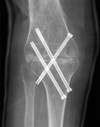
Arthroplasty Flashcards

1
Q

Options for this young (<50yo) patient with a painful right knee

A
  • Exhaust concervative treatment
    • PT, NSAIDS
    • off loading brace
    • Cortisone injections
  • Valgus producing tibial ostotomy
    • Contraindications
      • Inflammatory arthritis
      • Less than 90 deg flexion
      • Flexion contracture > 10 deg
      • Ligament instability (varus thrust)
      • Lateral tibial subluxation > 1cm
      • Medial compartment bone loss
      • Lateral compartment joint space narrowing
    • Predictors of failure
      • Smoking
      • > 60
      • Varus > 10
      • Other arthritides
  • Closing wedge problems
    • Patella baja
    • Loss of posterior slope
  • Opening wedge
    • Nonunion
    • Loss of valgus correction
2
Q

Contraindications to HTO

A

Inflammatory arthritis
Less than 90 deg flexion
Flexion contracture > 10 deg
Ligament instability (varus thrust)
Lateral tibial subluxation > 1cm
Medial compartment bone loss
Lateral compartment joint space narrowing

3
Q

Predictors of failure of HTO

A

Smoking
> 60
Varus > 10
Other arthritides

4
Q

Options for this 65yo male with painful right knee?

A
  • Exhuast non-operative
    • ​PT, NSAIDS
    • cortisone injection
    • offloading brace
    • Cane, mobility aids
  • UKA vs HTO vs TKA
  • UKA benefit over HTO and TKA
    • Smaller incision
    • Better knee function
    • Shorter stay with less pain
  • Technique
    • Do not overcorrect - can cause early failure
    • Varus - correct to 1-5 deg of valgus
  • Contraindications
    • ACL deficiency (strongest)
    • fixed varus or valgus deformity > 10 degrees
    • restricted motion
      • < 90° of flexion
      • fixed flexion contracture of > 10°
    • joint subluxation of 5 mm or greater
    • arthrosis of the additional compartment
      • modest Outerbridge Stage II chondromalcia of patella is acceptable
    • non-osteoarthritis arthritis
    • younger high activity patients and heavy laborers
    • overweight patients (> 90 kg)
  • Selection criteria
    • Pain must be localized to the compartment being replaced
    • Anterior knee pain means patellofemoral disease
    • Global pain means tricompartmental disease
  • Complications
    • Stress fractures
      • best visualized on bone scan
      • Usually on the tibial side
    • Tibial component collapse
      • poor mechanical properties of the bone
    • Failure
      • Overcorreciton
  • Undercorrection
  • Fixed-bearing (loosening)
  • Mobile bearing (diseae progression)
  • Patellar impingment (requires revision to TKA)
5
Q

Normal alignment of the knee

A

Lateral proximal femoral angle: 90 degrees

Mechanical Lateral distal femoral angle: 88 degrees

Anatomic Lateral distal femoral angle: 81 degrees

Medial proximal tibial angle: 87 degrees
Lateral distal tibial angle: 89 degrees

6
Q

What is this depicting and what are your considerations when measuring the deformity?

A

CORA - center of rotation of angulation

  • Draw a line threw the axis of the distal and proximal end
  • If there is only angulation - will occur at the apex of deformity
  • If there is combined translation - will occur at a distance equal to the amount of translation deformity
  • If angulation is seen on both AP and lateral, the true angulation will be larger than that seen on either XR
  • When you don’t see angulation in one plane, but you do on the other - this is the true angular deformity
7
Q

What is your appraoch to this patient?

A
  • History
    • Take a complete and ample history
    • Pain, functional issues, issues in other joints
    • Previous surgeries, trauma
    • PMHx, meds, all
  • Physical
    • Look
      • Gait, measure alignment and deformity
    • Feel
      • Assess stability of the hip, knee, ankle/foot
    • Move
      • ROM, contractures
    • Full NV exam
  • Imaging
    • Radiographs - full length standing AP/Lat
  • Indications for surgery
    • Ligamentous laxity on the concave side
    • LLD > 2cm
    • Uniconylar OA of the knee
    • Inability to place the foot in a plantigrade position
  • Conservative
    • Unloading brace
    • Shoe lift/orthoses
    • Appropriate analgesia
  • Considerations
    • Healing potential
      • Should be done in an area with better healing potential
      • Can accept some translation as long as the deformity is anticipated
    • Leg length discrepancy - affected by both closing/opening and varus/valgus, the affect is combined
    • Closing wedge can relatively lengthen ligaments and tendons
    • Opening wedge with lengthen = half the base of the triangle
  • Varus correction will produce lengthening
    • This will decrease as you go more distal
  • Valgus produces shortening​
8
Q

Technical goals of TKA

A
  • restore mechanical alignment (mechanical alignment of 0°)
  • restore joint line ( allows proper function of preserved ligaments. e.g., pcl)
  • balanced ligaments (correct flexion and extension gaps)
  • maintain normal Q angle (ensures proper patellar femoral tacking)
9
Q

You are planning a TKA for this patient. What are the order of releases

A
  • osteophytes
  • deep MCL (usually osteophytes and deep MCL is sufficient release)
  • Posteromedial corner
    • Semimembranosus
    • capsule
    • superfical MCL
      • can find as it blends into pes anserine complex
      • can not completely release or will have valgus instability (requires constrained prosthesis). Therefore perform subperiosteal elevation only
      • Differential release: performed with two component of superficial MCL
        • posterior oblique portion is tight in extension (release if tight in extension)
        • anterior portion is tight in flexion (release if tight in flexion)
  • PCL
10
Q

Order of release for a flexion contracture

A
  • Order of posterior release
    • osteophytes
    • posterior capsule
    • gastronemius muscles (medial and lateral)
  • You do not want to address by removing too much tibia
  • will change joint line and lead to patella alta
  • Performed with the knee flexed so there is less risk to the popliteal artery
11
Q

Important considerations for planning your TKA cuts

A

Femur

  • uses intramedullary guide, if can’t get this then use CT guided (post DFVO, trauma etc)
  • Distal femur valgus cut (5-7° from AAF )
    • jig measures 6 degrees from femoral guide (anatomic axis)
    • will vary if people are very tall (VCA < 5°) or very short (VCA > 7°)
  • Posterior referencing with femoral cut
    • 3 deg ER (normal DR is 3 deg IR)
    • otherwise will internally rotate your component
    • should be parallel to interchondylar axis
    • be careful with hypoplasia of the lateral femoral condyle, you can put the prosthesis into IR with a posterior reference system

Tibia

  • Cut should be perpendicular to mechanical axis
  • Can use intramedullary, unless there is deformity then need to use extramedullary
12
Q

This patient comes in with knee pain. What is the most common complications of TKA? How can you prevent it?

A
  • Abnormal patellar tracking, although not the most serious, is the most common complication of TKA.
  • The most important variable in proper patellar tracking is preservation of a normal Q angle (11 +/- 7°)
    • the Q angle is defined as angle between axis of extensor mechanism (ASIS to center of patella) and axis of patellar tendon(center of patella to tibial tuberosity)
  • Any increase in the Q angle will lead to increased lateral subluxation forces on the patella relative to the trochlear groove, which can lead to pain and mechanical symptoms, accelerated wear, and even dislocation.
  • Common errors include:
    • internal rotation of the femoral prosthesis
    • medialization of the femoral component
    • internal rotation of the tibial prosthesis
    • placing the patellar prosthesis lateral on the patella
13
Q

Where should the joint line be in TKA? What problems can you run into if you move it

A
  • Normal joint line
    • 1 cm above fibula
    • ​​2 fingerbreaths about tibial tuberosity
  • elevating the joint line (> 8mm leads to motion problems) and can lead to
    • mid-flexion instability
    • patellofemoral tracking problems
    • an “equivalent” to patella baja
  • lowering joint line
    • lack of full extension
    • flexion instability
14
Q

Saggital balancing. Go. All of it. You have 30 sec.

A
15
Q

You are planning a TKA on this patient. What is your order of release. What are some important considerations?

A
  • Classification
    • Stage 1 - not correctable
    • Stage 2 - > 10 deg, not correctable
    • Stage 3 - severe deformity, possibly incompetent MCL, severe bone loss
  • Order of Release
    • osteophytes
    • lateral capsule
    • iliotibial band if tight in extension (release if tight in extension)
      • with Z-plasy or release off Gerdy’s tubercle
    • popliteus if tight in flexion (release if tight in flexion
    • for severe deformities release both the iliotibial band and the popliteus
    • LCL
      • some authors prefer to release this structure first if tight in both flexion and extension
      • others prefer this should be the last structure to release, if you need to release it consider of constrained prosthesis
  • Considerations
    • ​Coronal balancing - older patient can use CCK, younger patient want to take less bone, but still want to do a primary knee
    • peroneal nerve palsy
16
Q

You do a TKA on this patient and surprise! He gets a peronal nerve palsy. What are some risk factors? How do you treat?

A
  • Prognosis
    • most resovle in 3 months
  • Risk Factors
    • use of epidural anesthesia;
    • previous spinal surgery (double crush);
    • valgus knee deformity
    • flexion contracture more than 20 deg
    • abarent retractors
    • pre-op neuropathy
  • Immediate
    • take of dressing
    • flex the knee
    • throrough documentation of physical exam
  • Post-op
    • AFO
    • PT for ROM
    • EMG with-in one month
  • At 3 months
    • Repeat EMG for improvement
    • Decompression with neuroloysis
      • 4 cm proximal
      • adherence to fibular head
      • 7-15 cm distal to fibular head
17
Q

Amount of antibiotics to put in antibiotic cement

A
  • Need to keep under 2g/40g of cement to preserve mechanical function
  • Safe loading dose
    • Vanco - 10.5g
    • Gent - 12.5g (although some report lower - 2g)
  • Masri recommends 3.6g tobra and 1g vanc per 40 mg
  • Powder is poured into liquid cement, vacum is not used - keeps porosity high to help with elution of cement
18
Q

What’s the most important factor in post-op TKA ROM?

A

Pre-op ROM

19
Q

Pros and Cons of a CR knee

A
  • Most common, relies on native PCL
  • Bone conserving
  • More consistent joint line preservation
  • Proprioceptive feedback
  • Disadvantages
    • Loss of PCL will lead to instability and failure
      • Tight PCL will cause tightness in flexion and cause lift-off of component
      • Excessive resection will cause failure from repetitive subluxation
      • Instability, pain, buckling
    • Harder to balance
      • Avoid in varus > 10, valgus > 15
    • PCL Rupture
      • Trauma
      • Osteolysis
    • Paradoxical movement - due to loss of ACL
      • Tibia slides forward under the femur instead of posteriorly
        Modern implants move center of rotation more posterior
20
Q

Pros and cons of anteriorly stabilized knee

A
  • Anterior lip prevents femor from rolling forward
  • PE is highly congruent, there is no cam
  • Advantage
    • Bone conserving
    • Easier balancing
    • Operative versatility
    • Regulated kinematics
  • Disadvantage
    • Increased PE surface
    • Minimal rollback
    • Flexion gap laxity = instability and pain - requires treatment to assess this
21
Q

Indications, pros and cons of a PS knee

A
  • Outcomes are the same for PS and CR knee
  • Indications
    • Previous patellectomy - weak extensor mechanism can lead to anterior dislocation
    • Inflammatory arthritis - leads to PCL rupture​
    • varus >10
    • valgus >15
  • Advantages
    • Easier balancing
    • No sliding
    • better flexion
  • Disadvantages
    • Cam jump - if flexion gap is loose, knee will hyperextend, rotate and jump over post and dislocate
      • Reduce with sedation, 90 deg of flexion and anterior drawer maneuver
      • Avoid in knees with >130 flexion
      • Ultimately needs to be revised to address loose flexion gap
        • Overreleased poplitues with saw blade
        • Overrelease anterior MCL
        • Anterior translation femoral component
    • Patella Clunk
      • Scar tissue superior to patella gets cause in box
      • Flexion - Ex at 45 deg
      • Treatment - arthroscopic or mini open debridement
    • PE Wear from tibial post
      • Causes aseptic loosening
      • If need if hyperextended will cause impingement anteriorly and increase wear rate
      • Flexed femoral component, excess tibial slope, anterior translation
    • Additional bone removed
      • For post
      • Large flexion gap
        • Due to PCL removable
        • Need to take more distal femur to account for this
  • Beware joint line elevation with patella baja** Max 8mm
22
Q

Indications for the use of this prosthesis?

A
  • LCL/MCL attenuation or deficiency
  • Flexion gap laxity
  • *Can be used more in revision TKA, but consider at times for primary
    • ie; severe valgus knee

the patient depicted had instability following CR knee and PCL rupture

23
Q

Indications for the following prosthesis

A

Constrained hinge with rotating platform

  • Tibia rotates with-in yoke to allow rotation during gait
    • Without rotation there was a high loosening rate
    • Intramedullary stem for high rotational loads
  • Indications
    • Global ligament deficiency
    • Hyperextension instability (polio or tumor resection)
    • Knee resection for tumor
    • Complete MCL (controversial)
24
Q

Why was this prosthesis choosen?

A

Tumour Prosthesis

  • tumour
  • significant bone loss
25
Q

You do this procedure on a healthy 65 yo male. What are all the complications?

A
  • Femoral Notching
    • Jig is placed too low on the femur
    • Lowers load to failure (fracture)
    • In bending the fracture extend from the notch creating an oblique fracture
  • Peroneal Nerve Palsy
  • PCL deficiency (in a CR knee)
    • Will become loose in flexion, knee will feel unstable and femur will start to slide forward
    • Will get anterior knee pain
    • Treat with revision to PC or CCK
  • Lateral Retinacular Release
  • Patella Fracture
  • Intra-operative MCL injury
    • Primary repair with 6 weeks NWB with full ROM (if young)
    • Recommended treatment is to covert to highly constrained prosthesis
  • Arthrofibrosis
  • Post-op Flexion Contracture
    • The most important factor of post-op range of motion is pre-op ROM
    • In a well balanced knee the gastroc is the cause of the flexion contracture
  • Severe extra-articular femoral deformity
    • Can do a combined osteotomy with TKA with long stem that goes past the osteotomy site
  • Osteolysis
    • Usually around 8-10 years
    • Gradual increase in effusion with mild warmth but no erythema
    • Normal lab results and aspiration
    • Most common place is the posterior femoral condyle
26
Q

What is usually causing a flexion contracture post-op TKA

A

gastroc

27
Q

When do you usually see osteolysis in TKA and where do you get it?

A

8-10 years

posterior femoral condyle

28
Q

You do the following procedure. They come back at 6 weeks and a stiff knee. How do you approach this?

A
  • Functional ROM
    • 90 degrees needed to go down stairs
    • 95 to get up from a chair
  • Factors to assess
    • Boney resection
    • patella baja/elevation of joint line
    • tibial slope (not enought)
    • posterior osteophytes
    • patient factors
  • Radiography
    • Assess joint line based on fibula and patella
    • Assess alignment of implants
    • presence of osteophytes
  • Treatment
    • depends on the surgeon, some will take them back if not to 90 by 6 weeks, can be done up to a year
    • Manipulation
      • usually at 6 weeks
      • Later manipulation as high rate of fracture
    • Scope
      • release adhesions, cement/loose bodies
    • Arthrotomy
      • poly exchange, patellar exchange, posterior release, tuberosity transfer, quads snip
      • Not highly recommended, often fails with recurrent pain and stiffness
    • Revision TKA
      • tibial tuberosity osteotomy, V-Y plasty
      • address tibial slope, patellar height
29
Q

TKA. Patient can no longer extend their knee. How do you approach this?

A
  • Can also occur with lowering of the joint line
  • Overview
    • patellar tendon rupture is a rare and devastating complication after TKA with an incidence reported ranging from 0.17% to 2.5%
    • Quadriceps tendon rupture extremely rare ~1%.
      • Higher risk with infection, multiple surgeries, hinged prosthesis
    • Patellar Fracture
      • Thickness <12mm
      • Lateral retinacular release (osteonecrosis)
        • Due to transection of superior genicular artery
      • Osteonecrosis
        • Theoretical risk with removal of fat pad, lateral release, quads turndown
        • Blood supply comes from the medial to lateral part of the knee
    • Can also be caused by maltracking or direct trauma
  • Quads or patellar tendon rupture
    • Overall very poor outcome
  • Treatment
    • Can treat conservatively if the component is solid and there is no extensor lag
    • Quads
      • Drill holes if no patella resurfacing
      • Suture anchors if there is less bone stalk
    • Patellar
      • Primary repair with drill holes or suture anchor does not do well
      • Component revision if there is enough bone, component resection if not
      • Allograft or autograft if there is significant extensor lag
      • Fresh frozen is the prefered choice
    • Use a detensioning wire (or mersiline tape) for 3 months
30
Q

Considerations when deciding whether to resurface the patella

A
  • High clinical variability in practice
  • Complications
    • patellar fracture
    • malailignment
  • Consequences
    • Anterior knee pain with higher revision rate
    • 50% re-operation rates and higher complication rates with no patellar resurfacing - at about 5 years outcomes become the same
  • Earlier designs had a high failure rate, newer designs have longer survivorship
  • Technique
    • Indicaitons to resurface
      • maliagnmnet
      • minimal wear
      • inflammatory disease
    • If you’re not resurfacing use a more anatomic design
    • Patellar cut
      • Use the caliper before and after
      • Under resection leads to pain and a tight compartment
    • Over resection leads to fracture - max thinness is 12mm
    • Medial-superior placement is ideal
      • Helps to restore Q angle with fewer tracking complications
    • Lateral Release
    • complications
      • fracture
      • component dislocation
      • wear
      • infection
31
Q

Diagnosis? Causes? Treatment considerations in TKA?

A

Patella Baja

  • Introduction
    • A condition manifested by a shortened patellar tendon
    • Leads to limited flexion due to patellar impingement on the tibia in extremes of flexion
  • Causes
    • proximal tibial osteotomy
    • tibial tubercle shift or transfer
    • proximal tibia previous trauma
  • Presentation
    • mechanical block to full flexion
  • Management
    • Operative with TKA
      • lowering joint line
        • distal femoral augmentation and cutting off more proximal tibia
        • avoid bone cuts that raise the joint line (raising the joint line will effectively increase the patella baja deformity)
      • elevating patella
        • use small patellar component and place superiorly
        • trim bone or polyethylene to reduce impingement
    • one option in severe deformity is to cut the patella but not to resurface it (this will reduce patellar impingement allowing for more knee flexion)
32
Q

This patient comes into your clinic with a painful knee. What is your approach and general priniciples?

A

RULE OUT INFECTION!!!!!!!!!

  • take a complete history around the pain, duration, time pain free, instability, PMHx, smoking
    • assess for knee stability, contractures, neurological issues, vascular issues
    • previous inctions
    • patellar tracking
  • Get all previous OR notes
  • ESR, CRP, possible aspiration
  • Bone scan - fracture, infection
  • Imaging - AP, lateral WB views, full length standing
    • compare to previous, assess for further lysis
    • assess opposite knee
    • Look at lysis around each component
    • component alignment, shift
      • posterior sag tibia = PCL
    • full alignment of knee
  • Goals of revison surgery
    • extraction of components with minimal bone loss and destruction
    • restoration of bone deficiencies
    • restoration of joint line
    • balance knee ligaments
    • stable revision implants
    • address patellar malalignment
    • soft tissue coverage
  • Exposure
    • Use most lateral incision with multiple incisions
    • Try to leave skin bridge 6cm if you can’t use old incision
    • Release all adhesions and scar tissue
      • do not pull off the patellar tendon
    • If the patella cannot be everted safely
      • Quads snip - snip across the quads
        • No need to protect WB
        • 45 deg angle to arthrotomy
      • Patellar turndown - V-Y
        • Associated with weakness
        • Protect WB with extension brace for 6 weeks
      • Tubercle ostotomy
        • Less weakness but can’t do if there is patellar baha
        • 2cm wide, 1cm thick, 6 cm long
        • start 1cm medial to tubercle with an osillating saw
        • finish laterally with an osteotome
        • stemed revision must bipass osteotomy
        • WBAT with ROMAT
  • Prothesis Extraction
    • Do with as little damage as possible
    • Contact rep for specific implant removal
      • ostotomes, punchs, slap hammer, clamps, saw, burr
    • Take poly out
    • Free femur with saw and osteotome
      • don’t lever
      • remove with punch or slaphammer
    • Similar technique with the tibia
      • may need to cut the stem
      • osteotomy for exposure
    • Patella
      • can leave, or cut the pegs off and then drill them out
    • Clear cement, if you are re-cementing you can leave the cement
  • ​​Bone Loss
    • Cause
      • _​_abrasion, infection, osetolysis, extraction
    • load sharing to the diaphysis (stem)
      • stem is cement often, unless you have such sever bone loss you need to go up to the diaphysis (not like a hip)
    • cavity defect filling
      • cement
        • for cavitary defect is < 1 cm
      • structural bone grafts
        • includes metal augments, or modular endoprosthetic devices
        • indicated for segmental defect > 1cm​​​
  • ​​Appropriate implants
    • Tibia first, establish joint line
      • use contralateral films
      • 2cm above fibular head
    • hinged
      • ​no ligamentous support
      • multiply revised
      • hyperextension seen in polio
      • tumor, infection
      • charcot (relative)
    • CCK/stem (constrained condylar knee)
      • ​MCL/LCL laxity
      • flexion gap
    • CR to PS or CCK
  • Soft tissue
    • Medial gastroc is the most reliable
    • Do not delay closure, should be done at the same time as revision
33
Q

Complications associated with Revision TKA?

A
  • Pain
    • pain scores less favorable than primary TKR
    • activity related pain can be expected for 6 months
  • Stiffness
  • Neurovascular problems
    • peroneal nerve subject to injury with correction of valgus and flexion deformity
  • Infection
  • Skin necrosis
    • prior scars should be incorporated into skin incision whenever possible
    • bloody supply to anterior knee is medially based, so lateral skin edge is more hypoxic
    • if multiple previous incisions, use most lateral skin incision
    • can use wound care, skin grafting, or muscle flap coverage (gastroc) for full thickness defects
  • Extensor mechanism disruption
    • can use extensor mechanism allograft using achilles tendon
    • Semi-T graft can be used to help augment
    • Can use this intra-op if you loose the tendon
34
Q

Indicaitons, contraindications and optimal position

A

KNEE ARTHRODESIS

  • Indications
    • painful ankylosis after infection or trauma
    • neuropathic arthropathy
    • tumor resection
    • salvage for failed TKA (most common)
    • loss of extensor mechanism
  • Contraindications
    • absolute
      • active infection
    • relative
      • bilateral knee arthrodesis
      • contralateral leg amputation
      • significant bone loss
      • ipsilateral hip or ankle DJD
  • Optimal Position
    • 5-8° valgus
    • 0-10° of external rotation (match other leg)
    • 0-15° of flexion
    • some limb shortening advantageous for patient self-care
35
Q

Options for fixation for the following? Complications?

A
  • Fixation
    • Intramedullary rod fixation
      • can be one long antegrade device or a two part device connected at the knee
      • patella can be left alone or incorporated into arthrodesis
    • External fixation
      • must allow compression of arthrodesis site
      • done with unilateral external fixation, Ilizarov, or Taylor Spatial Frame
    • Plate fixation
      • can be done alone in combination with intramedullary nailing
  • Complications
    • Nonunion
    • Infection
    • Low back pain
    • Ipsilateral hip degenerative changes
    • Contralateral knee degenerative changes
    • Fracture
    • supracondylar femur or proximal tibial metaphysis fractures
36
Q

Indications and contraindications; Advantages and disadvantages

A
  • Indications
    • patients with advanced arthritis and good proximal femoral bone stock
    • three types of patients for whom hip resurfacing is indicated (Amstutz, et al)
      • patients with proximal femoral deformity making total hip arthroplasty difficult
      • patients with high risk of sepsis due to prior infection or immunosuppression
      • patients with a neuromuscular diagnosis
  • Contraindications
    • absolute
      • bone stock deficiency of the femoral head or neck (e.g., cystic degeneration of the femoral head)
        • >75% femoral head
      • abnormal acetabular anatomy (small)
        • Associated with acetabular loosening
    • relative
      • coxa vara
      • increased risk for neck fractures
      • significant leg length discrepencies (resurfacing does not allow for leg length corrections)
      • female gender (controversial)
  • Advantages
    • preservation of femoral bone stock
    • improved restoration of hip biomechanics with lower risk of limb length discrepancy
    • lower dislocation rate
    • rapid recovery
    • revision is easier than an intremedullary THA
    • better stability compared to standard small head (22- to 32-mm) THA
    • ability to engage in high demand activities
  • Disadvantages
    • lack of modularity with inability to adjust length or correct offset
    • requires larger exposure than conventional THA
37
Q

Outcomes of hip resurfacing

A
  • variable outcome findings in the literature (79% to 98% success rate)
  • better results
    • young
    • larger males
    • excellent bone stock treated
    • osteoarthritis better than for dysplasia or osteonecrosis
  • some case series have shown survival comparable to conventional THA, while others have reported higher rates of early revision
  • some products have been removed from the market due to early failure
  • more recent prospective trials have shown few differences between resurfacing and THA
38
Q

Patient comes in one year following hip resurfacing for OA. Differential for associated complications.

A
  • RULE OUT INFECTION
  • Periprosthetic femoral neck fracture
    • incidence of 0% to 4% (more common than in THA)
    • frequent cause for revision in acute post-operative period (<20 weeks)
    • mechanism thought to be related to osteonecrosis
    • risk factors:
      • notching of the femoral neck
      • osteoporotic bone
      • large areas of pre-existing osteonecrosis
      • femoral neck impingement (from malaligned acetabular component)
      • female sex
      • varus positioning of femoral component
      • presents as groin pain
    • treatment
      • convert to a THA
  • Implant loosening (aseptic)
    • early loosening of the cemented femoral resurfacing component
  • Heterotopic ossification
    • higher incidence of heterotopic ossification (from wider exposure)
  • Elevated metal ion levels in blood and urine from metal debris (unknown significance)
    • Metallosis
    • pseudotumour
    • ALVAL (Aseptic Lymphocytic Vasculitis Associated Lesions)
      • Can see this on biopsy
    • Treatment is to replace with a THA metal on poly
39
Q

What are three ways to assess the rotation of your femoral component?

A
  • anteroposterior axis
    • defined as a line running from the center of the trochlear groove to the top of the intercondylar notch
    • a line perpendicular to this defines the neutral rotational axis
  • transepicondylar axis
    • defined as a line running from the medial and lateral epicondyles
    • the epicondylar axis is parallel to the tibial surface
    • A posterior femoral cut parallel to the epicondylar axis will create the appropriate rectangular flexion gap
  • posterior condylar axis
    • defined as a line running across the tips of the two posterior condyles
    • this line is in ~ 3 degrees of internal rotation from the transepicondylar axis, the femoral prosthesis should be externally rotated 3 degrees from this axis to produce a rectangular flexion gap
    • if the lateral femoral condyle is hypoplastic, use of the posterior condylar axis may lead to internal rotation of the femoral component
40
Q

Deformities associated with CAM (clinical/anatomical - not radiographic)

A

decreased head-to-neck ratio
aspherical femoral head
decreased femoral offset
femoral neck retroversion

41
Q

Deformities associated with Pincer (clinical/anatomical - not radiographic)

A

anterosuperior acetabular rim overhang
acetabular retroversion
acetabular protrusio
coxa profunda (deep socket)

42
Q

Radiological features of FAI

A
  • Technique
    • Hips in 15 deg IR with beam centered between symphysis and ASIS
    • Assess symmetry of obturator, tear drops; symphysis should be 1-2 cm from the coccyx
  • Findings
    • Overall
      • Tonnis grade - OA
      • Joint space < 2mm is a poor prognostic factor
      • Shenton’s line
    • Acetabulum
      • CEA > 40, Tonnis angle < 0
      • Version; cross over sign, ischial spine sign
      • Retroversion index > 33-50% are significant
      • Assess protrusio, coxa profunda
      • Posterior wall sign
    • Femoral side
      • Assess coxa vara
      • Sphericity and contour of femoral head - ‘pistol grip deformity
  • Dunn view
    • Alpha angle >50 (cam)
  • Cross-table lateral
    • Head neck off-set >8mm (cam)
  • false profile view
    • to assess anterior coverage of the femoral head
    • standing position at an angle of 65° between the pelvis and the film
43
Q

What is the tonnis grade for arthritis

A
  • Grade 0 - no signs of OA
  • Grade 1 - sclerosis with minimal joint space narrowing
  • Grade 2 - small cysts in head or acetabulum with joint space narrowing
  • Grade 3 - large cysts in the head with obliteration of joint space and severe head deformation
44
Q

Radiographic investigations for FAI or young hip

A
  • Differential
    • DDH (+/-cam)
    • FAI
      • cam
      • pincer/retroversion
    • Residual deformity
      • SCFE
      • LCP
  • AP View
    • Technique
      • Hips in 15 deg IR with beam centered between symphysis and ASIS
      • Assess symmetry of obturator, tear drops; symphysis should be 1-2 cm from the coccyx
    • Findings
      • Tonnis grade - OA
      • Joint space < 2mm is a poor prognostic factor
      • Shenton’s line
      • Acetabulum
        • CEA = 25-40
        • Tonnis angle 1-10
        • Version; cross over sign, ischial spine sign
          • Retroversion index > 33-50% are significant
        • Assess protrusio, coxa profunda
        • Posterior wall sign
      • Femoral side
        • Assess coxa vara/valgus
        • Sphericity and contour of femoral head - ‘pistol grip deformity’
  • Dunn view at 45deg
    • Alpha angle >50 (cam)
  • Cross-table lateral
    • Head neck off-set >8mm (cam)
  • false profile view
    • to assess anterior coverage of the femoral head
    • standing position at an angle of 65° between the pelvis and the film
  • Von Rosen
    • Flexion, abduction, IR
    • Helps to assess femoral version
    • Mimics a true PAO
  • Adduction or abduction views
    • Can help assess the congruency of the joint
    • mimics a valgus osteotomy
  • CT
    • can be used as adjunct to assess for structural abnormalities
    • Drawback is radiation in a young population
  • MRI
    • best modality to evaluate for articular cartilage, and labral degeneration and tears
      • 63% sensitive, 71% specific
    • can assess anatomy of femoral head/neck junction abnormalities
    • More accurate to assess the alpha angle
    • Cam - debonding anterior superior lesion in the cartilage
      • “outside-in”
    • Pincer - anterior superior as well, but can be more global
      • Posterior rim lesions, or ‘contrecoup’ lesions, should be assessed
  • dGEMRIC
    • Used to look at the integrity of the cartilage
    • Not commonly used
  • Flouroscopic guide hip injection
    • 4-6ml of anesthetic under flouro guidance can help
    • Have patient assess pain before and after injection
    • Can be very helpful to determine if pain in intra-articular
45
Q

Diagnosis? Treatment Options?

A

DDH

  • Conservative
    • first line of treatment
    • NSAIDS, activity modification
  • arthroscopic osteochondroplasty of acetabular rim and/or femoral head reshaping
    • reserved for mild to moderate lesions without posterior involvement of the femoral head or neck
    • Can be done via scope or surgical dislocation (trochanteric slide)
      • Risk of osteotomy non-union
  • periacetabular osteotomy +/- a femoral osteotomy
    • Salter osteotomy (Ganz/Bernese)
      • Most commonly used, allows medialization of the hip joint that reduces hip reaction forces
      • Lateral rotatation, anterior rotation, with medialization of femoral head
      • indications
        • Pain or progressive limp
        • Adequate ROM
          • >90 deg flexion
          • >30 deg abduction
        • concentrically reduces hip/congruent joint
        • good joint space (tonnis < 2)
      • advantages
        • provides hyaline cartilage coverage
        • posterior column remains intact and patients can weight bear
        • Does not change pelvic shape in women of weight bearing age
        • preserves hip abductors
    • salvage pelvic osteotomy (chiari, shelf)
      • Not used as frequently due to improvements in THA
      • indications
        • unreduced hip
        • incongruous joint with obliterated joint space
        • Can confirm with an abduction view whether the hip is reducible
      • major goal of procedure
        • cover femoral head with fibrocartilage (NOT articular cartilage)
        • Will often need a graft to cover the head anteriorly
      • Drawback of shelf is that you are not medializing the joint
  • Femoral Osteotomy
    • Often needed when you don’t get enough coverage of the head
    • Need to use flouro to decide where optimal position is
    • Don’t close the canal too much because it makes future THA difficult
  • Arthrodesis or resection arthroplasty
    • Options but not good ones for DDH
    • Resection arthroplasty might be better for patients with neuromuscular conditions
  • total hip arthroplasty (THA)
    • indications
      • treatment of last resort for those with severe arthritis
      • preferred treatment for older patients (>50) and those with advanced structural changes
      • in a patient with bilateral hip dysplasia, there are significant technical challenges that need to be addressed to ensure a successful total hip arthroplasty.
46
Q

Indications and Contraindcations to doing a PAO?

A
  • Indications - For DDH
    • Pain or progressive limp
    • Adequate ROM
      • >90 deg flexion
      • >30 deg abduction
    • concentrically reduces hip/congruent joint
    • good joint space (tonnis < 2)
  • Contraindicaitons for FAI (reverse PAO)
    • Posterior-wall overcoverage
    • Tonnis > 2
    • Combined pincer/cam
47
Q

Technique of a PAO

A
  • Smith-peterson
  • 4 cuts
    • anterior ischium below the acetabulum
    • superior pubic ramus
    • supra-acetabular ilium
    • posterior column (joining cut number one)
  • Re-approximate (for DDH)
    • Tilt laterally and anteriorly
      • Avoid excessive extension and retroversion
    • Rotate inwardly
    • Medialize
  • Fix with k-wires, assess ROM and position with flouro
  • Secrure with 4.5 cannulated screws
  • Arthrotomy
    • Assess head-neck junction and perform osteochondroplasty as necessary
48
Q

Complications associated with PAO

A
  • Most important factor affecting complications is the surgeons experience
  • Nerve dysfunction - most common
    • LFCN
    • Less commonly femoral, pudendal and sciatic
  • Vascular - related to ilioiguinal appraoch
  • Non-union
    • Usually the pubis, less commonly the ilium/ishium
    • Can be treated with bone grafting
  • Impingement
  • High incidence of HO
    • Give indomethecin
  • Femoral instability
    • If you don’t do associated femoral ostotomy can get recurrence of instability
  • Osteolysis
    • Can get osteolysis with excessive stripping
  • Inferior gluteal artery is the one to be concerned about, comes out below the piriformis
49
Q

Technique for a femoral osteotomy

A
  • PFO can be altered as per what you need
    • DDH - varus +/- ER
    • SCFE (Southwick) - flexion, abduction, IR
    • Femoral neck nonunion - southwick
  • Place a k-wire at the level of the ostotomy site
  • Place a k-wire where the blade should go
    • This will depend on the angle of the plate
    • Can also assess whether you will need flex or ex at this time
    • This should be at least 1.5cm away from k-wire1
  • 3rd kwire or chisel is used to determine the length of the blade
  • Use a chisel to make the blade cut along wire number 2
  • Mark your version with a k-wire
  • Osteotomize the femur at Kwire 1
  • Remove a wedge if you need to
  • Remove the chisel
  • Place the balde, watch this under flouro
  • Secure the blade to the proximal fragment
  • Secure the plate to the distal fragement
  • Check the position on flouro
  • Post-op
    • Start abduction at 6 weeks
    • TTWB until 8-12 weeks
    • Plate may be removed at 1-2 years
  • Complications are technical
    • Blade protrusion into joint
    • Fragmentation of distal fragment
    • Nonunion
  • Outcome
    • 25% conversion to THA at 15 years
    • Improved outcomes with less OA
50
Q

Young hip physical exam

A
  • ROM
    • Test in IR (20) ER (45) in seated position
    • Test AD (20), AB (45) and flexion (120) in supine
  • Gait
    • LLD
    • Trendelenberg
    • Weak adductors
      • DDH, anything that medializes the head
    • Pelvic obliquity
  • Arthritis
    • Anterior impingement
    • Roll test - tight and stiff
    • Patrick test - same as FABER with pain anteriorly
    • Stinchfeild test - Pain with resisted flexion
  • Trendelenberg Test
  • Hip contracture
    • Hyperextension of the lumbar spine
    • During gait lumbar will extend to compensate
  • Tompson test
    • Flex up both hips to neutralize spine
    • Then extend each hip indivicually
  • Rotation of hip is more accurately tested in prone
  • FADIR - anterior impingement
    • Flexion, adduction and IR
    • Will cause pain with anterior impingment
  • FABER
    • Look for SI pain
  • Anterior Apprehension
    • Extension with ER
  • Psoas snapping
    • Passive flexion to 90
    • ER with abduction extend the leg, the tendon will snap
  • Ober test
    • Patient in lateral
    • Abduct the hip with the knee flexed extend the hip
    • Pulls the IT band behind the GT
    • Tight IT - hip won’t adduct past neutral
    • Tight glut med - delay with hip in neutral
51
Q

Approach to residual LCP

A
  • Residual deformity
    • Coxa magna
    • Asphericity
    • Coxa plana
    • Short neck
    • Acetabulum remodels and becomes dysplastic
  • Usually requires
    • Osteochondroplasty, neck lengthening, distal trochanteric transfer
  • Approaches
    • Anterior approach combined with lateral for PFO
      • Can be extended up to do an acetabular osteotomy if they have instability
    • Surgical dislocation to do everything
  • If they need both POA and PFO you can stage it or do it at the same time
52
Q

Indications and contraindications to hip arthroscopy

A
  • Indications
    • FAI
    • labral tears
    • AVN (diagnosis and staging)
    • loose bodies
    • synovial disease
    • chondral injuries
    • ligamentum teres injuries
    • snapping hip
    • mechanical symptoms
    • impinging osteophytes
  • Contraindications
    • advanced DJD
    • hip ankylosis
    • joint contracture
    • severe osteoporotic bone
    • significant protrusio acetabuli
53
Q

Portals for hip arthroscopy

A
  • Anterolateral portal
    • function
      • primary viewing portal
      • anterolateral hip joint access
    • location and technique
      • located 2 cm anterior and 2cm superior to anterosuperior border of greater trochanter
      • typically established first under flouroscopic guidance
  • Posterolateral portal
    • function
      • posterior hip joint access
    • location and technique
      • located 2cm posterior to the tip of the greater trochanter
  • Anterior portal
    • function
      • anterior hip joint access
    • location and technique
      • located at intersection between
        • superior ridge of greater trochanter
        • ASIS
      • flexion and internal rotation of hip loosens capsule and assists scope insertion
54
Q

Complications of hip arthroscopy

A
  • Direct injuries
    • can occur from scope or cannula placement
    • most commonly reported complication
      • chondral injuries
  • Neurovascular injury
    • traction related
    • pudendal nerve injury
      • most common neurovascular complication
      • due to traction post in groin for traction
      • neuropraxia or compression injury
    • peroneal nerve injury
      • traction neuropraxia
    • may prevent traction injuries with
      • intermittent release of traction
      • adequate anesthesia
  • anterolateral portal
    • risks superior gluteal nerve
  • posterolateral portal
    • risks sciatic nerve
      • increased risk with external rotation of hip
  • anterior portal
    • risks lateral femoral cutaneous nerve injury
    • risks femoral neurovascular bundle
    • risks ascending branch of lateral femoral circumflex artery
55
Q

Most common complication of hip arthroscopy

A

chondral damage

56
Q

Risk factors

A

​AVN

  • Alcoholism
  • steroids (either endogenous or exogenous)
    • High the dose the higher the risk
  • dysbaric disorders (decompression sickness, “the bends”)
  • marrow-replacing diseases (e.g. Gaucher’s disease)
  • sickle cell disease
  • hypercoagulable states
    • May explain idiopathic disease
  • SLE
  • Inflammatory Bowel Disease
  • transplant patient
  • virus (CMV, hepatitis, HIV, rubella, rubeola, varicella)
  • protease inhibitors (type of HIV medication)
  • AVN associated with trauma
    • due to injury of femoral head blood supply (medial femoral circumflex)
    • AVN rates of specific injuries
      • femoral head fracture: 75-100%
      • basicervical fracture: 50%
      • cervicotrochanteric fracture: 25%
      • hip dislocation: 2-40% (2-10% if reduced within 6 hours of injury)
      • intertrochanteric fracture: rare
57
Q

Radiographic work-up for AVN

A
  • Think to get imaging of both hips, 50% of the time is a bilateral disease
  • Radiographs
    • AP hip
      • Double line sign associated with crescent sign
      • Present in 80% of cases
    • frog-lateral of the hip - more sensitive
      • classification systems based largely on radiographic findings (see below)
    • Size of lesion - % head on AP X % head on lateral
      • Small - < 15%
      • Medium - 15-30%
      • Large - > 30%
  • MRI - Gold standard
    • highest sensitivity and specificity
    • T1: dark
    • T2: bright (marrow edema)
      • Will have evidence of high signal from edema as well as low signal changes
    • Extent of femoral head involvement
    • Kerboul Angle
      • Combine necrotic angles on saggital and coronal
      • >200 associated with poorer outcomes
58
Q

Diagnosis? How do you know? Prognosis?

A

Transient Osteoporosis of the Hip

  • Classic picture is low signal on T1 and high signal on T2 with no evidence of low signal in T2
  • All the way threw the head, neck and trochanteric region
  • If there are risk factors or concerns of AVN you can do serial progression, TOH will resolve
59
Q

Ficat classification

A
  • stage 0:
    • plain film: normal
    • MRI: normal
    • clinical symptoms: nil
  • stage I:
    • plain film: normal or minor osteopaenia
    • MRI: edema
    • clinical symptoms: pain typically in the groin
  • stage II:
    • plain film: mixed osteopenia &/or sclerosis &/or subchondral cysts, without any subchondral lucency (crescent sign: see below)
    • MRI: geographic defect
    • clinical symptoms: pain and stiffness
  • stage III:
    • plain film: crescent sign & eventual cortical collapse
    • MRI: same as plain film
    • clinical symptoms: pain and stiffness+/- radiation to knee and limp
  • stage IV:
    • plain film: end stage with evidence of secondary degenerative change
    • MRI: same as plain film
    • clinical symptoms: pain and limp
60
Q

Options for treatment

A

AVN

  • Prognosis
    • 80% will collapse
  • Bisphosphonates
    • indicated for precollapse AVN (Ficat stages 0-II)
    • trials have shown that alendronate prevents femoral head collapse in osteonecrosis with subchondral lucency
  • Shock-wave, hyperbaric has been tried, but hasn’t been showed to work and there is no evidence behind it
  • Tantalum rod is an old procedure that was thought to provide support and perhaps revascularization but the outcomes have been shown to be poor
  • Core decompression with bone grafting
    • indications
      • for early AVN (FICAT 1, 2), before subchondral collapse occurs
      • Small lesions (<15%)
      • Reversible cause
    • Now will just do multiple tracks with a smaller drill bit
      • **Just don’t drill below the less troch region
    • Not very good results
  • Rotational osteotomy
    • indications
      • only for small lesions (<50%) in which the lesion can be rotated away from a weight bearing surface
      • Beaule paper
        • <45 deg
        • Kerboul < 200
        • off steroids
    • technique
      • typically performed through intertrochanteric region
      • Japan does a trans-trochanteric that has not become common in NA
      • Need to be careful because can make conversion to THA more difficult
  • vascularized free-fibula transfer
    • indications
      • pre-collapse in patients young and over 40
      • Moderate to large lesion
      • collapsed AVN in young patient
      • Reversible cause
    • technique
      • fibular strut is placed under subchondral bone to help prevent collapse or tamp up small areas of collapse
      • complications - are related to donor site morbidity
        • sensory deficit
        • motor weakness
        • FHL contracture
        • tibial stress fracture from side graft is taken
  • total hip replacement
    • indications
      • femoral head collapse
      • acetabular DJD
      • Advanced collapse in younger patients
    • Technique
      • Remember that the bone quality is poor - be gentle with acetabular reaming
      • Evaluate the femur for previous surgery - core decompression, osteotomy
      • Use a large head, use a lateral approach
      • Consider ceramic in younger patients
    • Outcomes
      • in young patients with osteonecrosis there is a higher rate of linear wear of the polyethylene liner and a higher rate of osteolysis than when compared to older patients who have THA for osteoarthritis
      • Should be using uncemented components so they can in-grow
  • total hip resurfacing
    • indications
      • in advanced DJD with small, isolated focus of AVN
    • contraindicated in underlying disease process or chronic steroid use causing AVN (poor bone quality) and renal disease (metal ions from metal-on-metal implant)
  • hip arthrodesis
    • indications
      • only consider in the very young patient in a labor intensive occupation
61
Q

Increased risk of progression with AVN

A
  • > 5mm less chance of progression
  • Lateral lesion in the weight bearing zone more chance of progression
  • Keboul angle > 200deg has increased chance of progression
  • Risk of contralateral hip (50%)
62
Q

Considerations for THA in patients with AVN

A
  • Use a large head, use a lateral approach
  • Consider ceramic in younger patients
  • Remember that the bone quality is poor - be gentle with acetabular reaming
  • Evaluate the femur for previous surgery - core decompression, osteotomy
63
Q

Steinberg classification AVN

A
  • 0 - normal
  • I - normal XR, abnormal MRI
  • II - cystic or sclerosis change
  • III - crescent sign (subchondral collapse)
  • IV - flattening of femoral head
  • V - narrowing of joint abnormal
  • VI - advanced degenerative changes
64
Q

Two classifications for DDH when considering THA?

A
  • Crowe
    • I
      • Less than 10% Proximal
      • Less than 50% lateral
    • II
      • 10-15% proximal
      • 50-75% lateral
    • III
      • 15-20% proximal
      • 75-100% lateral
    • IV
      • Greater than 20%
      • Greater than 100%
  • Hardofilakidis
    • Dysplasia (Type A)
      • Femoral head within acetabulum despite some subluxation. Segmental deficiency of the superior wall. Inadequate true acetabulum depth.
    • Low dislocation (Type B)
      • Femoral head creates a false acetabulum superior to the true acetabulum. There is complete absence of the superior wall. Inadequate depth of the true acetabulum.
    • High dislocation (Type C)
      • Femoral head is completely uncovered by the true acetabulum and has migrated superiorly and posteriorly. There is a complete deficiency of the acetabulum and excessive anteversion of the true acetabulum.
65
Q

What are you consderations for THA

A
  • Acetabulum
    • smaller cup, superior migration, false acetabulum
    • loss of anterior/superior and lateral coverage
      • Augment with screw fixation
      • Augment with implant augments
    • Small cup mean a small femoral head to accommodate sufficient poly
    • Offset-bore components are available that change the position of the head in the poly to reduce risk of instability
  • Femur
    • Previous surgeries
      • Loss of canal
    • Posterior GT
      • GT osteotomy or ETO
    • May need to shorten
      • 2-4 cm is max without shortening
    • Soft tissue changes associated with superior migration
      • Abductors become transverse
      • Psoas and capsule hypertrophy
      • Adductor, rectus and hamstring shortening
66
Q

Technical approach for each crowe classification

A
  • Technique for I
    • Acetabulum
      • can usually use the native acetabulum
      • size less of an issue
      • Just consider degree of bone loss
    • Femur - no need to shorten
      • Try to go cementless because these patients are young
      • Minimal deformity - metaphyseal fitting proximally coated stem
      • Larger deformity - small, DDH stem with minimal flare
        • Diaphyseal fitting, long coating
        • Allows correction of version
  • Technique for II/III and Hardofilikas B
    • Acetabulum
      • 1) Leave the cup superior (high hip center)
        • Means you will have a very small cup
        • May need less shortening
        • increased stress with early failure
      • 2) Medialize the cup (acetabuloplasty) at native hip center
        • Reduces joint reaction forces
      • 3) Augment with bone graft which is available from the femoral head
      • Poor results have been published when > 30% of head is uncovered requiring graft/augment (this is when you would consider leaving the cup more superior)
    • Femur
      • Will need a diaphyesal fitting stem with modular components to assess verion
        • fully porous coated
        • fluted?
      • May need to be shortened
        • >2-4 cm
        • GT osteotomy with sequential femoral resection
        • Canal that is left is poor quality
        • Need to use a small DDH cemented stem
      • Subtroch osteotomy is better but harder to do - do this from a posterior approach
        • make subtroch osteotomy and leave hip in pseudoacetab
        • prepare the acetab
        • shorten your femur as necessary, release the head and soft tissues
        • size your stem
        • reduce secure the stem with k-wires/allograft as necessary
      • Risk of nonunion
  • Technique IV
    • Put in the native acetabulum
      • Extra-small cup
      • The bone will be soft, don’t over ream
      • Do the last reamer on reverse
    • ​Posterior approach for femoral shortening osteotomy via GT or subtroch osteotomy
67
Q

Indications and contraindications to hip arthrodesis

A
  • Indications
    • salvage for failed THA (most common)
    • young active laborers with painful unilateral ankylosis after infection or trauma
    • neuropathic arthropathy
    • tumor resection
  • Contraindications
    • active infection
      • delay until no active sinus and normal lab values
    • severe limb-length discrepancy greater than 2.0 cm.
    • bilateral hip arthritits
    • adjacent joint degenerative changes
      • lumbar spine
      • contralateral hip
      • ipsilateral knee
    • severe osteoporosis
    • degenerative changes in lumbar spine
    • contralateral THA
  • increased failure rate (40%) in THA when there is a contralateral hip arthrodesis
68
Q

What is true about the biomechanics and pathology of this patient?

A

Hip Arthrodesis

  • Pathomechanics
    • reduces efficiency of gait by ~50%
    • increases pelvic rotation of contralateral hip
    • increases stress at adjacent joints
  • Biochemistry
    • increases oxygen consumption
    • requires 30% more energy expenditure for ambulation
  • Prognosis
    • provides pain relief and reasonable clinical results in most patients
    • success may be limited by adjacent joint degeneration in 60% of patients
      • lumbar spine, ipsilateral knee or contralateral hip may be affected
      • low back pain and arthritic ipsilateral knee pain are the most common symptoms
      • may start within 25 years of hip arthrodesis
        *
69
Q

This is a 16yo male with a combined pelvis and femoral neck fracture. He developed infection post-op. After I&D, 6 weeks IV abx and 6 weeks PO abx, he has resolving ESR/CRP with an abx holiday. What is your plan now? Explain your technical approach.

A

Hip arthrodesis

  • goals
    • achieve apposition of arthrodesis surfaces, obtain rigid internal fixation and promote early mobilization
  • plan for future THA
    • preserve the abductors
    • preserve bone stalk; address missing bone stalk
  • optimal position
    • 20-35° of flexion
      • Appropriate flexion for sitting, reduce lumbar strain
      • Too much can exaccerbate LLD
    • 0°-5° adduction
      • avoid abduction as it creates pelvic obliquity and increased back pain
      • Too much adduction can make sexual activity and toileting difficult in women
    • 5-10° external rotation
      • Facilitates putting on shoes and foot care
    • Leg length
      • < 2cm difference is acceptable
        • Can use abduction to correct 2cm (cannot abduct further than that)
        • Do not abduct > 6 deg
      • >4 cm stage the procedure and do a second lengthening procedure
  • Approach
    • lateral approach with trochanteric osteotomy is preferred
      • important to preserve the abductor complex (used to strip the abductors off the pelvis, now elevate them)
      • avoid injury to the superior gluteal nerve
    • anterior approach to hip is also popular
      • Less risk to the abductors
      • supine on the operative table makes positioning hip easier
      • Screw augments the plate - lag screw done first, the plate onto crest up near SI
  • Address bone deficiet
  • Instrumentation
    • cobra plating
    • anterior plating
    • double plating
      • trochanteric approach
      • better with excessive bone loss, provides more support
      • 2 stage - first prepare the joint, second fix the GT back down
        • improves fusion rates
70
Q

This patient comes back to you at 25yo with low back pain. What is you assessment/approach now?

A
  • Prognosis
    • provides pain relief and reasonable clinical results in most patients
    • success may be limited by adjacent joint degeneration in 60% of patients
      • lumbar spine, ipsilateral knee or contralateral hip may be affected
      • low back pain and arthritic ipsilateral knee pain are the most common symptoms
      • may start within 25 years of hip arthrodesis
  • ​Conservative treatment
    • Consider other causes of low back pain
    • Do a full hx and neurological exam
    • ask about progression of the pain and any attempt at conservative treatment
    • ask about pain in other joints
    • try to prolong take-down
  • Conversion of fusion to THA
    • Indications - pain in adjacent joints
      • severely debilitating back pain (most common - 70%)
      • severe ipsilateral knee pain with instability
      • severe contralateral hip pain
      • Relative:
        • LLD > 3 cm
        • previous nonunion of arthrodesis
    • ​Contraindications
      • unable to cope with large procedure (relative)
      • active infection - stage procedure)
  • Pre-op
    • Full History
      • Pain
      • Indications and age at initial procedure
      • Spontaneous vs surgical
      • Complications following the procedure
    • Exam
      • Incisions, palpate abductors
      • Assess hip position, LLD
      • Lumbar spine, contralateral hip, ipsilateral knee
      • Full NV exam
    • Rule out infection
      • ESR, CRP
      • Neutrophil count intra-op
      • If concerns, take down with prostalac with staged procedure
    • Imaging
      • AP/lateral pelvis, full length standing (LLD), judet views
      • Assess for position of the plate and bone stalk
    • CT for bone, and alignment
    • MRI or ECG to access abductors​
  • Outcomes
    • clinical outcome is dependent on abductor complex function
    • Good outcome patient can expect
      • Improved back/joint pain
      • Increased mobility
      • Resolved leg length discrepancy
    • the presence of hip abductor complex weakness or dysfunction
      • requires prolonged rehabilitation (2years)
      • severe lurching gait may develop
71
Q

Technique and considerations to perform THA on these patients

A

Hip Arthrodesis conversion to THA

  • Goals
    • (1) identify and preserve the hip abductor muscles
    • (2) accurately identify the hip rotation center of the acetabulum
    • (3) perform concentric reaming of the acetabular bone bed to achieve medialization and sizing of the component
    • (4) avoid placement of the acetabular cup in an excessively cephalic position
    • (5) optimize leg length equalization
    • (6) restore ideal femoral offset to avoid impingement and instability
  • Supine on a radiolucent table
    • supine easier assessment of acetabulum
    • will need flouro guidence
  • Approach
    • GT osteotomy (more common)
      • Fix with circlage wires or claw plate for weaker bone
    • Posteroateral
      • Avoid issues with nonunion of osteotomy site
  • Take down osteotomy
    • Junction between the ilium and femoral neck is the osteotomy site
    • Use flouro
    • Anterior acetabular rim and sciatic notch can also be used
    • Transverse acetabular ligament if present
  • Have allograft or augments available in case of poor bone stalk or deficiency
  • Acetabulum
    • Define the acetabulum
    • Be cognicent of the position of the other hip
      • Flexion and adduction of the other hip might cause pelvic obliquity and you may antevert the cup
    • Cup
      • Porous coated, multi-hole cup or porous metal (TM)
      • Large cup to allow for a larger poly/head
  • Femur
    • Want more offset to improve abductor function
    • Large head (>36)
      • Increase ROM, decrease instability
    • Proximal stem usually ok
      • Have a revision stem on hand if required
  • Post-op
    • PWB for 6-8 weeks
    • Will require can for excessive period of time
    • Hip precautions or abduction splint
      • Because poor soft tissue/abductors
      • At risk of dislocation
  • Complications
    • Intra-op
      • pelvic discontinuity
      • greater trochanteric fracture
      • femoral perforation
      • calcar fracture
    • Post-operative
      • trochanteric osteotomy nonunion
      • infection
      • dislocation
      • nerve palsy
      • HO​
  • Outcomes
    • clinical outcome is dependent on abductor complex function
    • Good outcome patient can expect
      • Improved back/joint pain
      • Increased mobility
      • Resolved leg length discrepancy
    • the presence of hip abductor complex weakness or dysfunction
      • requires prolonged rehabilitation (2years)
      • severe lurching gait may develop
72
Q

Indications and contraindications to convert hip arthrodesis to THA

A
  • Indications
    • severely debilitating back pain (most common - 70%)
    • severe ipsilateral knee pain with instability
    • severe contralateral hip pain
    • Relative:
      • LLD > 3 cm
      • previous nonunion of arthrodesis
  • ​Contraindications
    • unable to cope with large procedure (relative)
    • active infection - (stage procedure)
73
Q

Hip Biomechanics and joint reaction force

A
  • Center of rotation is the center of the femoral head
  • Forces threw the hip
    • 2.5 X in single leg stance
    • 3 X with walking
    • 6 X with running
  • Cane - takes weight away from the hip
  • Decrease joint reaction force - increase in ratio of A/B (shift center of rotation medially)
    • moving the acetabular component as far medial, inferior, and anterior
    • shifting body weight over affected hip
      • this results in Trendelenburg gait
      • reduces abductor pull
    • increasing offset of femoral component
    • long stem prosthesis
    • lateralization of greater trochanter
    • varus neck-shaft angulation
      • increases shear across joint
    • cane in contralateral hand
      • reduces abductor muscle pull and decreases the moment arm between the center of gravity and the femoral head
    • carrying load in ipsilateral hand
      • produces additional downward moment on same side of rotational point
  • increase joint reaction force
    • valgus neck-shaft angulation
      • decreases shear across joint
    • Lateralize cup/liner
    • decreased offset
74
Q

THA Press Fit Stem Design and Complications

A
  • Design
    • Pressfit
      • Usually taper with proximal fit
      • .5-1mm smaller than stem
      • Fractures due to underreaming
      • Don’t need screws for acetab
    • Line to line
      • long cylindrical stem, isn’t really used except for revision
        • Fails distally due to modulus mismatch
      • Ream to same size
      • Depends on scratch fit for stem
      • Need to use screws for acetabulum
  • Fixation
    • Bone ingrowth - porous coating
      • Poor size - 50-150 um
      • Poor depth - deeper is better
      • Porosity 40-50%
      • Circumferential coating
    • Bone ongrowth - grit blasted
      • Micro-divots created by blasting the surface
      • Define by surface roughness (depth of pores created)
      • Bone growth in valleys, microinterdigitation
      • Always with a press-fit technique, usually with a wedge taper
  • Optimizing fixation
    • Minimal Gap
      • <50um - osteoblast jumping distance
    • HA coated
      • Will get ingrowth, but this helps
      • Osteoconductive
      • Decreases time to stability
  • Complications
    • Intra-op fracture if underreamed
    • High loosening in irradiated bone due to lack of ingrowth
      • Will last for about 2 years if you don’t have ingrowth
    • Stress sheilding
      • Can be due to distal fixation and long porous coating
      • More related to different in modulus
        • Hoek’s law - stiff spring sees more load
        • Better to have a less stiff component so you get less sheilding
      • Increased stiffness
        • Stem diamter (Large stem)
        • Geometry (Solid stem)
        • Metallury (cobalt)
75
Q

THA Cementing design and complications

A
  • Cement interdigitates into metaphyseal bone, not diaphyseal bone
  • Indications
    • Better interdigitation into OP bone - mantal should be >2mm
    • Limited remodeling potential
    • Better for irradiated bone
    • Type C femur - hard to get a tight fit with the pressfit (DORR)
  • Technical aspects leading to poor results
    • Reduce porosity
    • Pulse lavage canal
    • Pressurization
    • Centralization
    • Stiff Stem
  • Complications
    • Cement mantel fracture
    • Loosening - a stiff stem is better because it will bend less in the cement mantel
  • Smooth vs rough
    • Smooth will slide threw the cement as the cement undergoes creep and will remain stable over time
    • Rough although seems like it would be stable at first, will not slide as the cement creeps and can act as sandpaper
76
Q

Cement generations

A
  • First
    • hand mixed
    • hand packed
  • Second
    • ​Canal lavage/preparation
    • cement plug
    • cement gun
  • Third
    • vacuum-mixing to reduce cement porosity
    • cement pressurization
    • femoral canal preparation
      • pulsatile lavage
77
Q

Optimization of cement fixation

A
  • limited porosity of cement
    • leads to reduced stress points in cement
  • cement mantle > 2mm
    • increased risk of mantle fractures if < 2mm mantle
  • stiff femoral stem
    • flexible stems place stress on cement mantle
  • stem centralization
    • avoid malpositioning of stem to decrease stress on cement mantle
  • smooth femoral stem
    • sharp edges produce sites of stress concentration
  • absence of mantle defects
    • defined as any area where the prosthesis touches cortical bone with no cement between
    • creates an area of higher concentrated stress and is associated with higher loosening rates
  • proper component positioning within femoral canal
    • varus or valgus stem positioning increases stress on cement mantle
78
Q

Radiographic signs a stem has ingrown (cement vs biologic)

A
  • Barrack and Harris grading system - Cement
    • grade A
      • complete filling of medullary canal
      • “white-out” of cement-bone interface
    • grade B
      • slight radiolucency of cement-bone interface
    • grade C
      • radiolucencies > 50% of bone-cement interface or incomplete cement mantles
    • grade D
      • gross radiolucencies and/or failure of cement to surround tip of stem
  • signs of a well-fixed cementless femoral component
    • spot-welds
      • new endosteal bone that contacts porous surface of implant
    • absence of radiolucent lines around porous portion of femoral stem
    • proximal stress shielding in extensively-coated stems
    • absence of stem subsidence on serial radiographs
  • signs of a well-fixed cementless acetabular component
    • lack of migration on serial radiographs
    • lack of progressive radiolucent lines
    • intact acetabular screws
79
Q

Complications of THA component fixation

A
  • Aseptic loosening
    • causes
      • poor initial fixation
      • mechanical loss of fixation over time
      • particle-induced osteolysis
    • clinical presentation
      • acetabular loosening
        • groin/buttock pain
      • femoral loosening
        • thigh pain
        • start-up pain
    • evaluation
      • sequential radiographs
      • bone scan
    • treatment
      • revision of loose components
  • Stress shielding
    • definition
      • proximal femoral bone loss in the setting of a well-fixed stem
    • risk factors
      • stiff femoral stem
        • most important risk factor
      • large diameter stem
      • extensively porous coated stem
      • greater preoperative osteopenia
    • clinical implications
      • clinical implications of proximal stress shielding unknown
  • Intraoperative fracture
    • risk factors
      • use of press fit technique
    • treatment
      • acetabular fracture
        • stable cup
          • add screws for additional fixation
        • unstable cup
          • remove cup, stabilize fracture, and reinsert cup with screws
      • femur fracture
        • stable prosthesis
          • consider cerclage cables/wires
          • limit weight-bearing
        • unstable prosthesis
          • remove prosthesis, stabilize fracture, reinsert new stem that bypasses fracture by two cortical diameters
80
Q

Stages of osteolysis and types of wear

A
  1. particulate debris formation
  2. macrophage activated osteolysis
  3. prosthesis micromotion
    • ​micromotion leads to more wear
    • elevated N-telopeptide
  4. particulate debris dissemination
    • ​moves to effective joint space
    • hydrostatic pressure increases with inflammation
  • Radiostereometric analysis is the most accurate way to assess wear
  • particulate size < 1micron
  • Adhesive wear
    • most important in osteolytic process
    • microscopically PE sticks to prosthesis and debris gets pulled off
  • abrasive wear
    • cheese grater effect of prosthesis scraping off particles
  • third body wear
    • particles in joint space cause abrasion and wear
  • volumetric wear
    • main determinant of number of particles created
    • directly related to square of the radius of the head
    • volumetric wear more or less creates a cylinder
      • V=3.14rsquaredw
      • V is volumetric wear, r is the radius of head, w is linear head wear
    • head size is most important factor in predicting particles generated
  • Linear wear
    • is measured by the distance the prosthesis has penetrated into the liner
81
Q

Wear characteristics by material

A
  • polyethylene
    • non-cross linked UHMWPE wear rate is 0.1-0.2 mm/yr
      • linear wear rates greater than 0.1 mm/yr has been associated with osteolysis and subsequent component loosening
    • highly-cross linked UHMWPE generates smaller wear particles and is more resistant to wear (but has reduced mechanical properties compared to conventional non-highly cross-linked)
    • factors increasing wear in THA
      • thickness < 6mm
      • malalignment of components
      • patients < 50 yo
      • men
      • higher activity level
      • femoral head size between 22 and 46mm in diameter does not influence wear rates of UHMWPE
  • Ceramics
    • ceramic bearings have the lowest wear rates of any bearing combination (0.5 to 2.5 µ per component per year)
    • ceramic-on-polyethylene bearings have varied, ranging from 0 to 150 µ.
    • has a unique complication of stripe wear occurring from lift-off separation of the head gait
      • recurrent dislocations or incidental contact of femoral head with metallic shell can cause “lead pencil-like” markings that lead to increased femoral head roughness and polyethylene wear rates.
  • Metals
    • metal-on-metal produces smaller wear particles as well as lower wear rates than those for metal-on-polyethylene bearings (ranging from 2.5 to 5.0 µ per year)
    • titanium used for bearing surfaces has a high failure rate because of a poor resistance to wear and notch sensitivity.
    • metal-on-metal wear stimulates lymphocytes
    • metal-on-metal serum ion levels greater with cup abduction angle >55 degrees and smaller component size
82
Q

Factors increasing wear in THA

A
  • thickness < 6mm
  • malalignment of components
  • patients < 50 yo
  • men
  • higher activity level
  • femoral head size between 22 and 46mm in diameter DOES NOT influence wear rates of UHMWPE
83
Q

Describe macrophage activation by particulate debris (<1micron)

A
  • Macrophage activation
    • results in macrophage activation and further macrophage recruitment
    • macrophage releases osteolytic factors (cytokines) including
      • TNF- alpha
      • TGF-beta
      • osteoclast activating factor
      • oxide radicals
      • hydrogen peroxide
      • acid phosphatase
      • interleukins (Il-1, IL-6)
      • prostaglandins
  • Osteoclast activation and osteolysis
    • increase of TNF-alpha increases RANK
    • increase of VEGF with UHMWPE inhances RANK and RANKL activation
      • RANKL mediated bone resorption
        • an increase in production of RANK and RANKL gene transcripts leads to osteolysis
84
Q

Factors contributing to periprothetic fracture of the knee

A
  • poor bone quality
    • age
    • steroid use
    • rheumatoid arthritis
    • stress-shielding
  • mechanical stress-risers
    • screw holes
    • local osteolysis
    • stiffness
  • neurological disorders
    • epilepsy
    • Parkinson’s disease
    • cerebellar ataxia
    • myasthenia gravis
    • polio
    • cerebral palsy
85
Q

Su classfication of periprosthetic fractures

A
  • Type I
    • Fracture is proximal to the femoral component
  • Type II
    • Fracture originates at the proximal aspect of the femoral component and extends proximally
  • Type III
    • Any part of the fracture line is distal to the upper edge of anterior flange of the femoral component
86
Q

options for fixation of periprosthetic fracture around femoral compoent of TKA?

A
  • antegrade intramedullary nail
    • supracondylar fracture proximal to the femoral component (Su Type I)
  • retrograde intramedullary nail
    • intact/stable prosthesis with open-box design to accommodate nail
    • fracture proximal to femoral component (Su Type I)
    • fracture that originates at the proximal femoral component and extends proximally (Su Type II)
  • ORIF with fixed angle device
    • intact/stable prosthesis
    • Su Types I or II (described above) unable to accommodate intramedullary device
    • fracture distal to flange of anterior femoral component (Su Type III)
    • techniques
      • condylar buttress plate (non-locking)
      • locking supracondylar plate
      • blade plate
      • dynamic compression screw
    • complications
      • nonunion
        • increased risk in plating via extensile lateral approach compared with submuscular approach
  • revision to a long stem prosthesis
    • indications
      • loose femoral component
      • Su Type III (described above) with poor bone stock
  • distal femoral replacement
    • indications
      • elderly patients with loose or malpositioned components and poor bone stock
    • advantages
      • immediate weight-bearing
      • decreased operative time of procedure
87
Q

risk factors for tibial TKA fracture

A

prior tibial tubercle osteotomy
component loosening
component malposition
insertion of long-stemmed tibial components

88
Q

Risk factors for patella fracture TKA

A
  • patellar osteonecrosis
  • asymmetric resection of patella
  • inappropriate thickness of patella
  • implant related
    • central single peg implant
    • uncemented fixation
    • metal backing on patella
    • inset patellar component
89
Q

Options for treatment of patella fracture following TKA

A
  • Casting in extension
    • stable component
    • intact extensor mechanism
  • Operative
    • indications
      • loose patellar component
      • extensor mechanism disruption
    • techniques (indications for each have not been clearly defined)
      • ORIF with or without component revision
      • partial patellectomy with tendon repair
      • patellar resection arthroplasty and fixation
      • total patellectomy
90
Q

variables that lead to catastrophic poly wear

A
  • PE thickness
    • <8mm
  • articular surface design
    • flat PE increases contact load
    • solution
      • high congruency with less roll back is less anatomic but more load sharing
  • kinematics
    • excessive femoral rollback
    • solution
      • _​_less femoral rollback
      • nadir has been move more posterior in most knees
  • PE sterilization
    • Oxygen rich environment
      • subsurface delamination
      • pitting
      • fatigue cracking
    • Solution
      • sterilize in nitrogen gas or vacum
  • PE machining
    • Ram bar extrusion = machine shear forces = stretched PE bands
      • delamination and fatigue
    • Solution
      • direct compression moulding
91
Q

What are the 4 important variables that determine THA stability

A

component design
component position
soft-tissue tensioning
soft tissue function

92
Q

How can you improve stability in THA

A
  • Component design
    • large femoral heads
      • head-neck ratio increased
        • allow greater arc range of motion prior to impingement
      • skirts can be avoided
        • ​decrease the head-neck ratio
      • jump-distance is increased
        • amount of translation prior to dislocation
    • femoral offset
    • elevated rim liner
      • a posteriorly placed elevated rim liner may increase joint stability
    • lateralized liner
      • increasing offset
  • Component Postition
    • Acetabular position
      • ​anteversion - 20° ± 10°
      • abduction - 45° ± 10°
    • Femoral stem position
      • ​10°- 15° of anteversion
  • Restore offsett
    • increasing length of femoral neck
    • decreasing neck-shaft angle
    • medializing the femoral neck while increasing femoral neck length
    • trochanteric advancement
    • alteration of the acetabular liner (see “component design” above)
93
Q

What is the consequence of a malpositioned component

A
  • excessive retroversion
    • posterior dislocation
  • excessive anteversion
    • anterior dislocation
  • excessive abduction (high theta angle, vertical cup)
    • posterior superior dislocation
    • eccentric polyethylene wear and late instability
  • excessive adduction (low theta angle, horizontal cup)
    • impingement in flexion
    • inferior dislocation
94
Q

How does offset affect THA

A
  • increased offset leads to
    • increased soft-tissue tension
    • decreased impingement
    • decreased joint reaction force
  • deceased offset may lead to
    • instability
    • abductor weakness
    • gluteus medius lurch
    • increasing offset improves hip stability
95
Q

What are contraindications to hip joint preserving surgery

A
  • >50
  • >1 Tonnis
  • inflammatory arthritis;
  • bipolar lesions
  • uncontained lesion;
  • inability to perform rigorous postoperative physical therapy regimen
96
Q

What are the indications and advantages of mosaicplasty in the hip

A
  • unipolar lesion on the femur < 2cm
  • pros
    • elimination of the need for a second procedure (as in ACI)
    • contains hyaline cartilage
    • near-immediate weight bearing
  • cons
    • requires hip dislocation
    • donor site morbidity
97
Q

What are you options for cartilage replacement of the hip

A
  • Femur
    • <2cm = “apple bite” following cam resection
      • microfracture
      • mosciacplasty
      • allograft
    • 2-6cm
      • micorfracture if found
      • allograft if known
    • >8cm
      • THA
  • Acetabulum
    • <6cm
      • microfracture
    • >6cm
      • THA
98
Q

complications of a constrained liner

A
  • Acetabular loosening
  • Dissociation of the constrained liner from the shell
  • Material failure
  • Disengagment of the constraining ring
  • Excessive wear due to thin liner
99
Q

What are the pros and cons of ceramic bearings

A
  • Pros
    • best wear characteristics
    • particles are non-oncogenics
  • Cons
    • risk of fracture
      • 1/5000-1/12000
      • 70% with-in 12 months
      • higher with small, neutral offset heads
    • Chipping 1.2%
    • Squeak 0.45%
    • Stripe wear with edge loading
    • Fewer prosthetic options
    • More technically demanding
100
Q

What is the treatment of fractured ceramic on ceramic

A
  • complete synovecotmy
  • can’t but ceramic head on damaged trunion
    • some options are available
  • best option is ceramic on ceramic
  • Metal on poly most commonly used but carries risk of accelerated wear from 3rd body debris
101
Q

What are good prognostic factors following PPJI

A
  • THA rather than a TKA
  • causative bacteria is known
  • gram-positive
  • antibiotic therapy tailored to the causative bacterium is administered for 12 weeks
  • infection is not polymicrobial ie. single bug
  • patient factors are optimal (ie. no comorbidities, no immunosuppression)
102
Q

4 necessary criteria when choosing antibiotic cement for spacer

A
  • water soluble
  • thermodynamically stable
  • must have a bactericidal effect
  • released gradually over an appropriate period of time
  • evoke minimal local inflammatory reaction
103
Q

Benefits of a spacer over beads for treatment of PPJI

A
  • substantially shorter surgical time
  • shorter hospital stay
  • reduced blood loss (during 2nd stage)
  • decreased transfusion requirements (during 2nd stage)
  • better interim hip function
104
Q

Paprosky acetabulum

A

Type I - Minimal deformity, intact rim

Type IIA - Superior bone lysis with intact superior rim
Type IIB - Absent superior rim, superolateral migration
Type IIC - Localized destruction of medial wall
Type IIIA - Bone loss from 10am-2pm around rim, superolateral cup migration
Type IIIB - Bone loss from 9am-5pm around rim, superomedial cup migration

105
Q

Paprosky Femur

A
  • Components
    • ischial bone loss
    • tear drop lysis
    • femoral head center migration
    • kohlers line (ilioischial line)
  • Type I Minimal metaphyseal bone loss
    • proximal fitting wedge taper stem
  • Type II Extensive metaphyseal bone loss with intact diaphysis
    • fully porous coated diaphyseal stem
  • Type IIIa Extensive metadiaphyseal bone loss, minimum of 4 cm of intact cortical bone in the diaphysis
    • fully porous coated dephyseal stem
  • Type IIIb Extensive metadiaphyseal bone loss, less than 4 cm of intact cortical bone in the diaphysis
    • fully porous coated modular tapered stem with derotational spines
  • Type IV Extensive metadiaphyseal bone loss and a nonsupportive diaphysis
    • APC
      • step cut into native femur reinforced with circlage wires to help creeping subsitution
      • risk of disease transmission, benefit is bone in young patients
    • impaction grafting
      • clean canal of cement and fibrous tissue, reinforce with mesh
      • morsalized allograft then cement in revision stem
    • proximal femoral replacement
      • instability, loosening, can’t reattach soft tissues
106
Q

Options for acetabulum reconstruction in revision THA

A
  • Paprosky 1/2
    • porous metal, hemispherical, multi-hole revision cup
    • can use metal augmets to bolster the cup if there is significant bone loss
    • allograft is no longer recommended
  • Paprosk 2c/3a/3b
    • porous metal cup with metal augments
    • antiprotrusio cage with cemented liner
      • if you think the acetabulum won’t heal (cancer, pagets) then you don’t need to put a cup in because you’ll never get ingrowth
      • does not have good long term outcomes
      • advantages of cementing liner
        • can cement liner in proper position
        • can use antibiotic cement
    • Distraction cup
      • jumbo cup is used to distract the disontinuity, need to be careful in poor quality bone
      • only really good results from paprosky
    • cup-cage construct
      • cage has a phalange that goes into the ishium and then secured with screws onto the ilium
    • custom triphlange cup
      • made to bridge the defect
      • porous titanium, HA coated
      • phalange for ischium, ilium and pubis
      • expensive, stiff, question of whether it can incorperate