Gross 1 Unit 1 diagrams Flashcards
Supraobital Nerve
Supertrochliar Nerve
External Nasal Nerve
Infratrochlear Nerve
Lacrimal Nerve
Zygomaticotemporal Nerve
Zygomaticofacial Nerve
Infraorbital Nerve
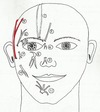
Auriculotemporal Nerve
Buccal Nerve
Mental Nerve
Ophthalmic Nerve, Suproarbital N., Supratrochlear N., External Nasal N., Infratrochlear N., Lacrimal N.
Maxillary Nerve, Zygomaticotemporal N, Zygomaticofacial N., Infraorbital N
Mandibular Nerve, Auriculotemporal N., Buccal N., Mental N
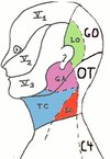
Ophthalmic N. dermatome division, Trigeminal N. (V1) 1st division
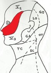
Maxillary N. dermatome division, Trigeminal N. (V2) 2nd division
Mandibular N. dermatome division, Trigeminal N. (V3) 3rd division
Dorsal Primary Rami C2-C4, Greater Occiptial N. (dorsal ramus of C2), Occipitalis Tertius N. (dorsal ramus of C3) and Dorsal Primary Ramus of C4 N.
External Jugular V
Posterior External Jugular Vein
Transverse Cervical Vein
Suprascapular Vein
Posterior Auricular Vein
Retromandibular Vein
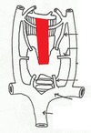
Anterior Jugular Vein * deep to SCM *
Subclavian Vein
Greater Occipital N., Dorsal Primary Rami of C2; supplies the occipital region
Occipitalis Tertius N., Dorsal Primary Rami of C3/ supplies the upper part of the back of the neck
Dorsal Primary Ramus of C4 supplies the lower part of the back of the neck
Ventral Primary Rami of C2-C4, Lesser Occipital N. (C2), Great Auricular N. (C2,C3), Transverse Cervical N, (C2,C3) and Suptraclavicular N. (C3,C4)
Lesser Occipital N. Ventral Primary Rami of C2; supplies the lateral part of the occipital region, posterior to the ear
Great Auricular N. Ventral Primary Rami of C2,C3; supplies the angle of the mandible, and the area inferior to the ear. it accompanies the external jugular V.
Transverse Cervical N. Ventral Primary Rami of C2,C3; supplies the anterior surface of the neck (the throat)
Dermatome for C2 Dorsal Primary Rami: Greater Occipital N. Ventral Primary Rami: Lesser Occipital N, Great Auricular N., Transverse Cervical N
Dermatome for C3 Dorsal Primary Rami: Occipitalis Tertius N., Ventral Primary Rami: Great Auricular N., Transverse Cervical N., Supraclavicular N
Dermatome for C4, Dorsal Primary Rami for C4, Ventral Primary Rami: Supraclavicular N
Great Auricular N., Ventral Primary Rami C2,C3
Lesser Occipital N., Ventral Primary Rami C2
Transverse Cervical N., Ventral Primary Rami C2,C3
Supraclavicular N., includes 3 branches: Lateral, Intermediate and Medial Supraclavicular N.
Descending Hypoglossi N. aka: superior root of the ansa cervicalis; C1 or C1,C2; descends from the hypoglossal N. to join the inferior root (descendens cervicalis N.)
Descending Cervicalis N. aka: inferior root of the ansa cervicalis; C2,C3; descends from the Cervical N. branches to join the superior root (descendens hypoglossi N.)
Ansa Cervicalis C1-C3; supplies the sternohyoid, sternothyroid and omohyoid muscles
Nerve to the thyohyoid muscle exclusively from C1 fibers through the hypoglossal N.
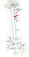
Phrenic N.
Platysma very weak; very thin; affected by Bell’s Palsey
Sternocleidomastoid (SCM) 2 heads; affected by torticollis (wryneck)
Trapezius
Anterior Triangle superior boundary: inferior border of the mandible; anterior boundary: anterior midline; posterior boundary: anterior border of the SCM
Posterior Triangle anterior boundary: the posterior border of the SCM; posterior boundary: the anterior border of the trapezius; inferior border: the superior border of the middle third of the clavicle
Submental Triangle only unilateral triangle; boundaries: right and left anterior bellies of the digastric and the body of the hyoid bone; floor: mylohyoid M.; contains minor veins and lymph nodes
Submandibular Triangle boundaries: inferior border of the mandible, anterior belly of diggastric and the posterior belly of digastric; floor: mylohyoid and hyoglossus muscles; contains: submandibular gland, internal carotid A., facial A., internal jugular V., glossopharyngeal N. (CN IX) and vagus N, (CNX)
Carotid Triangle boundaries: posterior belly of the digastric, superior belly of the omohyoid and the anterior border of SCM; floor: portion of the thyrohyoid, hyoglossus and inferior and middle constrictor muscles; contains parts of the common and internal carotid A., external carotid A. and 3 of its branches and corresponding tributaries of the internal jugular V.
Muscular Triangle boundaries: superior belly of omohyoid, anterior midline of the neck and the anterior border of SCM; floor: posterior layer of pretracheal fascia; contents: sternohyoid and sternothyroid muscles, thyroid gland, trachea and esophagus
Posterior Belly of Digastric Muscle
Anterior Belly of Digastric Muscle