Gluteal region and posterior thigh week 4 Flashcards
sciatica
The term sciatica refers to radiating pain, weakness, numbness, or tingling down the back of the leg and potentially into the foot which is rleated to injury or impingement of the sciatic nerve. Sciatica is a sx and not a medical condition itself.
Baker’s cyst
cysts containing synovial fluid that develop on the back of the knee (popliteal region). typically occur in conjunction with some other knee pathology (knee OA, meniscal tears)
Where is the gluteal region anatomically located? How does the gluteal region communicate with the pelvic cavity and perineum?
the gluteal region lies posteriorlateral to the bony pelvis and proximal end of the femur. Inferiorly, the gluteal region is continuous with the posterior thigh. communicates with pelvic cavity and perineum through the greater and lesser sciatic foramina, respectively
What are the 2 groups of gluteal muscles and what are their general fxns? What muscles are in those groups?
deep group: consists of small muscles that mainly fxn to laterally rotate the thigh (piriformis, obturator internus, superior gemellus, inferior gemellus, quadratus femoris)
superficial group: mainly fxn to abduct and extend the thigh (gluteus maximus, gluteus medius, gluteus minimus, tensor fasciae latae)
What tendon creates the sciatic foramina? What structures pass through the greater and lesser sciatic foramina?
The sciatic foramina are created by the sacrotuberous ligament
structures passing through the greater sciatic foramen: superior gluteal vein, artery, and nerve
piriformis
sciatic nerve
inferior gluteal vein, artery, and nerve
posterior femoral cutaneous nerve
nerves to the obturator internus, superior gemellus, inferior gemellus, and quadratus femoris
pudendal nerve, internal pudendal vein and artery (will be discussed in GU block)
structures passing though the lesser sciatic foramen:
tendon of the obturator internus muscle
pudendal nerve and internal pudendal vessels
Identify the ligaments in this picture. What foramina does the ligament labeled 1 create?

greater and lesser sciatic foramina

What is the origin, insertion, innervation, and function of the tensor fasciae latae?
origin: anterior superior iliac spine and anterior iliac crest
insertion: iliotibial tract (thickening of fascia lata). note that the iliotibial tract attaches to the lateral condyle of the tibia
fxn: abducts, medially rotates, and flexes thigh at the hip joint
innervation: superior gluteal nerve
What is the origin, insertion, innervation, and function of the gluteus maximus?
origin: sacrum, coccyx, ilium posterior to the posterior gluteal line, throacolumbar fascia, sacrotuberous ligament
insertion: iliotibial tract (which attaches to lateral condyle of tibia) and posterior proximal lateral femur (gluteal tuberosity)
fxn: extends the thigh at the hip joint especially from a flexed position (e.g. rising from sitting or during stair climbing). Laterally rotates and abducts the thigh at the hip joint when forceful contraction is required
innervation: inferior gluteal nerve
What is the origin, insertion, innervation, and function of the gluteus medius and gluteus minimus?
gluteus medius:
origin: ilium btwn anterior and posterior gluteal lines
insertion: lateral greater trochanter of femur
gluteus minimus:
origin: ilium btwn anterior and inferior gluteal lines
insertion: antero-lateral greater trochanter of femur
the gluteus medius and minimus have the same fxn and innervation.
fxn: abducts the hip and maintains a level pelvis in the coronal plane during locomotion when one foot is off of the ground.
anterior fibers: medially rotate and flex the hip joint
posterior fibers: suggested role of lateral rotation and extension
innervation: superior gluteal nerve
What is the origin, insertion, innervation, and function of the piriformis?
origin: anterior sacrum
insertion: superior greater trochanter
fxn: laterally rotates thigh at hip joint
innervation: nerve to the piriformis
note: the piriformis passes through the greater sciatic foramen
What is the origin, insertion, innervation, and function of the obturator internus?
origin: internal surface of the obturator membrnae and surrounding bone
insertion: medial greater trochanter
fxn: laterally rotates thigh at the hip joint
innervation: nerve to the obturator internus
What is the origin, insertion, innervation, and function of the superior gemellus?
origin: ischial spine
insertion: tendon of the obturator internus
fxn: lateraly rotates thigh at the hip joint
innervation: nerve to the obturator internus
What is the origin, insertion, innervation, and function of the inferior gemellus?
origin: ischial tuberosity
insertion: tendon of the obturator internus
fxn: laterally rotates the thigh at the hip joint
innervation: nerve to the quadratus femoris
What is the origin, insertion, innervation, and function of the quadratus femoris?
origin: lateral ischial tuberosity
insertion: intertrochanteric crest of femur
fxn: laterally rotates thigh at the hip joint
innervation: nerve to quadratus femoris
What muscles steady the head of the femur in the acetabulum?
piriformis, obturator internus, gemelli, quadratus femoris
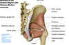
Identify these gluteal muscles.


Identify these muscles of the gluteal region.

Identify this muscle of the gluteal region.

What are the 6 lateral rotators of the hip? Where do they receive innervation from?
obturator externus (in anterior compartment of thigh)
obturator internus
piriformis
superior gamellus
inferior gamellus
quadraus femoris
As a group, these muscles (with exception of the obturator externus which is innervated by the femoral nerve made up of ventral rami of L2-L4), these muscles receive innervation from the ventral rami of L5, S1, and S2
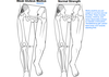
What is the main fxn of the gluteus medius, minimus, and tensor fasciae latae?? What fxn do thesemuscles have in gait? What sign may be observed when these muscles are weak and what conditions may they be seen in?
The primary fxn of the gluteus medius, minimus, and tensor fasciae latae is abduction of the thigh at the hip joint. During walking, their fxn is to stabilize the pelvis and prevent pelvic drop away from the stance limb as the contralateral limb swings through for limb advancement. In a position of hip flexion, the gluteus medius and minimus muscles also fxn as medial rotators of the thigh. These muscles are innervated by the superior gluteal nerve. When weight is supported on one leg, the gluteus medius and minimus of the stance limb contract to fix the pelvis so that it remains level. If these muscles are weak or paralyzed, when asked to stand on the affected limb the pelvis will sag toward the unsupported side, called Trendelenburg Sign. This sign is most commonly seen associated with stroke or in pts with hip OA who have a weak gluteus medius due to hip pain and disuse atrophy
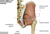
What structures pass above and below the piriformis? What supplies cutaeneous innervation to the posterior thigh and posterior knee and what is its relation to the piriformis?
The superior gluteal artery, vein, and nerve pass above the piriformis to supply the gluteus medius, minimus,and tensor fascia latae.
The inferior gluteal artery, vein, and nerve pass below the piriformis. The inferior gluteal nerve innervates the gluteus maximus.
The sciatic nerve also passes inferior to the piriformis. Some ppl do not have a sciatic nerve (the 2 nerves split right away) and one pierces the piriformis which can cause piriformis syndrome, pain
The posterior femoral cutaneous nerve passes inferior to the piriformis and medial to the sciatic nerve to supply cutaneous innervation of the posterior thigh and posterior knee
Identify the neurovasculature of the gluteal region.


What muscles are in the posterior compartment of the thigh? What muscles are considered “the hamstrings”? What common origin and fxn do the hamstrings share? What are the sources of blood supply and innervation?
- the hamstrings are comprised of the long head of the biceps femoris, semitendinosus, and semimembranosus. the short head of the biceps femoris is also in the posterior compartment of the thigh but is not a hamstring muscle.
- the hamstrings have a common origin off of the ischial tuberosity and their major fxn are extension of the thigh at the hip and flexion of the leg at the knee. the short head of biceps femoris does not corss the hip joint and therefore has no action at the hip joint
- the major source of blood supply to the posterior compartment are the perforating arteries (branches of the profunda femoral artery). the tibial nerve innervates the hamstrings and the common fibular nerve innervates the short head of biceps femoris
Describe how the perforating arteries travel to supply the posterior compartment of the thigh.
the perforating arteries are branches of the deep femoral artery as it descends anterior to the adductor brevis muscle. the first perforating artery originates superior to the adductor brevis, the second anterior to the adductor brevis, and the third inferior to the adductor brevis. all 3-4 perforating arteries pierce through the adductor magnus near its attachment to the linea aspera and supply the posterior compartment of the thigh.

What is the origin, insertion ,fxn, and innervation of the semimembranosus?
origin: ischial tuberosity
insertion: posterior medial tibia
fxn: entends the thigh at the hip joint, flexes the leg at the knee joint, medially rotates the leg at the knee when the knee is flexed
innervation: tibial nerve
What is the origin, insertion ,fxn, and innervation of the semitendinosus?
origin: ischial tuberosity
insertion: medial superior tibia
fxn: extends the thigh at the hip joint, flexes the leg at the knee joint, medially rotates the leg at the knee when the knee is flexed
innervation: tibial nerve
What is the origin, insertion ,fxn, and innervation of the biceps femoris long head?
origin: ischial tuberosity
insertion: lateral head of the fibula
fxn: flexes the leg at the knee joint, laterally rotates leg at the knee joint when the knee is flexed, extends the thigh at the hip joint
innervation: tibial nerve
What is the origin, insertion ,fxn, and innervation of the biceps femoris short head?
origin: lateral linea aspera in the middle third of the femur
insertion: lateral head of the fibula
fxn: flexes the leg at the knee joint, laterally rotates the leg at the knee joint when the knee is flexed (note it does not act at the hip bc it does not cross it)
innervation: common fibular nerve
Identify the muscles of the posterior thigh.


Identify the muscles of the posterior thigh.


What 2 nerves is the sciatic nerve composed of? What do its nerves innervate? Where do these nerves split? What paths do they take in the thigh and leg?
The sciatic nerve descends from the gluteal region into the posterior thigh. It lies on the adductor magnus and is crossed by the long head of the biceps femoris. The sciatic nerve (through it 2 branches) innervates posterior aspect of thigh and most of the leg. The sciatic nerve itself is not described as innervating anything. It is two nerves bound in one epineurim. Its branches split ino the tibial nerve and common fibular nerve on the posterior aspect of the thigh (but sometimes happens in gluteal region). Tibial nerve innervates all of the muscles of the posterior thigh (except for the short head of the biceps femoris). It then decends down the middle of the popliteal fossa and passes to the posterior compartment of the leg and then on to the sole of the foot. Common fibular nerve innervates the short head of the biceps femoris in the posterior thigh and then courses around the head of the fibula (comes down lateral side of popliteal fossa). It then divides into superficial and deep branches. The supericial branch innervates the lateral compartment of the leg and the deep innervates the anterior compartment of the leg. The common fibular nerve also innervates the foot.

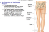
What is the location of the patella determined by?
Tightness of the iliotibial band, quadriceps femoris. Vastus lateralis is stronger than vastus medialis so patella is pulled more laterally.
Identify the structures in this picture. What is significant about the structures in red boxes?

pes anserinus is point of attachment of the gracilis, sartorius, and semintendinosus. can get bursitis in this area of the knee

