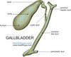
Gallbladder images Flashcards
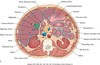
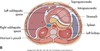
anatomy of gallbladder image

GB & biliary sysem

common bile duct image
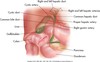
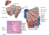
image hepatic lobules and sinusoids
normal relational anatomy image

GB anatomy image

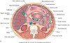
cross sectional- level of the 2nd lumbar vertebra

sectional anatomy level of 3rd lumbar vertebra

Sagittal section of the abdomen, 8 cm from the midline.

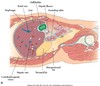
Sagittal section of the abdomen, 7 cm from the midline.


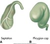
anatomic variants

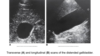
Sonographic Evaluation of the Biliary System

the main lobar fissure (MLF) is seen as an echogenic linear echo within the liver (L) connecting the right portal vein (PV) to the neck of the gallbladder (GB).

Main Lobar Fissure

-string connecting to PV; GB is the balloon; connects to GB neck

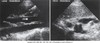
sag. GB


Trv. GB


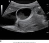
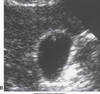
distended GB image

Distended GB
hydropic GB


contracted GB

Hydrops of GB

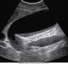
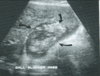
common bile duct image
Transverse view of the common bile ducts. This view shows the portal triad with the portal vein posterior, the common duct anterior and lateral, and the hepatic artery anterior and medial.


common bile duct image

Sagittal image. The hepatic artery (HA) is shown anterior to the common ducts (CD). The portal vein (PV) is anterior to the inferior vena cava (IVC). GB, Gallbladder.

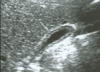
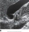
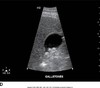
cystic duct image

Coronal decubitus view. The cystic duct seems to arise from the neck of the gallbladder (GB) (arrows). AO, aorta; IVC, inferior vena cava; PV, portal vein; L, liver.

sludge patterns
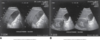

sludge patterns

sludge patterns

how do we measure GB wall thickness?

The gallbladder should be measured on the transverse image at the anterior wall that is perpendicular to the transducer (see markings

how do we measure GB wall when we see focal areas of thickening?

The sagittal image of the gallbladder is often at an angle to the transducer and may be used to measure wall thickness when the sonographer can achieve a perpendicular angle.
thickened GB wall image
we see - thick GB wall w/ some focal thickening
-calculous cholesystitis

acute cholecystitis

seen in this image:
hypoechoic area around GB wall=pericholecystic fluid
distended GB lumen > 4 cm
GB wall thickening > 3 mm
acute cholecystitis
we see: GB packed w/ sludge & wall thickening
what does pericholecystic fluid look like on US?
hypoechoic area around GB
we see acute cholecystitis w/ pericholecystic fluid collection around the GB

acute cholecystitis image
we see stones cause posterior shadowing

acute cholecystitis image

acute cholecystitis

- swollen edematous GB in middle aged man w/ cirrhosis & ascites*
- -shrunken liver floating in ascites*

Emphysematous Cholecystitis w/ image
What is Emphysematous Cholecystitis?

- Rare & fatal (15%) complication of acute cholecystitis affects mor men, 50% diabetic. gallstones may not be present(30-50%)
- Associated w/ a gas-forming bacteria in the gallbladder wall and lumen with extension into the biliary ducts
- This condition is a surgical emergency.

what does emphysematous cholecystitis look like?

looks like a curtain of gas w/in GB
- if gas is intraluminal look for a prominent bright echo along the anterior wall w/ ringdown or comet-tail reverberation directly posterior to the echogenic structure (arrow)

Gangrenous Cholecystitis common appearance is:

diffuse med.-coarse echogenic debris filling the GB lumen w/out bile duct obstruction
3 characteristics:
- no shadowing 2. not gravity dependant 3. no layering effect
GB and Biliary system

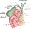
common bile duct


portal triad

Portal triad, portal vein, hepatic artery, common bile duct
Caroli’s Disease

Caroli’s disease

what is Acalculous Cholecystitis?

complication of acute cholechystitis w/out cholelithiasis
shown: Acute cholecystitis with thickening of the gallbladder wall secondary to edema and inflammation.
what causes acalculous cholecystitis?

decreased blood flow through the cystic artery
2° to trauma, burns, postoperative patients, HIV,
Extrinsic compression of the cystic duct by a mass or lymphadenopathy may also cause this condition.
acalculouscholecystitis w/ thickening of GB walls 2° to edema & inflammation

cholelithiasis

shown: many tiny stones (higher frequency transducer would improve clarity of image)
cholelithiasis

- A single, large gallstone is lodged into the neck of the distended gallbladder. The sharply defined shadow is noted.*
- (single layer of gallstones)*
cholelithiasis

A single, large gallstone is demonstrated near the neck of the gallbladder. The thickening of the gallbladder wall is noted.
cholelithiasis
Tiny gallstones are demonstrated within the sludge layered along the posterior margin of the gallbladder.
what pathology is present?

cholelithiasis w/ sludge, GB wall thickening, & ascites

cholelithiasis
Several medium-sized stones (without a shadow) are seen along the posterior margin of the gallbladder. The patient should be rolled into a decubitus position to watch the movement of these stones

Describe what we see: echogenic foci present in GB lumina, non shadowing
sludge and calcs (cholelithiasis)

cholelithiasis
Multiple stones with posterior shadow
wht we see: thick GB wall, sludge, cholelithiasis w/ shadowing
**always try to show shadowing**
cholelithiasis
shown: Solitary stone with posterior shadow.

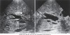
cholelithiasis: floating stones

shown: Longitudinal and transverse scans of the gallbladder (GB) with a layer of stone “floating” (arrow) along the thick bile layer of sludge (Sl).
Cholelithiasis: Wall Echo Shadow (WES)Sign indicates what?
that the gallbladder is a packed bag. The sharp posterior shadow is noted.
- This appearance is different from that of the porcelain gallbladder because the anterior wall is not as bright or echogenic.

how is the WES sign different from the porcelain gallbladder?
because the anterior wall is not as bright or echogenic.

WES sign image

porcelin GB image

polyp image
what is a polyp?
4mm adenomatous polyp image
a type of neoplasm⇒small, well defined non mobile, soft tissue projections from the GB wall
malignancy suspected if: wall thickening at point of attachment, or > 1 cm

Porcelain GB - calcium hard crust on GB wall, more common in older females; incidental finding, and is associated w/ cholelithiasis

25% will develop CA of the GB
differential includes a packed bag or WES sign
porecelain GB
bright wall from calcified calcium w./ shadowing

cholesterolosis
do not move when pt changes position

cholesterolosis
cholesterol polyps are small, ovoid wall projections seen to arise from the GB wall

adenomyomatosis

Multiple papillomas are demonstrated along the anterior wall of the gallbladder, causing a “ring down” of echoes to occur.
adenomyomatosis

GB Carcinoma facts:
GB carcinoma-rare w/ rapid profression- infiltrates the GB locally or diffusely and causes a thick & rigid wall.
mortality rate = 100%
Associated w/ cholelithiasis in 80% to 90% of patients
2x as common as CA of bile ducts, common in women >60
Arises in the body of the gallbladder or rarely in the cystic duct

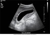
choledochal cysts

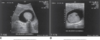
Transverse and longitudinal scan of a young patient with a choledochal cyst (Ccy) in the right upper quadrant.
choledochal cysts
appear as true cysts in RUQ w/ or w/out communication w/ biliary system, the cystic structure may contain intrenal sludge, stones, or solid neoplasm

caroli’s disease

caroli’s disease
dilated biliary ducts
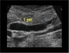
dilated biliary duct
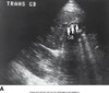
long. imgage of common biliary duct dilated to 1 cm

levels of obstruction
intrapancreatic & subpancreatic
levels of obstruction
porta hepatic

Mirizzi Syndrome

images shows stone lodged in cystic duct
Mirizzi Syndrome

other causes of obstruction
shown:Inflammation of the pancreas may cause the common duct to dilate. This patient has acute pancreatitis (P) and dilation of the common duct (crossbars).
A, Aorta; IVC, inferior vena cava

Carcinoma of the head of the pancreas with obstruction of the common bile duct (CBD) is demonstrated.other causes of obstruction
other causes of obstruction:
Dilated intrahepatic ducts secondary to a mass in the area of the porta hepatis
other causes of obstruction:
Dilated intrahepatic ducts secondary to a mass in the area of the porta hepatis

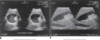
Choledocholithiasis

A, Transverse plane demonstrates gallstones (arrow). GB, gallbladder.
Choledocholithiasis
A, Transverse plane demonstrates gallstones (arrow). GB, gallbladder.
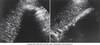
Ascariasis
on US we may see an enlarged duct w/ a moving “tube” or parallel echogenic lines w/in the biliary duct

Ascariasis
on US we may see an enlarged duct w/ a moving “tube” or parallel echogenic lines w/in the biliary duct

inrahepatic Cholangiocarcinoma
a large heterogeneous hypovascular solid hepatic mass may be seen w/ variable texture that ranges from hypoechoic to hyperechoic
biliary duct dilation may be associated w/ these obstructive masses

intrahepatic Cholangiocarcinoma
a large heterogeneous hypovascular solid hepatic mass may be seen w/ variable texture that ranges from hypoechoic to hyperechoic
biliary duct dilation may be associated w/ these obstructive masses
intrahepatic
Hilar Cholangiocarcinoma facts:
Includes jaundice, pruritus, and elevated cholestatic liver parameters.
Tumor may extend outside of the ducts to involve the adjacent portal vein and arteries.
Chronic obstruction leads to atrophy of the involved lobe.
Majority of patients die within 1 year of diagnosis

Hilar Cholangiocarcinoma facts:
Includes jaundice, pruritus, & high cholestatic liver parameters.
Tumor may extend outside ducts & involve adjacent portal vein and arteries.
Chronic obstruction leads to atrophy of the involved lobe.
Majority of patients die within 1 year of diagnosis

distal cholangiocarcinoma
difficult to distinguish from hilar cholangiocarcinoma; progressive jaundice in majority of patients.
Sonographic findings
**Sclerosing tumor is nodular with focal irregular ductal constriction and wall thickening.**
Has a hypoechoic and hypovascular appearance with poorly defined margins

distal cholangiocarcinoma
difficult to distinguish from hilar cholangiocarcinoma; progressive jaundice in majority of patients.
Sonographic findings
- Sclerosing tumor is nodular with focal irregular ductal constriction and wall thickening.
- Has a hypoechoic and hypovascular appearance with poorly defined margins

Metastases to the Biliary Tree

Most common tumor sites that can spread to the biliary system are from the breast, colon, or melanoma.
Metastases to the Biliary Tree

Most common tumor sites that can spread to the biliary system are from the breast, colon, or melanoma.
“FAST” Scan Protocol
spine is most posterior w/ aortaand IVC directly anterior
celiac axis arises from the antreior wall of the aorta.
LLL is anterior to the pancreas, which is directly anterior to the prevertebral vessels (aorta, IVC, CA, SV)
“FAST” Scan Protocol

Right upper quadrant. Inferior vena cava (IVC).
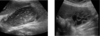
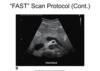
FAST Scan protocol
Right upper quadrant. Inflamed gallbladder

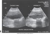
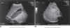
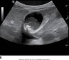
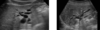
Acute Cholecystitis versus Cholelithiasis
Sagittal (A) and transverse (B) images of the distended gallbladder with edema and wall thickening secondary to acute cholecystitis and sludge.

Epigastric Pain: Pancreatitis
shown: Acute epigastric pain may be secondary to pancreatitis. This image also shows the enlarged pancreatic duct.

