Eye disease Flashcards
part 1,2,3
WHta do we need to be able to see?

Some processing visual signals ar ein retina but most is in the brain

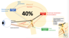
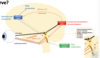
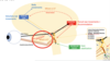
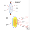
The eye and what happens where
- We need to get clear retinal image so need smooth refractive surface and transparancy of cornea with clarity of lens and then healthy optic nerve to transmit electrical signals ot brian so it can begin to interpret what we are seeing.
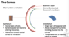
The cornea
5 layers
- Outmost layer is epitheliam cells
- Responsible for majoirty of refraction of light
- Needs to be smooth for clear image to be fgormed
- Transparancy maintained in part of epithelium (stoped tears enterign cornea) and endothelial cells which pump aqueous out of cornea to maintain clear cornea and supply nutrients.
- Can get oxygen form atmsophere to meet metsbolic needs.
- Endothelial cells do not reproduce
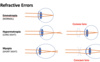
Refractive Errors
- SHape and size has impact on refraction of light but axial length has greater effect
- Hypermetropia/ myopia - due to difference in axial length then image may be formed behind or in front of retina not retinal plane lik emmetropria.
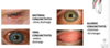
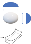
Astigmatism
- Cornea is not spherical
- Different radii of curvature in different axes
- In this case the verticsl radius is hsorter than horizontal radius so the curvature of the cornea is steeper in the vertical axis than horixontal axis
- To correct this a cyclindrical lens is needed with different powers in different axis
- Often described as rugby ball eyes
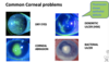
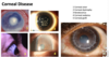
Corneal Disease
- Corneal ulcer - massive white area in middle and would be unabel to see through this. often by poor hygiene of contact lenses wearer.
- Corneal dystrophy - rare and some have genetic inheritance. Depends on opacity as to if they can see well.
- Keratoconus - cornea becomes distored

The lens
- SOme refraction
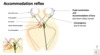
- Accomdoation/ fine focus - circumferential ciliary muscle contracts allowing lens capsule to relax, lens beocmes more spherical
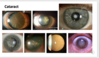
- Cataract = clouding of the lens -> age, diabetes, corticosteroids, congenital, trauma. Can be treated by surgery.

Cataract on image
Clouding of the lens
Age, diabetes, corticosteroids, congenital, trauma
Csn be treated by surgery

Retina layers
Centrla retinal arteru - to inner layers
chroid artery - to outer layer
Retina =

Optical Coherence Tomography
Allows oyu to see the exact layer where disease has occured

Foveal dip
Wher elight entering eye csn cause sharpest image as highest visual acuity

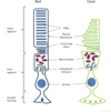
Concentration of Rods and Cones in Retina

Photoreceptors
Where phototrasnduction tkes place
Light stimulates chemical cascade -> hyperpolarisation
- Rods scotopic vision
- Cones photopic vision
- Disks hold opsin molecules
- Rhodopsin in rods, iodopsin in cones
- Light stimulates the isomerisation of retinal which activates opsin leading to hyperpolarisation of the photoreceptor.
- 3 different opsins present in all cones but one predominates in each of three different cones:
- “Blue” cones containing mostly blue-sensitive opsin are excited chiefly by a wavelength of around 420 nm,
- “Green” cones by a wavelength around 530 nm, and
- “Red” cones by a wavelength near 560 nm.

Phototransduction
Photons -> electrical signals that leave eye within optic nerve

On and off switches in phototransduction
Need to know: cells of inner retina are used in modifcation of the electrical signal created by transduction

Retinal Disease
- Colour blindness
- Retinal vascular occlusion
- Diabetes
- Macular degeneration
- Retinal detachment

Colour blindness
Multiple forms depending on which cones affected and if total loss or problem with functioning.
Red-green colour blindness is most common and usually mild (anomaly)
Uusually tested with ishihara test plates

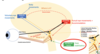
Coronar retinal artery occlusion (CRAO)
If the retina loose blood supply then the retina thins as cells die
Vision is very poor - usually present with sudden/total visual loss and not much can be done

Diabetic Retinopathy
Small blood vessels leak or get blocked
Loose pericytes so vessels expand and leak
Leads to oedema, exudation and haemorrhage in inner retinal layers
Vascular occlusion -> vascular dilation -> haemorrhage
Can see large amount exudste surrounding fovea area and lot shaemorrhages. Can treat with intervitreous injections

Wet Age -related macular degeneration
Often present with distortion

Why do you get distorition with Wet ARMD

Retinal Detahcment
Mor eprone if trauma to eye or diabetic. As we age vitreous retracts as scaffold so retina can move around and can detach from back of eye. but if vitreous remains slightly attached the traction can caue tear to form and fluid from vitreous cavity can lead behind retina and cause detachment.
Retina usually stays in place form vitreous scaffold and continuous pumping of retinl pigment epitheliun keeping potential space fluud free.
Symptoms - flashign lights,lots floaters and potentiall shadow appearing in vision.
Need to reatch retina as soon as possible so the photoreceptors can receive adewuate blood supply

The optic nerve
Fibres from superior part retina enter superior part optic nerve and vise versa.
A nerve fibre layer defect in superior part retina will cause inferior field defect and vise versa
Central retinal artery passes through centre optic nerve but doesnt supply blood to optic nerve, jsut to retina. Optic nerve relies on poserterior ciliary arteries for blood suppl.

Optic Nerve Problems
- Inflammation (optic neuritis)
- Glaucoma
- Ischameia
- Compression
Optic nerve problems can cause:
- Reduced visual acuity
- Altered colour visual
- Visual field defects

Optic Neuritis
Presents as progressiv eloss of vision over few days and then improves next 6-8weeks
Red desaturation - red objects appear more browny colour
If oyu examine optic nerve it csn still look normal if informaiton in posteiror prt nerve but if in anteiror part nerve then can see swelling
Can be reccurrent so may see optic nerve looks pale when examining.

Glaucoma
Complicated gorup diseases
Intraocular pressur e- usually maintianed at level between 14-21mmHg and ciliary body produces aqeous whih flows into anterior chamber annd out through trabecular meshwork. There is feedback mechanism but this isnt well udnerstoof.
As nerve fibres are lost, you can see central cup of optic disc enlarges and this is due to less nerve fibres entering optic nerve and you will get correpsonding field loss accoridng to area of optic nerve damaged most.
Treatment - various topical drops that either reduce aqeous production or improve outflow. Laser and surgery also options for more severe cases

Anteiror Ischaemic Optic Neuropathy
Most commonly due to micrvascular occlusion related to disbetes, hypertension, atherscelrotic disease, but can be due to inflammatory causes including Giant cell arteritis. Patients with this usually present with several weeks of being unwell, pain in jaw, loosing appetite, scalp tender etc and if not caught early then visual loss can occur which is irreversible and more seriously once one eye blidn then without treatment v likely other eye will become blidn to. Need high dose steroids which have own side effects so balancing act.
Posteiror ciliary arteries affected

Optic nerve compression
- Thyroud eye disease - causes inflamamitoe xtraocular muscles and lmyphocytic infiltration of orbital fat and causes ptoptosis and forward movement of eye (out of line) and can cause double viison. If swelling contunues then optic nerve compression. Might need steroids, immunsuppresion, surgery etc
- Orbital cellulitis- mostly in children due to sinus infection, bones of sinuses are fragile, sinus/infections can break through into orbit causing optosis optic nerve compression. Need simaging and then appropriate surgery to clesn out sinuses and relieve pressure of optic nerve

Whta stops us from seeing?

Visual Pathway
- Optic nerves meet and decussate at optic chiasm
- Dmaage to right side brain cause defect in left visual field vise versa

Visual field defect
Pituitary adenoma - bitemporal hemianopyias

WHta do we do with electrical signals from optic nerve?
*

Occipital lobe in visual pathway -
Occipital lobe - Persuit eye movements and accomodation
Eye movements controlle dby which optic nerves
3rd 4th 6th optic nerves
Eye muscles
Nuclei serving nerves in close proximity so communication occurs between the optic nerves

In persuit eye movements
- In persuit eye movement - eyes move in same direction. Medial rectus and contralateral lateral rectus
- In accomodation movements - eyes turn in. Both medial recgti
Accomdoaiton reflex
- Causes eyes to converge by 3rd nerve and get pupil constriciton and accomodation of the lens

Pupil constrictiona nd accomodation of the lens
Occur by short cilliary nerve from ciliary ganglion

Pupil Responses
In responds to accomodation and also light
In this reflex, no invovlement of visual cortex. Fibres form optic tract peel of into pre tectal ganglion and decussate at Edinger-Wesphal ganglion and fibres then travel to ciliary ganglion via short icliary nerve an dcause constriction of pupil which is bilateral due to decussation of fibres.
In absence light, pupil will then dilate
Active dialtion pupil aslo due to sympathetic stimulation as shown.
Different NTs used - Ach in Para-S and NA in symapthetic system

Parasympathetic vs Sympathetic in eye
Dilation can be very useful in examining the fundus

Pupil Responses - PERLA
- Often see PERLA acronym for cranial N II for examininignm. Pupils equal reacting to light and accomodation.
- To light - Pupil constriction
- To accomodation - Pupil constriction, convergence, lens accomodation
- If react to light then will react to acocmodation so tens to do just light on in practice. Only check acocmodation usually if dont react to light.
- If no response to light tho usually no response to accomodation
How to keep an image still
We do this from input from vestibular nuclei
Ear infections or little too much to drink and room spins

What is it part -

- We need to detemrine what we are seeing na dhwta it is.
- ventral stream (occipital lobe -> temporl lobe) = repsonsible for interpreting what we see.
- Mmeory is vital for vision,
- Recongition objects, facial recognition, colour mmeory
Dorsal stream responsible for telling us hwere object is

Dorsal stream = occipital lobe -> frontal lobe. Tells us where object is
Spatial awareness and the ability to find things -> thos ewith damaged to dorsal strema struggle with some activites relating to spatial awareness. But vision may be totally normal.
Spatial awareness and the ability to find things
Response

Response to do about hwta we see etc. Frontal lobe important. Eg, can choose which way we will run. Hvaign received visual input the frontal lobe connects to motor cortex to enable us to repsond appropriately.
Summary of visual pathway etc

Blindsight
We can shut of occipital lobes an dinfo from eyes go straight to frontal cortex and our bodies take appropriate action eg driving someone without really beign ocnciously aware as know where going.
Bilaterla occipitla lobe damage can also show similar form blindsight eg, can copy from board to pen and paper without being ocnciously able to see what they are doing and navigate aroudn room because visual signals can go straight to frontal lobe
Human echo location
Ability of brain to turn sound into visual image.
eg sharp clicking then sound bounces of object and then this one guy an then see the objects from echos bouncing off
What causes pain in eyes?
- Foregin body
- Trauma
- Reduced tear film
- Corneal epithelial disturbance
- Inflammation
- Raised IOP (intraocular eye pressure)
Cranial nerves
Involved in eyes
- II Optic Nerve
- III Oculomotor Nerve
- IV Trochlear nerve
-
V Trigeminal nerve:
- Opthalmic division - Upper eye lid including palpebral conjuctiva. Also bulbar conjuctiva, cornea, ciliary body and iris
- Maxillary division - Lower eye lid including palpebral conjuctiva
- Mandibular division
- VI ABducens nerve
- VII Facial nerve
Mechanism of pain
- Nociceptive - damage to non-neural tissue
- Neuropathic - damage to nerve
- Inflammatory - activation of nociceptors by inflammagory mediatora
- Sudden raised intraocular pressure - activation of nociceptors by ATP
Retina has no nerve endings, receptors are on surface of eye
- Noxious mechanical forces -> mechano-receptors
- Heat, exogenous irritants, endogenous inflammation -> polymodal receptors
- Dryness, temperature changes -> cold receptors
The eylid
Eyelid is just like any other part human body: has muscle, fat, skin, cartilagenous plate, lots oil glands, sweat glands.
Glands are really importnat. help you understand lumps and bumps on eyelid.

Blepharitis and Meibomianitis -
conditiosn of eyelashes and eyelids which can cause itching,rednes,s burning, blurred vision etc.
- Blepharitis - inflammation of eyelids, usually affects both eyes along edges of eyelids when tiny oil glands near base eyelashes become clogged.
- Meobomianitis - inflammation of meobomian glands
Top pic - squamous type
Bottom pic - Oily form - sebaceous type
- Inflammation of eyelids/ meibomian glands
- Veyr common
- Multiple type
- Treatment: Warm compresses and Lid hygiene
Meibomianitis - lots little white dots, (secretions from meiobomian glands). If they become inflamed or block then excess secretions form which can disturb tear film.

Lumps and bumps on eyelid
Chalazion - gland sblocked - still get secretions and so tarsal swells. Try massage eylid to open blocked gland and warm compresses.

Entropion, Ectropion and Trichiasis - eyelash problems
Entropion/Ecrtopion - as people age, fst shrinks,muscles actign on eyelid under less tension, eyelids turned in etc.

Summary of eyelid diseases

The conjuctiva - problems
Most common cause is conjuctivitis
Batceirla last 3/4 days without treamtnet but viral can last many weeks.

Foreign bodies in the eye

Cornea

Common corneal problems
*

Anterior chamber of the eye
Ciliary body produces aqeuous which travel forward and into anteiror chamber circulates and leave sby trabecula rmeshwork into canal of schwenn into venous system. If outflow for this fluid blocked then ontraocular pressure will increase and acutely the eye can become very painful.

Acute angle closure glaucoma AACG
- Sore red eyes, severe pain (ATP mediated), vomiting, reduced vision (as aqeous no where to go)
- Rainbow haloes (corneal oedema altered refraction of light)

Mechanism of acute angle closure glaucoma - Hypermetropia mechanism
(long sight smaller eyes)
- Shallower AC
- Enlarged lens
- Pupil dilates in low light
- Iris sticks to enlarged lens
- Aqeous cannot flow in to anteiror chamber and pushes iris forward
- Iris closes off already narrowed angle
- IOP rises.
Tag laser Iridotomy - for treatment Acute angle closure galucoma AACG
- Iridotomy - small holes in iris that will allow aqeous to always be bale to get into anteiror chamber
- Often have catarcats surgery
The iris (Uvea tract) and uveitis
Uveitis = inflamamiton of any or sll of these structures. Can be relate dothe rysstemic inflammatory diseases

Acute Anteiror Uveitis - commonest form uveitis
- Inflammation of the iris snd causes eye to become red and can see inflamamtory cells floating in anterior chamebr in aqeous. I lots cells then can form fluid level which you can see.Can also cause iris to stick to lens so funny shape when try dilate pupils sometimes cna be flower shape
- Treatment - topical steroids for eye and dialting drops to tyr breakdown wher eiris got stuck to lens.

Red eye summary

WHta is opthalmology?
- Opthalmology is a branch of medicine delaing with the diagnosis, treatment and prevention of diseases of the ye and visual system. It can be either a surgical or medical speciality. Most opthalmologits are super specilaised.
- In UK over 400,000 cataract operations are performed annually. This is most common operation performed in UK
- Opthalmologitst carry out more outpatient appointments than any other speciality