eye and ear Flashcards
Outer hair cells are damaged by what medication?
aminoglycosides
What is the pathway of hearing?
inner hair cells synapses on spiral ganglion in the modialus -> axons of the spiral ganglion cells form the auditory nerve of CN VIII -> enter brainstem at cerebellopontine angle -> terminate in ipsilateral coclear nuclei in the inferior cerebellar peduncles in the medulla -> project to ipsilateral and contralateral superior olivary complexes -> lateral lemniscus -> inferior colliculus -> medial geniculate nucleus in thalamus -> heschl’s gyri (not via the internal capsule)
What organ senses linear acceleration? What organ senses angular acceleration?
linear - otolith organs
vertical linear acceleration - saccule
horizontal linear acceleration - utricle
angular - semicircular canals
What is the crista amupllaris?
Sensory epithelium of the semicircular canals containing hair cells and supporting cells
Where are the hair cells in the semicircular canals? Where are the hair cells in the cochlea? Where are the hair cells in the otolith organs
semicircular canals - gelatinous cupula
cochlea - tectorial membrane
otolith organs - otoconia (calcium carbonate crystals imbedded in gelatinous matrix)
How does acceleration get transmitted into electrical signals traveling in the vestibular portion of CN VIII?
Vestibular pathway - The stereocilia of hair cells in the semicircular canals are lodged in a gelatinous cupula. Angular head accelerations cause endolymph fluid movement that moves the cupula, thereby exerting a shear force on the stereocilia. If the stereocilia are displaced toward the kinocilium, the membrane of the hair cell depolarizes. If the stereocilia are displaced away from the kinocilium, the membrane hyperpolarizes.

Where is the horizontal component of sound analyzed? Where is the vertical component of sound analyzed?
horizontal - superior olivary complex
vertical - pinna of outer ear
What is the cocktail party effect?
outer hair cells change height to make certain frequencies less stimulatory so you can focus on certain sounds and ignore other sounds
what are otoacoustic emissions?
noises that the outer hair cells make while changing height. Is used as part of auditory screening in newborns to test for deafness
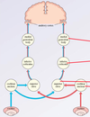
What does the vestibulo-thalamo-cortical pathway mediate? What is the pathway?
Conscious perception of equilibrium and head orientation in space
hair cells of the semicircular canals -> vestibular portion of CN VIII -> vestibular nuclei in medulla-> decussate and ascend to -> contralateral ventral posterior nucleus of the thalamus -> primary vestibular cortex (located just posterior to the face area of the postcentral gyrus)
What does the vestibulo-ocular reflex mediate? What is the pathway of the VOR?
Mediates compensatory eye movements to maintain visual fixation on a stationary object while the head is moving.
Afferent arc: afferent fibers in CN VIII -> synapse on the medial vestibular nucleus -> projects to the
- contralateral abducens nucleus -> inhibiting one pair of medial and lateral rectus muscles
- ipsilateral abducens nucleus -> exciting the other pair of medial and lateral rectus muscles

What reflex does the vestibulo-spinal pathway mediate? What is the pathway?
maintains postural equilibrium by activating anti-gravity muscles
Lateral vestibular nucleus -> descends ipsilateral through the lateral vestibulospinal tract -> motor neurons at all levels of the spinal cord

What reflex does the vestibulo-colic pathway mediate?
Maintains head stability during body movement
Medial vestibular nucleus -> descends through the medial longitudinal fasciculus (MLF) -> cervical spinal nerves

What reflex does the vestibulo-sympathetic pathway mediate?
Adjustments in blood pressure, heart rate, respiration and digestion during changes in position and posture
otolith organs -> vestibular nuclei -> brainstem parasympathetic control centers -> converges with baroreflex
What is the pathway of the pupillary light reflex?
Afferent arc - CN II -> synapse in ipsilateral pretectal nucleus -> sends axons to ipsilateral and contralateral Edinger-Westphal nuclei -> sends parasympathetics to both eyes -> both pupils constrict
When fixation is shifted from far to near, what three things happen? What nerve are they controled by?
1) accommodation - ciliary muscles change lens shape to increase refraction of light
2) convergence: both medial rectus muscles activate to direct both eyes inward towards the nose
3) pupillary constriction via the pupillary sphincter muscles
All three muscles (ciliary, medial recuts and pupillary spincter) are controlled by CN III
What is the visual pathway of neurons?
Rods/cones -> bipolar cells -> ganglion cells -> axons of ganglion cells for the optic nerve -> decussation of nasal retinal fibers (temporal visual fields) at the optic chiasm -> optic tract -> lateral geniculate nucleus -> superior visual field fibers go into Meyer’s loop and inferior visual field fibers go into parietal radiations -> visual cortex -> “where” pathway to parietal lobe or “what” pathway ro temporal lobe
What is the pathway of conjugate (saccade) eye movement?
“Burst” neurons in the paramedian pontine reticular formation (PPRF) synapse on motor neurons and interneurons in the abducens nucleus
- > motor neurons from the abducens nucleus cause the lateral rectus to contract
- > interneurons in the abducens nucleus travel through the medial longitudinal fasciculus (MLF) to synapse on the contralateral oculomotor nucleus and causes contraction of the medial rectus
What lesion causes internuclear ophthalmoplegia? How does it present?
INO lesion is a unilateral lesion in the medial longitudinal fasciculus (MLF).
Presents as impaired conjugate gaze. The ipsilateral eye cannot adduct and the contralateral eye has nystagmus when abducting.
Vergence is intact.

Where is the lesion in lateral (horizontal) gaze palsy? How does it present?
Unilateral lesion of the paramedian pontine reticular formation.
Impaired conjugate gaze in both eyes towards the side of the lesion. Conjugate gaze in both eyes away from the side of the lesion is normal.
Where is the lesion in one-and-a-half syndrome? What causes this syndrome? How does it present?
Lesion of one paramedian pontine reticular formation and ipsilateral medial longitudinal fasciculus. Can be due to a stroke in the dorsal pons or multiple sclerosis.
Presents as a combination of ipsilateral horizontal gaze palsy (can’t move eyes to the side of the lesion) AND ipsilateral INO (ipsilateral eye can’t adduct and contralateral eye has nystagmus)

What are the pupil abnormalities?
CN III palsy, Horner’s syndrome, Tonic pupil, Argyll-Robertson pupils, Marcus Gunn pupil
What are the two muscles of the middle ear? What do they do? What nerve innervates them?
Tensor tympani - stretches the tympanic membrane to facilitate high frequency vibrations. Innervated by CN V
Stapedius muscle - pulls the stapes off the oval window, attenuating the impact of loud sounds (acoustic reflex). Innervated by CN VII
Why can loop diuretics result in hearing loss?
There is a potential difference between the endolymph and the perilymph that maintain acoustic thresholds. Loop diuretics that block the Na+/K+/2Cl- transporter decrease the potential difference between the two fluids


