Exam 7 Flashcards

1
Q

What are the actions of insulin?

A

STOPS:

  • Gluconeogenesis
  • Glycogenolysis
  • Lipolysis
  • Ketogenesis
  • Proteolysis

Go:
-Glucose uptake in muscle
-Glycolysis
-Glycogen synthesis
-Protein Synthesis
Uptake of Ions specially K+, PO4

2
Q

How does Insulin affect carbohydrates metabolism?

A

Decrease blood glucose by helping glucose leave blood and limiting the raise in blood glucose

Increases glucose transport and adipose into muscle

Promotes gycogen formation in liver and muscle

Aids GLUCOKINASE: phosphorylation of glucose

Inhibits gluconeogenesis and glycogenolysis

3
Q

Actions of Insulin in Lipids metabolism

A

Overall, Inhibits mobilization and oxidation of FA

Inhibits Ketogenesis

Ketogenesis: breakdown of FA in liver, during fasting produces Ketoacidosis.

Promotes FFA storage as Triglycerides. LIPOPROTEIN LIPASE-Storage of triglycerides

Inhibits uptake of FFA in muscles, in favor of glucose uptake in muscles

Inhibits Lipolysis: HORMONE SENSITIVE LIPASE

4
Q

The effects of protein are overall cataolic or anarobic?

A

ANABOLIC

Decreases blood AA

Increases AA and protein uptake by tissues

Increases protein synthesis

Inhibits protein degradation

5
Q

What are the other actions of insulin?

A

Increases activity of Na/K ATPase pump = Uptake of K+ into cells

Promotes Phosphate, Mg++ uptake into cells

Decreases appatite via the SATIETY CENTER in HYPOTHALAMUS

6
Q

What happens when there is an excess of insulin in the blood/body?

A

Causes HYPOGLACEMIA

Cortisol release is stimulated to increase appetite

7
Q

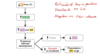
What or how does Insulin Resistance occur?

A

When blood insulin remains high (over-produced) and the cells fail to respond normally. This is a defensive mechanism during illness to protect brain’s glucose supply.

Resistance can occur due to receptors alterations, decreased affinity or number of receptors.

Post-receptor changes in the intracellular action of insulin.

8
Q

How do Hormones cause insulin resistance?

A

Cortisol, GH, Thyroid hormones, Epinephrine, Estrogen/Progesterone:

Epi: antagonizes insulin and stimulates the release of glucagon, thus increasing blood glucose

Progesterone: during pregnancy favors the transfer of nutrients to fetus.

9
Q

How does obesity cause insulin resistance?

A

Type II diabetes

Impared insulin signaling

Decrease GLUT4 expression in adipose and muscle tissue or it can be normal, but Decrease Transport of clucose occurs due to TRANSLOCATION/DOCKING of GLUT4 into plasma membrane.

10
Q

How does liver or kidney failure cause insulin resistance?

Sepsis and Insulin Antibodies?

A

Defect in kidney results in defect of Post reception pathway and possible decrease in GLUT4 in skeletal muscle.

Sepsis: Insulin signaling defect with infection. Stress causes hypoglacemia, release of glucose.

Insulin antibodies affect the normal effects of insulin

11
Q

Characteristics of Diabetes Mellitus type 1 and type 2

A

Type 1: insulin defficiency due to destruction of pancreatic Beta cells, autoimmune issue usually

Type 2: Insulin resistance: can be associated with down-regulation of insulin receptors in muscle and adipose tissues or issues with insulin signaling.

12
Q

What is Insulinoma?

A

Excessive production of insulin by Beta cells in the pancreas

13
Q

What are the results of lack of insulin or lack of insulin action?

A

Hyperglacemia due to decrease in hepatic output: Type 2 diabetes and decreased glucose uptake by cells. Cell starvation

Increase in glycogenic AAs liver keeps making it

Increase glucagon release because carbohydrate ingestion does not suppress it

Blood Hyperosmolality: glucose draws water and doesn’t leave

Osmotic diuresis in kedney: beyond threshold ~300 mOsm/L, glucose excess in urine and water follows.

Hyperlipidemia: Increased oxydation of fat, Fat accumulation in liver, Increase Ketoacidosis.

Increase glycolysis and Increase Lipid uptake

Peripheral tissue catabolism: Muscle wasting and weight loss

Increase gluconeogenesis and Decrease AA uptake by cells

14
Q

Glucagon

What cells secret it?

What stimulates its release?

What inhibits its release?

A

Secreted by alpha cells in the pancreas

Synthesized as preproglucagon untils its release

Sequence is identical in all species

Stimulation: Hypoglacemia, Protein, and AA intake/ingestion

Fasting, Stress (specially infection), Intense excercise, CCK (CHOLECYTOKININ is released when protein and fat is ingested)

Inhibition: Glucose, Insulin, SOMATOSTATIN

15
Q

Actions of Glucagon

Pathophysiology

A

G-protein/cAMP

Opposite of Insulin actions

Mobilizes energy, Increases glycogenolysis, gluconeogenesis, lipolysis, ketoacid formation

Little to no effect on glucose utilization by peripheral tissues.

Pathophysiology

Tumor of Alpha cells Glucagonoma

Results in Diabetes Mellitus and Necrolytic Migratory Erythma (blisters, swelling and pressure in areas of body)

Hyperglucagonemia/diabetes mellitus with infection

Glucagon:Insulin ratio increases

16
Q

Where is Somatostatin secreted?

A

Delta cells of pancreas, Hypothalamus, and GI cells

SS-28: GI tract

SS-14: Pancreas and Hypothalamus

Stimulated by all nutrients

Inhibits Insulin, Glucagon, GI Hormones, GI motility, enzymes, gastric acid secretion.

AAs are needed for liver to perform gluconeogenesis

17
Q

Adipose tissue as an Endocrine organ

Leptin and Adiponectin

A

Leptin

Inhibits appetite by inhibiting Neuropeptide Y

Increase BMR

Leptin resistance may contribute to Obesity

Adinonectin

Improves insulin sensitivity

High adinonectin = low risk of Type II diabetes

Low adinonectin = obesity and diabetes in cats by increasing TYROSINE PHOSPHORYLATION in insulin receptor in skeletal muscle

18
Q

What are the processes and regulatory systems that depend on calcium and phosphorus?

A

Vitamin D

Parathyroid Hormone

Calcitonin

Neurotransmission, learning, memory, muscle contraction, mitosis, mobility, secretion, fertilization, blood clotting, structure of bones and teeth.

Intestine: absorbs

Kidney: Reabsorption

Skeleton: Reservoir

Skin: Makes it

Liver: Makes it

19
Q

What is they biologically active form of calcium?

Where is the highest concentration?

What cells are involved in calcium homeostasis?

A

Highest: Extracellular

50% Ionidized (active) free form. The rest bound in albumin, complex anions.

10% complexed in other forms

Phosphate bicarbonate

Intracellular: lowest concentration

Cytosolic Ca++ can be increased as needed- fine balance control. Concentration depends on membrane permeability and motility. Intracellular storage.

The endocrine cell receptors are involved in Ca++ homeostasis

Endoplasmatic Reticulum: Calcium Channels, Na/Ca exchanger.

20
Q

Hypercalcemia & Hypocalcemia

What causes alterations in the forms of Calcium in plasma?

A

Increase in plasma Ca concentration

Constipation, polyuria, polydipsia, lethargy, coma, death.

Hypocalcemia: decrease in plasma Ca++.

Twitching, cramping of skeletal muscle. Seizures, sensory and motor neurons highly excitable due to Lower threshold for excitation. Lower ECF Ca++

Numbness/tingling (paresthesia) seizures.

  • Changes in plasma protein concentration
  • Changes in complex anion concentration
  • Acid-base disturbances
  • Changes in HCO3 cuases changes in Calcium
  • Lactation, Renal failure, Vitamin D disorders.
21
Q

Acedemia and Alkalemia

What is the difference in Albumin-bound-calcium in each state?

A

Acedemia: More H in the blood

  • High concentration of Ionized Ca++ (free form), so less is bound to Albumin

Alkalemia: Less H in the blood

-Low concentration of Ionized Ca++ bound to Albumin.

22
Q

Calcium Homeostesis

What role do kidneys, intestines, parathyroid, and vitamin D have on this?

A

Bone is constantly remodeled to that Ca can be released into the blood or absorbed from blood.

Intestine absorbs Ca, the amount is regulated by Vitamin D.

Kidneys reabsorbed Ca and this is regulated by the PTH calcitonin

23
Q

Calcitonin

A

Calcitonin works to control calcium and potassium levels. It does this by inhibiting the activity of the osteoclasts, the cells that break down bone. When the osteoclasts break down bone tissue, the calcium enters the bloodstream.

Osteoclast: bone reabsorbing cells Clast:collapsing

Osteoblast: bone forming cells Blast: building

24
Q

Phosphate metabolism

A

Components of ATP, DNA, Lipids, Cofactors, RNA, bone

It regulated through urinary secretion

It is stored in Muscle

The percentage absorbed from diet is fairly constant

Balances many Cations and it is important for buffering and Magnesium absorption.

25
Magnesium metabolism
Necessary for Neuromuscular transmission Cofactor in enzyme reactions Dietary Mg absorbed by gut is enhanced by Vitamin D Excreted in Urine
26
Parathyroid Hormone Chief Cells and Oxyphil Cells
1. Regulates plasma Ca and Phosphate 2. PTH stimulates bone resorption. Increases kidney reabsoprtion Ca and Phosphate Excretion in Urine. 3. P receptor acts when high Ca is present in blood, then degradation of PTH ganules and inhibits its release (PTH) Phosphate high in blood, then PTH secretion Increases via P receptor PTH also stimulates synthesis of Vitamin D Chief cells: Oxyphil cells: not present until puberty and decrease with aging
27
Stimulation and Inhibition of PTH
Stimulation: Decreased calcium (Bone resorption and calcium reabsorption in Kidney) Increased Blood phosphate (Phosphate excretion in urine) Decreased Mag Inhitition: Vitamin D negative feedback (calcium absorption in SI) Decrease PTH release Increased blood calcium
28
Action of PTH Receptor Pathway
Binds to plasma membrane receptors and activates G-proteins/cAMP pathway Bone: Increases bone resorption to relase Ca and Phosphate into blood Kidney: Stimulates calcium reabsorption DCT, while it inhibits Phosphate reabsorption in PCT causing excretion of Phosphate Increases Mg reabsorption and stimulates synthesis of vitamin D in kidneys Intestine: Increases Ca absoprtion via vitamin D
29
Hyper and Hypoparathyrodism
Hyperparathyroidism: Tumors or hyperplasia Primary Form Cs: Increased PTH and hypercalcemia, Hypophosphotemia, Renal calculi, Bone pain/Fractions Secondary Form Hypocalcemia, Increased Phosphate in blood, increases in PTH, may result from renal failure or increased dietary phosphate **Decreased blood Ca = Increased PTH** **Hypothyroidism** Caused by accidental removal of gland, Autoimmune destruction, Idopathic Cs: Decreaed PTH, Hypocalcemia, Hyperphosphatemia
30
Calcitonin synthesis and Role
**Parafollicular cells of Thyroid Gland** synthesize calcitonin Decreases blood Ca and Phosphate by: - Inhibiting bone resorption - Increases urinary P excretion - Inhibits renal reabsorption of Ca Stimulation: Increase blood Ca Vitamin D (via feedback) Ingested food, Ca absorbed in SI, so don't need to release it or reabsorb it from kidney
31
Role and Vitamin D synthesis
Required for bone formation and Increases Ca absorption from GI tract Hormone synthesized and Vitamin from diet Skin: synthesizes precursor **7-dehydrocholesterol** After absorption from GIT goes to liver and it is converted to **25-hydroxycholecalciferol** Kidney: most converted to **1,25-dihydrohycholecalciferol** Intermediate and active forms circulate bound to protein carriers
32
Vitamin D actions
Fat-Soluble, so it can be stored in adipose and liver -Toxicity results in **hypercalcemia, renal failure, elevated phosphate** Metabolism: excreted in bile Most Actions occur in the intestine - Stimulate Ca absorption via **calbindin** - Stimulates Mg absorption and phosphate absorption Weakly stimulates Ca and P reabsorption in Kidney
33
Vitamin D and Ca absorption Intestines Effects of vitamin D deficiency
Active -Intake Low, via transcellular process (dominates) Passive (no assistance needed) -Intake High, paracellular (dominates) Acts through cytosolic receptor and increas es production of CALBINDIN Calbindin binds Ca inside the cells and fascilitates transport to bsolateral membrane Other effects of Vitamin D Stimulates bone resorption in the prescence of PTH Increases Ca transport and uptake by SR in skeletal muscle cells Decreases PTH synthesis feedback effect Deficiency = muscle weakness, abnormal contractions, Ricketts in young, Ostomalecia in older animals, Cardiac disfunction
34
Melatonin and Eicosanoids What is the major role of melatonin? Where is it produced and synthesized from? What supresses melatonin release? where is the photic information transmitted and processed?
Major role in sleep and wakefullness Produced by the **Pineal Gland** and synthesized from **Tryptophan (N-acetyl-5methoxytryptamine)** with SEROTONIN as the intermediate. Blue light supresses melatonin release. Photic information from the RETINA is processed/recieved in the HYPOTHALAMUS and SNC via **Supra-Chiasmatic Nucleus, then to Pineal Gland**
35
Regulation of Melatonin What stimulates and inhibits secretion of melatonin? What is is related to?
Stimulated by darkness. It is related to the length of the night Retinal photoreceptors release **norepinephrine** which activates **beta-adrenergic** receptors in the pineal gland. Seratonin is converted into Melatonin by darkness stimulation Inhibited by light Retinal photoreceptors become hyperpolrized, which inhibits norepinephrine release. Blind people can still have ligth-induced sdupression of melatonin
36
Circadian Rythm of melatonin secretion What is it controlled by? When does secretion begins and peaks?
Controlled by **endogenous pacemaker in the supra-chiasmatic nucleus** Environmental lightining alters timing of the circadian rythm - Day-night cycles can modify the rythm - Brief pulses of light can supress release of melatonin Secretion usually begins at DUSK and pekas between 2-4 am Shifts in melatonin secretion after flights across time zones and in night-shift workers *Tryptophan-seratoning-melatonin*
37
Actions of Melatonin and Receptors
Activates Receptors **MT1 & MT2** Regulates/Affects - Sleep - Circadian rythm - Mood - Sexual maturation and reproduction - May have anti-inflamatory effects on immune system - May have beneficial effects on cancer (removing pineal gland enhances tumor growth) - Aging (decreases with age) may reduce cell damage by reducing free radicals
38
Biological functions and SAD
SAD: Seasonal affective disorder - Abnormal circadian rythms involved in mood disorders - Bright light therapy can decrease SAD **Sexual maturation and reproduction** Hours of light/darkness associated with reproductive activity May affect sex steroid synthesis and modulate ovarian function Horses seasonal estrous cycle. Spring decreases melatonin, which allows GnRH to increase and stimulate progesterone synthesis. Supressing melatonin in mares with light, babies born in January Decrease melatonin in puberty leads to increase in GnRH
39
How is Melatonin used in VetMed?
Treatment for Alopecia X in dogs -May decrease GnRH, which decreases FSH/LH effects on adrenal androgen precursors Tx for anxiety and seizure activity Short-term side effects are minimal -Sedation and incoordination
40
Eicosanoids What are the two major pathways for synthesis?
Group of signaling molecules synthesized by oxidation of 20-carbon essential fatty acids (EFA) **Arachidonic acid and Eicosapentaenoic acid** 1. Omega-3 EFAs from Eicosapentaenoic acid LESS-inflamatory 2. Omega-6 from arachidonic acid PRO-inflamatory **Omega-3 diet reduces inflamation** **Balance between each type determines actions**
41
Derivation of Eicosanoids
Omega-3 FAs: yields eicosapentanoic acid (5 double bonds) # 3,5 Omega-6 FAs: yields arachidionic acid (4 double bonds) # 2,4 - Prostanoids with 2 double bonds - Leukotrienes with 4 bonds
42
Nomenclature for Eicosanoids
- 2 letter abbreviation = PG - One A-C sequence letter = PGE - Subscript indicating the # of double bonds = PGE2 Prostaglandins A-I differ in subtituents on the cyclopentane ring - PGAs are alpha and beta-unsaturated ketones - PGEs are Beta-hydroxyl ketones - PGFs are 1,3-diols
43
EPA Cascade (eicosapentaenoic acid)
Formed from Omega-3 EFAs Forms mostly prostanoids Major function is to dampen inflammatory effects of arachidonic acid prostanoids Less inflamatory pathway
44
Arachidonic acid (AA) Cascade
Formed from Omega-6 FAs - Prostanoids: Prostaglandins (PG), Prostacyclins (PGI), Thromboxanes (TX), Major effects - Leukotrienes (LT) - Lipoxins (LX): stimulate inflammatory responses, modulate pain and fever, reproductive functions, INHIBIT GASTRIC ACID SECRETION (usually paracrine local action), Blood pressure regulation, Platelet activation/inhibition
45
Synthesis of Arachidonic Acid
Dietary precursors: - Linoleic Acid (18 carbon EFA) - Gamma linoleic acid - **Cats can't convert linoleic acid to arachidonic acid due to low Delta-6-desaturase enzyme** **Phospholipase A** releases arachidonic acid from phospholipids in cell membrane -Arachnidonic acid is oxydated by \*Cyclooxygenase (COX1, COX2) to make prostanoids \*Lipooxygenase (5LOX) to make leukotrienes
46
Major actions of prostanoids derived from Arachidonic Acid
-Local hormones with autocrine or paracrine action. Short half-life (seconds to minutes). Mediated by specific receptors. Mediate inflammation (except lipoxins) PGE2,Smooth muscle contraction, bronchoconstriction, heat, fever. PGI2vasodilation, inhibits platelet aggregation. TAX2 Vasoconstriction, stimulates platelet aggregation.
47
Inhibiting prostanoid formation NSAIDs
Non-steriodal anti-inflammatory drugs. - Decrease inflammation, redness, swealling, heat. - Inhibit COX1, COX2: **Aspirin, Carprofen, Fluxinin, Phenylbutazone, Ibuprofen, naproxen** - NSAIDs that inhibit only COX2: Less side effects, **Firocoxib, deracoxib, Melaxicam, Piroxicam, Celecoxib.**
48
Reproductive events after fertilization
**First Trimester**: 1. Migration of primordial germ cells from yolk sac 2. Sex cords develop in gonad, paramesonephric ducts develop 3. Sex evident from structures 4. Begening of development of male ducts and testes or Development of female ducts and ovaries **Second Trimester:** 4. Development of male ducts and testes or Development of female ducts and ovaries 5. Formation of braod ligament Bull and Ram 6. Testicular descent **Third trimester:** 6. Testicular descent Boar & human earlier Colt Later in the 3rd trimester
49
What is an Embryo? What is a Fetus?
Embryo: An organis in the early stages of development **Placentation has not yet taken place** Generally this embryo has not acquired an anatomical form that is readily recognizable in apperance as member of a specific species Fetus: Potential offspring within the uterus that is generally recognizable as a member of a given species **Marked by development of placentation and organogenesis**
50
What are the Primary Embryonic Layers? Embryology How does the embryo start?
The embryo starts as a mass of cells that eventually form cell layers and it will differentiate into embryo and proper and placenta **Endoderm** Digestive system, lungs, endocrine system **Mesoderm** Muscle, skeleton, Cardiovascular, _Reproductive System_ - Gonads (testicles and ovaries) - Uterus, Cervix, cranial vagina - Epididymis, ductus deferencs - Accessory sex glands **Ectoderm** Nervous system, Skin, Hair. Reproductive tract: - Vagina and Vestibule - Penis and clitoris
51
Embryology and placentation What is blastocyst? What is the Inner Cell Mass (ICM) and Trophoblast? What do the Trophoblastic cells eventually give rise to? What is the Chorion?
The embryo is called blasstocyst when a cavity is recognazible The ICM and trophoblast corresponds to the two distinct cellular populations that result from the embryo becoming partitioned. Tight junctions: found in the outer cells Gap Junctions: found in the inner cells **Trophoblast = Chorion** The Chorion: will become the fetal component of the placenta
52
What are the four steps to be achieved before the embryo can attach to the uterus? What is SYNGAMY? What is an ootid? What is a Zygote? What is a Blastomere? What is a Morula? What is a Blastocyst? What is a hatching Blastocyst?
1. Development within the confines of ZONA PELLUCIDA 2. Hatching of the BLASTOCYST from zona pellucida 3. Maternal recognition of pregnancy 4. Formation of extraembryonic membranes **Syngamy:** fusion of the male and female PRONUCLEI **OOTID:** cell name when a female and male pronuclei can be observed -It is one of the largest cells and has subsequent cell divisions within the cinfines of the zona pellucida **Zygote:** Single-celled embryo that undergoes mitotic divisions called CLEAVAGE DIVISIONS First cleavage division: generates two-celled embryo, which are called **Blastomeres** **Morula:** After the eight-celled embryo stage the ball of cells fromed is called Morula Early blastocyst: after the morula continues to divide a blastocyst is formed. **Blastocyst consists of:** - ICM: inner cell mass - Blastocoele: cavity - Trophoblast: single layer of cells **Hatching blastocyst:** grows in zona pellucida and free floats within the uterus
53
What do the extraembryonic of the preattachment embryo consists of ?
- Yolk Sac: from primitive endoderm. Feeds the embryo until placentation - Chorion: from Trophoblast, primitive endoderm, and mesoderm - Amnion: filled with fluid and serves to hydrolically protect the embryo - Allantois: comes out of the hindgut joins and fuses with chorion to become **Allanto-Chorion** **1. Trophoblast cell is the placenta** **2. Blastocoel** **3. Inner cell mass: become fetus**
54
Origen of the Pituitary Gland
Posterior Lobe: **Neurohypophysis** - Contains axons and nerve terminals of neurons from hypothalamus - Formed from a **diverticulum from floor of brain = *infundibulum*** Anterior lobe: Stomodeal Ectoderm **Adrenohypophysis** - Tissue from the root of the mouth - Glandular epethilieal cells produce GLYCOPROTEIN hormones - Formed from an evagination from the oral cavity = ***Rathke's Pouch***
55
What happens to Rathke's pouch?
Stalk of Rathke's pouch regresses and separates from Stomodeal Ectoderm -It becomes closely associated with cells of INFUNDIBULUM Adrenohypophysis loses attachment to mouth
56
What hormones are released by the anterior and posterior pituitary gland? Where are posterior pituitary hormones released into? Hormones released into circulation require a larger exogenous amount to be effective than locally released hormones
Anterior: - ACTH: adrenocorticotropic hormone - TSH Thyroid stimulating hormone - Gonadotropins FSH, LH - GH Growth Hormone - Prolactin PRL Posterior: **Deposited directly into circulation, Hypothalamic hypophyseal Portal System** - ADH Antidiuretic hormone - Oxcytocin
57
Sexual Differentiation | (Involves specific substances)
**Sex differentiation**: process... group of unspecified cells develop into a functional recognizable group of cells (ex: male and female reproductive tracts) **Sex Determination:** A system that determines the sexual characteristics.. GENES, ALLELES or Hormonal parameters. Ex: vulva, penis **Karyotype:** This describes the chromosomal complement of an organism, X, Y Dog: 78 XX (female), 78 XY (male) **Phenotype:** observed characteristics that depend on the genotype and affected by the environment **Chimera:** produced by the fusion of two different zygotes **Mosaic:** an individual with two different cell lines that originated from the same individual
58
SExual differentiation 3 Stages
1. Chromosomal Sex (Karyotype): determined at fertilization , XX or XY 2. Gonadal Sex: **Sry gene induces testes formation** 3. Phenotypic sex: determined by substances produced in the male testes to cause regresssion on the female tract
59
Sexually indifferent Stage Microanatomy of Sexually indifferent Stage
**Primordial germ cells:** (primary undifferentiated stem cells that will differentiate toward gametes) - Originate in the yolk sac - Migrate trough the hindgut to the undifferentiated gonad within the dorsal body wall (a.k.a genital or gonadal ridge) **Pronephros** Primitive Kidney **Mesonephros** male (female regress) **Metanephros** functional kidney **Mesonephric ducts (Wolffian ducts)** male (female regress) **Paramesonephric ducts (Mullerian ducts)** Female (male regress) **Primitive sex cord** **Primitive germ cells**
60
Sex Determination Male Key Players
Initially, Testis determining Factor (TDF) and Sex Determining Region-Y (SRY) causes production of TESTOSTERONE - Antimullerian Homrnone secreted by **Sertoli Cells** (AMH) causes degeneration of the paramesonephric duct - Dihydrotestosterone causes development of penis
61
Sex Determination female Key Players
XY chromosomes with Sex Determining Region-Y and Testis determining factor - No SRY protein - Ovaries develop - No Anti-Mullerian Hormone - Paramesonephric ducts become the oviducts, uterus, cervix, and part of the vagina - Complete female tract
62
Developmental Sequence of the Testis
Level of Gonadal Ridge: - Anti-Mullerian Hormone produced by Sertoli Cells - Testosterone prescence causes regression of female duct system - Paramesonephric duct (Mullerian duct) Regress (Antimullerian Hormone) - Mesonephric ducts join Rete Tubules (future Rete Testis) becomes Efferent ducts, **Epedidymis**, and **Ductus Deferens** - Mesonephric tubules become **Seminiferous Tubules** - Undifferentiated sex cords become Seminiferous tubules - Tunica albuguinea remains
63
Male Fetal Maturation: Testes Descent
Prior to descent, they are in the retroperitoneal position **The gubernaculum** connects the fetal testis to the peritoneum After the Gubernaculum goes through the inguinal ring, ther is a rapid growth of the **distal gubernaculum** **O**nce the testes are through the **inguinal region** they are pulled through because the gubernaculum shrinks. Abdominal visceral growth pushes them too. **The Scrotum** has vaginal tunics where the testes are kept.
64
Female Differentiation
Absence of Testosterone, Anti-Mullerian Hormone and Dihydrotestosterone - Rete tubules dissapear - Paramesonephric (Mullerian) duct develops and enlarges - Regressing epithelial cords - Distinct clusters of follicles at periphery of ovary - The gonad resembles an ovary - Mesonephric ducts have completely regressed - Fused Paramesonephric ducts (future cervix and uterine body) and continue to fuse/elongate to from a more defined cervix and vagina - Cranial vagina: - Caudal vagina: originates from urogenital sinus **Final Stages** - The broad ligament develops (from peritoneum, genital fold) - The ovaries magrate caudally to fetal growth
65
Endocrine Reproduction ## Footnote **Simple and Neurocrine Reflexes** **Both start with Stimulus, Sensory Neuron, Transmission, and go different ways via Efferent neurons.**
**Simple Neural Reflex:** Employs nerves that release simple neurotransmitters directly onto target tissue. Ex: muscles for sexual behavior are stimulated by thermal, tactus, or visual factors. Efferent neurons release neurotransmitters to sweat glads, scrotm, etc. **Neuroendocrine Reflex:** Requires a neurohormone (released by neuron) to enter into blood and act on remote target tissue -signal goes to brain, release neurohormone into bloodstream, target tissue Ex: The hypothalamus is the neural control center for Repro Hormones. Calf suckling stimulates release of oxytocin and mammary gland is the target tissue
66
Hypothalamus Hypothalamo-Hypophyseal Portal System (Anterior Pituitary) Posterior Pituitary
Paraventricular Nucleus: has direct effect on reproduction The pituitary is positioned in depression of the Sphenoid bone, **The Sella Turcica** **The third ventricle** separates the lateral portions of the hypothalamus **Hypothalamo-Hypohyseal Portal system** allow minute quantities of hormone to act before being diluted in the systematic circulation **Posterior Pituitary** does not have a portal system. Neurohormones are deposited directly into **Systematic Circulation**
67
Characteristics of Reproductive Hormones
- Act in small quantities - Possess short half-lives - Bind to specific receptors - Can cause sexual promotion (ex: steroids) - Can help maintain pregnancy - Luteolysis (destruction of the Corpus Luteum)
68
Hormones classification according to source
Hypothalamic: Gonadotropin Releasing Hormone (Neurohormone) Pituitary: Follicle Stimulating Hormone, Luteinizing Hormone, Oxytocin (Neurohormone), Prolactin. Gonadal Hormones: - Prostaglandin F2 Alpha (uterus) - Progesterone (ovary, Corpus Luteum) - Estrogen (ovary, follicle) - Equine Chorionic Gonadotropin (placenta)
69
Hormones Mode of Action Biochemical classification
Neurohormones: -GnRH, FSH, LH, Oxytocin Realising Neurohormones: GnRH Gonadotropins: synthesized by gonadotrope cells in anterior pituitary gland -FSH, LH Sexual Promoters: secreted by the gonads -Estrogen, Progesterone, Testosterone, eCG, hCG Pregnancy maitenance hormones: - Progestins - Placental Lactogen Leutolytic Hormones: -PGF2a **Peptides** very small protein. GnRH, Prolactin, Relaxin, Decapeptides. **Glycoproteins** Large proteins. Polypeptide protein with carbohydrate moieties. FSH, LH, Inhibin **Steroids** Progesterone, Testosterone, Estradiol **Protaglandins** Lipid base. PGF2a, PGE2 \*\*\*To make an animal ovulate we can use gycoproteins, but they are large and cause antibodies to be formed in response\*\*\*
70
Protein Hormones Steroid Hormones
They bind to transmembrane receptor and cause activation of P**rotein Kinase Chain** **G-coupled** mechanism **Steroid Hormones** can bind to membrane and nuclear receptors -Fast response binds to membrane receptor: **Estradiol** increases myometrial contractions **Progesterone** decreases myometrial contractions -Slow Response: binds to nuclear receptor **Estradiol & Progesterone**
71
CNS Regulation of the Reproductive System Hypothalamic Nuclei
Receives, processes, and interprets sensory inputs **Ultimately regulates GnRH** - Tonic Center in Female - Surge Center in Male (only paraventricular) **Hypothalamic nuclei** -Nerve cell bodies found in the hypothalamus, Axons terminate in the H-HPS **-**GnRH, TRH, CRH - Picogram amounts released - Neurohormones released into blood
72
Hypothamalo-Pituitary Gonadal Axis
Regulates production of hormones based primarily upon a positive or negative feedback system Ex: **Estrogen** from a growing follicle causes positive feedback on the surge center, but as negative feedback on the anterior pituitary ++GnRH --Itself Key players: - GnRH - FSH, LH, Prolactin (anterior pituitary) - Oxytocin (paraventricular nucleus and transported to posterior pituitary for release into the bloodstream - Gonadal hormones: Testosterone, Estrogen, Progesterone.
73
Mechanism of defeminization of Hypothalamus Female ESTRADIOL
Testosterone produced by the testes penetrates the BBB. - Once testosterone is in the brain, it is converted to ESTRADIOL - ESTRADIOL defiminizes the hypothamlamus because the surge center does not develop \*\*\*Steroid hormones go back and forth in form\*\*\* **Estradiol bound by Alpha-Fetoprotein** which prevents estradiol from crossing the BBB **Aromatase Enzyme** converts testosterone to Estradiol Alpha-Feto-Protein (¤E2)
74
Average age of puberty Determine more by weight than by age
75
Puberty Hypothalamic Nuclei and Release of GnRH Ultimately causes spermatogenesis and ovulation Hypothalamic Nuclei Gradually Matures
- Puberty is not limited by the potential response of the pituitary or gonads - Prepubertal animal given exogenous GnRH will produce FSH and LH ***It is the failure of the hypothalamus to produce sufficient quantities of GnRH to cause gonadotropin release that is known to be the najor factor limiting onset of puberty*** The development of thsese nuclei are dependent on: - Threshold body size - Nutritional factors - Environmental cues - Photoperiod - Genetics
76
Possible Influence of Metabolic Signals Upon GnRH Neurons Influence of Social Cues
- Blood glucose might stimulate GnRH neurons - Adipocytes secrete Leptin, which may stimulate neuropeptide Y and GnRH - Kisspeptin neurons thought to directly act on GnRH neurons - Blood FAs may stimulate neurons **Social Cues** Exposing female swine to a boar decreased the onset of puberty by 4 weeks. In lambs puberty requires decreasing day length, so the time of the year when they are born can influence their onset of puberty
77
Definitions female cyclicity
Estrous (adj.) – ex: The length of the estrous cycle in the bovine is 21 days. Estrus (n.) – period of sexual receptivity. Heat. Estrous Cycle – consists of a series of predictable reproductive events beginning at estrus and ending at the subsequent estrus. Provides females with repeated opportunities to copulate and become pregnant. Anestrus – not cycling Estrual (adj.) – used to identify a condition related to estrus Parturition – giving birth Uterine involution – acquisition of normal uterine size and function (gross and histologic have different time frames)
78
Estrous Cycles Categorization Different type of breeders
• Estrous cycles are categorized according to the frequency of occurrence throughout the year. There are 3 types: cattle I21 ) Swine Rodents dogs , , 1. Polyestrus - • Have a uniform distribution of estrous cycles throughout the entire year • Cattle, Swine, Rodents 2. Seasonally Polyestrus • Have “clusters” of estrous cycles that occur only during a certain season of the year Elk • Short-Day Breeders – they cycle as the day length is decreasing (fall) sheep goat, deer , , * Sheep, Goats, Deer, Elk * Long-Day Breeders – they cycle as the day length increases (spring) • Mares 3. Monoestrus • Have only one cycle per year • Dogs, Wolves, Foxes, Bears • Domestic dogs typically have 3 estrous cycles per 2 years, but are classified monoestrus
79
Estrous Cycle 2 Major Phases
1. Follicular Phase - Primary Ovarian Structure: Large Follicle (s) - Hormone: **Estradiol** (secreted by follicles) - Period: from Corpus Luteum regression to ovulation 2. Leutal Phase - Corpus Luteum - **Progesterone** (secreted by CLs) blocks Estradiol - Period: from ovulation to CL regression - Follicles continue to grow/regress, but do not produce high quantities of Estradiol
80
Estrous Cycle 4 Stages Follicular phase Proestrous Estrus
**1. Proestrous:** Begins when **progesterone declines** as a result of **Luteolysis** Ends at the onset of **Estrus** 2-5 day period depending on the species ***_Transition from Progesterone to Estradiol_*** FSH & LH Antral Follicles mature for ovulation and tract prepares for estrus and mating **2. Estrus:** Peak Estradiol secretion by dominant follicles Duration varies between species **3. Metestrous** Corpus Luteum formation (Luteinization) Begining of Progesterone secretion Transition from Estradiol to Progesterone Dominance **4. Diestrus** Sustained secretion of high levels of Progesterone from mature Corpus Luteums Longest stage of the cycle (2/3 of the time) Ends with Luteolysis
81
Behavioral Estrus Signs in different species
82
Estrous Cycle Phases (Follicular & Luteal) Estrous Cycle Stages (PEMD) Exceptions BITCH & QUEEN
Anestrus: 5 months **Follicular Phase** - Proestrous: 9 days; drop of blood FSH (**Inhibin** secreted by *developing follicles* inhibits FSH secretion) - Estrus: 9 Days; decreasing Estradiol (secreted by *Granulosa Cells* of Follicle) and rising Progesterone Ovulation 2-3 days after LH Surge (primary oocyte needs 2 days to mature) Fertilization 48-72 hours after ovulation. The delay allows **Superfecundation** (multiple ovulations produce multiple oocytes during a single estrus period that are fetilized by spermatozoa from different males) in canids. **Luteal Phase** Diestrus: 2 months Both pregnant and open bitches are considered to be in Diestrus. Pregnancy status does not alter the length of diestrus. Bitches that do not become pregnant are **pseudopregnant**
83
Reproductive Cyclicity of Queens with or without Copulation
**Proestrous** **Estrous** **Proestrous:** Interestrus period in queen not mated (ovulates only if stimulated) Period of about 7-10 days Growing follicle, but it dies if not stimulated **Diestrous** about the same length as gestation **Anestrus:**"Without cyclicity" Females that do not exhibit regular estrous cycles Causes of anestrus: -Pregnancy (NOT IN CATS) Quees come back in heat within days after parturition Cattle wait 40-45 days to get pregnant again - Presence of offspring - Season (photoperiod) - Stress - Pathology
84
What is Anestrus? What are possible reasons for the lack of reproductive cycles in female animals?
Anestrus: "Without cyclicity" Females that do not exhibit regular estrous cycles Causes of anestrus: -Pregnancy (NOT IN CATS) Quees come back in heat within days after parturition Cattle wait 40-45 days to get pregnant again -Presence of offspring TRUE ANESTRUS: Insufficient hormonal stimulus Poor nutrition, Stress, Pathology APPARENT ANESTRUS: Failure to detect Estrus Failure to recognize that a female is pregnant
85
Gestational Anestrus Presence of Offspring Anestrus
Normal condition brought about by inhibition of Gonadotropins Releasing Hormone by **Progesterone** (Secreted by Corpus luteum and Placenta) **Presence of Offspring:** Suckling can't be totally responsible for lactation anestrus, but the presence of the calf has some effet - Nerve damage from mammary gland, afferent pathway severed, LH frequency and amplitude increases, the cow returns to cyclicity. - When calves are weaned from cows with intact mammary glands. - Mammary stimulation is not totally responsible for anestrus.
86
Lactation Anestrus
Cyclicity is completely supressed in SOWS during lactation In suckled cows, it is delayed as much as 60 days (Supression of LH) -Duration influence by suckling sessions. **3 or more** sucklings per day causes **partum anestrus** **-Dairy cows** typically do not display lactational anestrus because the calf is removed **-Beef cows** about 60 days
87
Seasonal Anestrus Normal Condition
Usually allows animals to carry fetus during a **seasonally favorable time** Ex: Spring for lambs Modified by **photoperiod** to come back to cyclicity, similar to onset of puberty
88
Possible role of **Kisspeptin Neurons** in regulation of cyclicity in long and short day breeders
1. Increased day length = excitation of retinal neurons 2. **Retinal neurons** synapse in **suprachiasmatic nucleus** 3. Inhibitory neurons convert excitatory reponse to an inhibitory response 4. Postsynaptic adrenergic fiber- decreases norepinephrine secretion 5. **Decreased norepinephrine = reduced melatonin** secretion from pineal gland (pinealocyte) 6. Low melatonin results in excitation to the RFRP neurons and they increase secretion of neurotransmitter RFRP-3 7. Increased RFRP-3 - Short-day breeders: Decreased Kiss-10 causes decreased GnRH = Decreased FSH, LH - Long-day breeders: Increased Kiss-10 causes increased GnRH = Increased FSH, LH
89
Anestrus Induced by Stressors Estrous Cycles parameters Mare, Cow, Ewe, Bitch, Queen
Negative energy balance can induce Anestrus Starvation leads to shutdown of Repro system first **Primiparous females** have high metabolic demans bc still growing and lactating. Mare: 19-22d, Estrus 4-7d, Diestrus 14-16d Cow: 18-22d, 6-30 hrs, 16-17d Ewe: 14-16d, 24-36 hrs, 12-13d Bitch: 4-8 mts, 7-9d, 60-80d Queen: Induced ovulator, inter-estrus period 7-14d if not mated
90
Folliculogenesis How may do exist at birth? What kind of pattern does it have? **Reminders** • GnRH – Gonadotropin Releasing Hormone * Gonadotropins * FSH – Follicle stimulating Hormone • LH – Luteinizing Hormone * Progesterone (P4) * Estrogen (E2) * Corpus Luteum (CL), Corpora Lutea (CLs) • Estrous – the CYCLE * Estrus – the STAGE of the cycle
Defined as the process whereby immature follicles develop into more advanced follicle and become candidates for ovulation **A few hundered thousand PRIMORDIAL follicles exist at birth** The majority of the primordial follicles present at birth will not be selected to develop Follicles grow and regress even before puberty **Wave-like patter** process
91
Folliculogenesis Primary Ovarian Structures
Primary Follicles: from primordial follicles, begining of development Secondary Follicles: Zona pellucida present Antral (Tertiary) follicle (s): - Developing antral follicle, Cortex present - Antral (dominant) follicle: Antrum present (filled with follicular fluid) Big Ovulating Follicle: Erupts and Oocyte exits Corpus Luteum: right after ovulation Corpora Albicans \*In general all types of follicles are present within the ovary at any point in time\* \*Developing and functional Corpora lutea may or may not be present depending on the stage of the Estrous cycle\* **Exception Mare**
92
Follicular Phase Governed by? Four significant events? What are they key players in Follicular Phase? What role does Inhibin play? What role does Estradiol have? Which hormone exerts positive feedback on Hypothalamus to cause LH Surge? Which hormone produces negative FSH feedback on Hypothalamus? Where and what cells secret Inhibin and Estradiol?
Governed by Ovary, Hypothalamus and Anterior Pituitary through secretion of ESTRADIOL in the absence of Progesterone Events: 1. **FSH** (primarily) & LH released from anterior lobe of Pituitary 2. Follicular preparation (growth) for ovulation 3. Sexual receptivity 4. Ovulation (**LH** highest peak) Dominant Hormone is ESTROGEN during follicular phase - Changes in reproductive tract - Behavioral changes - Controls onset of preovulatory LH Surge **Key Players in Follicular Phase** -**Tonic Center of the Hypothalamus**: releases Gonadotropin Releasing Hormone, which stimulates FSH\> and LH release from anterior lobe of pituitary gland FSH\> and LH cause the growth and development of follicles on the ovaries The follicles produce ESTROGEN -**Surge Center of the Hypothalamus:** responds to Positive Feedback to increase levels of ESTROGEN in the absence of Progesterone \**Positive feedback* causes hypothalamus to release large quantities of GnRH\* -GnRH causes **LH Surge**, which results in OVULATION \*\*The Surge Center is turn ON once ESTROGEN reaches a threshold level\*\* **Inhibin** is secreted from Granulosa cells of Antral Follicle (largest) and inhibits FSH secretion by targeting Gonadotrophs of anterior lobe of pituitary gland **Estradiol (also secreted from Granulosa Cells) levels** reache threshold and it stiulates secretion in Large Quantities of GnRH, which leads to LH SURGE preovulation
93
What are the two groups of GnRH neurons in the hypothalamus? What are the results of stimulation to those neurons?
1. The Surge Center - Preovulatory increased GnRH = LH surge (Estradiol positive feedback = Lots of GnRH, thus FSH and LH secreted) 2. The Tonic Center - Inhibin secretion from granulosa cells leads to decrease FSH release. Summary of Preovulatory Hormones - Decrease Progesterone due to Luteolysis - Release of negative feedback of Progesterone at level of hypothalamus increases GnRH - Increase GnRH increases **FSH** and LH - Increasing Estrogen production by developing follicle - Positive feedback of Estrogen on GnRH SURGE CENTER hypothalamus - LH Surge triggered
94
Antral (Tertiary) Follicle (s) Pictures
Granulosa Cells OOcyte Hilus Theca cells
95
Follicular Dynamics Follicular Growth and Degradation
1. Recruitment: phase in which a small cohort of Antral Follicles begin to grow (emerge) and secrete ESTROGEN 2. Selection: Follicles are selected from previously recruited follicles - Either die (atretic) or progress further 3. Dominance: one or more large preovulatory follicles that will undergo ovulation - Monotocous spp or Polycocous 4. Atresia: Antral Follicles in which the antrum dissapears (they die) \*\*Most follicles are recruited during atresia.. very few advance to ovulation\*\* Atresia occurs continously during folliculogenesis
96
Follicular Dynamics Hormones during Recruitment Selection Dominance
Recruitment: - High FSH - Low LH No inhibin, No Estrogen Selection: - Low FSH - Moderate LH - Low Inhibin - Low to Moderate Estrogen Dominance: - Low FSH - High Inhibin - High LH - High Strogen
97
How does the Follicle Produce Estrogen on a cellular level?
2-Cell, 2-Gonadotropin Model 1. When LH binds to **Theca Interna Cells** it causes synthesis of enzymes that convert Cholesterol to Testosterone. cAMP protein Kinase system 2. The T**estosterone** travels from Theca Interna Cells to **Granulosa Cells**. When FSH binds to the Granulosa Cells it causes synthesis of enzymes that convert **Testosterone to Estrogen**. cAMP protein Kinase system 3. The Estrogen leaches into the capillaries and become systematic 4. The systematic Estrogen has effects on the brain, mating posture, phonation, and physical activity.
98
**OOGENESIS**
Begins with the **development of primordial germ cells** in the embryo Primordial cells divide **mitiotically** into OOGONIA OOGONIA divide into PRIMARY OOCYTES to enter into the first Meiotic prophase At the end of meiotic phase the nuclear material is arrested. This arrest is call **Dictyate**, a form of **Nuclear Hibernation** PUBERTY At puberty, the female begins to cycle and ovulate. The LH surge allows the meiotic arrest to be lifted and the first meiotic division takes place LH SURGE = Meiotic Inhibition Lifted **Secondary Oocyte** The meiotic division results in the formation of secondary oocyte which possesses the first polar body (1/2 of genetic material). Primary oocyte 4N to 2N first polar body **First Polar Body 2N** **Second Polar Body 1N (ovulation)** ***The bitch ovulates a primary oocyte, eggs not fertile until two days later***
99
Ovulation, Luteinization, and Luteolysis
**Ovulation** - LH Surge dependent - Most species 24 hrs except Mares (3-5 d) - The bitch ovulates 2-3 d after LH surge - **Cow Ovulates after Estrus** Ovulated follicle: Collapses, fills with blood, becomes a **Corpus Hemorrhagicum** (CH) **Secondary Oocyte is ovulated** except canids (primary oocyte) **Preovulatory LH Surge** It is critically important bc sets in motion a series of biochemical reactions that lead to ovulation. Ovulation is a complicated process that includes the purposeful destruction of follicular tissue. \*Ovulation is brought on by elevated blood flow, break down of connective tissue, and ovarian contractions\*
100
**Induced Ovulators**
They don't respond to GnRH and LH Surge -Require stimulation of the vagina, cervix, and/or uterus for ovulation to occur Ex: Cats, rabbits, ferrets, Giant Fruit Bat, 13-Lined Ground Squirrel, Sumatran Rhinos. -Camelids in addition to stimulation require a substance in seminal plasma for ovulation (**Ovulation Inducing Factor-OIF or Beta-Nerve Growth Factor-B-NGF)** **Copulation and ovulation** 1. Copulation stimulates sensory nerves in the vagina and cervix 2. Impulses are then relayed to the spinal cord 3. Impulses are then relayed to the Surge Center in the Hypothalamus 4. If sufficient stimuli is provided, neurons in the perovulatory center fire, causing a large quantity of GnRH to be secreted, which stimulates an LH Surge
101
Luteal Phase: Metestrus and Diestrus Three main events
1. Luteinization of follicular cells to luteal cell - Granulosa Cells to Large Luteal cells and Theca Cells to Small Luteal Cells 2. Growth and development of the Corpus Luteum and production of Progesterone 3. Luteolysis **Luteal Phase** - Begins right after ovulation - CL formation first - Progesterone increases - Diestrus: CL is fully functional and Progesterone Plateaus - During the last 3 days of the Luteal Phase the CL Lysis and the Proestrus Phase is initiated - Follicular Phase is resumed
102
Formation of the Corpus Luteum What membrane begins to breakdown? Corpus Hemorrhagicum Functional Corpus Luteum
CL originates from an ovulatory follicle The basement membrane begins to breakdown as ovulation nears The Corpus Hemorrhagicum forms when small blood vessels rapture and Theca and Granulosa Cells mix
103
Formation of the Corpus Luteum What membrane begins to breakdown? Corpus Hemorrhagicum **Functional Corpus Luteum** **Luteinization**
When the Corpus Luteum is a mixture of Large Luteal Cells (originate from Granulosa Cells) and Small Luteal Cells (originate from Theca Cells) -Remnant of Follicular Antrum present sometimes **Luteinization** **-Theca & Granulosa Cells (**produced Estrogen prior to Ovulation) transformed into Luteal Cells -Progesterone Produced by **Luteal Cells** **Corpus Luteum** **-Oxytocin and Relaxing** hormones produced by CL
104
Corpus Lutem in Mares and Sows Mares can't palpate a CL because of ovulation fossa. Ovary is inside out.
105
The Physiologic Effects of Progesterone (produced by the CL) Hypothalamus Anterior Pituitary Uterus Mammary Glands
**Hypothalamus:** -Negative feedback: Reduces basal GnRH amplitude and frequency Supression of FSH & LH Prevents behavioral estrus Stops preovulatory LH Surge **Anterior Pituitary** -Negative Feedback: **Uterus** -Positive influence on uterine glands to secrete "Uterine Milk" (histotroph) for potential concepts Reduces myometrial tone (Except in mare) **Mammary Glands** Prior to parturition causes final **alveolar development**
106
LUTEOLYSIS Source of PGF2-alpha Mechanism of Luteolysis (Systematic vs. Local)
Destruction of the CL Essential in order to return to a new follicular phase - Dramatic drop in blood Progesterone 1-3 days - Releases negative feedback of Progesterone on the hypothalamus and anterior pituitary **Prostaglandin F2 Alpha** from the **Uterus** - Cessation of Progesterone secretion - Regression to form a Corpus Albicans - Removal of Negative Feedback from Progesterone on GnRH resultion in new follicular phase _Mechanism of Luteolysis_ Vascular Contecurrent Exchange mechanism: allows PGF2a secreted by the uterus to be transported directly to the ovary and CL without dilution by the systematic circulation. Ruminants 90% PGF2a denatured in one circulatory pass Cow 5ml, Mare 1ml dose Sow: Systematic and Countercurrent Exchange. CL not responsive until day 12 post ovulation. **\*\*\*Systematic Pathway only in Mare\*\*\*** **Ovarian artery** **Utero-ovarian vein** **Exogenous PGF2a** most effective after day 5 post ovulation. One administration usually does not cuase luteolysis. Repeated adms will eventually cause luteolysis.
107
Hormonal Induction of Luteolysis
Oxytocin receptors appear in Endometrium in the late Luteal Phase (~10-12d when progesterone can't block formation of oxytocin receptors) The CL contains large amounts of Oxytocin Oxytocin Release stimulates a PULSE of PGF2a release PGF2a stimulates more Oxytocin (positive feedback system) **A critical number of PG**F2a **pulses within a given timespan are required to comple luteolysis** \*\*One injection of PGF2a will induce the positive feedback loop and cause luteolysis\*\* \*\*NSAID blocks luteolysis if administered before or shorlty after PGF2a administration\*\*
108
Reproductive Physiology of the Male and Fertilization Tonic Center No Surge Center **Endocrinology of the Male**
_Males don't have Surge Center_ in **Hypothalamus** **Tonic Center** -Basal release of GnRH in frequent, intermitent bursts througout the day and night Trigger release of LH and FSH **Testis** Leydig **LH** (interstitial cells): analogous to the Theca interna cells in females - Contain receptors for LH - Produce Testosterone - Secrete Inhibit Sertoli (Nurse) cells: analogous of Granulosa Cells in females - Contain receptors for FSH - Convert Testosterone to Estradiol - Secrete Inhibin \*\*\*FSH longer half-life than LH\*\*\*
109
Where does formation of the spermatozoa starts? What do they divide into?
Formation of Spermatozoa starts near the basal membrane of **seminiferous epithelium** The spermatogonium divide to form other spermatogonia and ultimately **primary spermatocytes** The early sperm cells all develop in the space between two or more **Sertoli Cells (bind FSH and secrete testosterone)** and are in contact with them. The intracellular bridges between adjecent germ cells in the same cohort or generation - Spermatids - Secondary spermatocytes - Primary spermatocytes - Spermatogonia \*\*Sertoli cells convert Testosterone to Dihydrotestosterone and Estradiol\*\* Dihydrotestosterone, Estradiol, and Testosterone is transported in the blood and exerts NEGATIVE FEEDBACK on Hypothalamus GnRH neurons, supressing FSH. **Both Sertoli cells and Leydig secrete Inhibit** (inhibits FSH secretion)
110
Where are Spermatozoa produced? What is Spermatogenesis and the goals? What are the phases of Spermatogenesis?
Process of producing spermatozoa in **the seminiferous tubules** Provides continual supply of male gametes (billions of sperm each day) Provides immunologically priviledge site (Blood Testes Barrier) Developmental cells are not destroyed by male's immune system Three Phases 1. Proliferation: primary spermatocytes, mitotic division (BASAL) 2. Meiotic Phase: involves primary and secondary spermatocytes (ADLUMINAL) **Genetic diversity DNA replication** 3. Differentiation Phase: No further cell division (ADLUMINAL) Transformation from spermatid to spermatozoa **From spherical shape to having a head, midpiece, and flagellum**
111
Lumen of Seminiferous Tubule
Sertoli Cells Basal Lamina Tight Junctions Leydig Cells
112
What are the stages of the Differentiation Phase in Spermatogenesis?
1. Golgi Phase - Acrosomic vesicle formation - Spherical spermatid has a well developed golgi apparatus - Sitting on top of the nucleus - Pro-acrosomatic granules 2. Cap Phase - Acrosomic vesicle spreading over the nucleus 3. Acrosomal Phase - Nuclear and cytoplasmatic elongation 4. Maturation Phase - Final assembly that forms a spermatozoan
113
Differentiation-Maturation Phase What parts of the spermatozoa develop/change during this phase? How does Spermatozoa Move?
Mitochondria form a spiral assembly around the flagellum that defines the midpiece. - Head - Neck - Middle piece: gives tail flexibility when it becomes mobile - Principle piece: Makes up a majority of the tail - Terminal piece: end piece where only the microtubules end **Capitulum:** Fits into the depression in posterior nucleus Movement **Flaglellum:** beats in a snake-like fashion to propel forward **Geometric Clutch Model** 9 pairs of microtubules arranged radially around two central filaments
114
What is Spermiation? What is the cycle of the Seminiferous Epethelium? What are Spermatogenic waves? 13 Cycles, 8 Stages
Spermiation is the process of continual release of spermatozoa into the lumen of the siminiferous tubules Spermatozoa in different stages of development **Cycle of the Seminiferous tubules** The time it takes for progression through all stages The time frame differs between species Bull and Dog: 61 days Ram: 47 days Boar: 39 days Stallion: 55 days **Spermatogenic Waves** - Spiral movement, like a corkscrew towards the inner part of the lumen - They are able to do this because they have **gap junctions** which allow communication between developing cells At any given cross sectional location along the seminiferous tubule, once can observe the different stages of the cycle (**Stage I, I, III, IV, V, VI,VII, VIII)**
115
Sperm Pathway to Fertilization Boar, Alpaca, Stalllion & Bull, Buck, Ram, Dog, and Cat.
**Boar, Alpaca, Stalllion** Uterus to Capatitation **Bull, Buck, Ram, Dog, and Cat.** Vagina, Cervix, and Capacitation **After Capacitation** Oviducts, Bind to Zona Pellucida, Acrosome Reaction, Penetrate Zona Pellucida, Bind to oocyte plasma membrane, Enter Oocyte Cytoplasm, Decondensation, Male Pronucleus. **For fertilization to occur both sperm and oocyte must meet at a time when they are both viable.** **Fertelization Cow** 1. Immediate transport: entrance into cervix/uterus 2. Cervix: priviledge pathways. Removal of non-motile sperm 3. Uterus: capacitation initiated. Phagocytosis 4. Oviduct: capicitation completed: hyperactive motility 5. Fertilization: Acrosome reaction. Spermatozoon penetrates oocyt. Male and female **pronuclei form**
116
Sperm transport in Female Tract Rapid Phase and Sustained Phase Ovioductal Reservoir
**Rapid Phase** - Oviduct in minutes - Await arrival of oocyt (s) - +/- liable \*Result of muscle contraction following copulation **Sustained Phase** -Sperm delivery to oviducts "continually" Cervix, Uterotubal Junction - Extend time over which fertelization can occur - Selected so that are viable and morphologically normal **Oviductal Reservoir** Spermatozoal reservoir in the **Oviductal Isthmus** Regulates the number and motility of spermatozoa attached
117
Sperm and Seminal Plasma Capacitation and Spermatozoa What has to happen before Spermatozoa reaches maximun fertility? What is the sequence of events leading to Fertilization? What are the barriers to fertilization (oocyte related)?
Mixing sperm and seminal plasma coats the sperm with proteins that must be removed for maximal fertility Capacitation: stripping these plasma membrane proteins by **Uterine factors** The removal of these surface molecules exposes portions of the molecules that can **bind to the Zona Pellucida** of the Oocyte. \*\*Spermatozoa must reside in the uterine tract before they acquire maximum fertility\*\* **Fertilization Sequence of Events** 1. Hyperactive motility (disco dancing, in one spot) 2. Binding to Zona Pellucida 3. Acrosomal Reaction 4. Penetration of Zona Pellucida 5. Sperm-oocyte membrane fusion 6. Sperm engulfed 7. Decondensation of sperm nucleus 8. Formation of male pronucleus **Barriers to fertilization** Cumulus Cells Zona Pellucida Oocyte membrane (oolema)
118
Acrosome Reaction What happens if the sperm enters prematurely? What happens during Cortical reaction?
If Acrosome reaction occurs prematurely, sperm can't penetrate the Zona Pellucida If the Acrosome reaction fails to occurs, sperm can't penetrate ZP Before the reaction occurs all the membranes of the head are intact. **During acrosome reaction** The plasma membrane overlaping the acrosomal membrane begins to fuse with the outer acrosomal membrane. The fusion creates pores that allow acrosomal enzymes to pass, which leads to ZP penetration - Spermatozoa enters **perivitelline space** - The membrane fuse and the cortical reaction is induced - The equatorial segments and the post nuclear cap are intact **During the Cortical Reaction** -The sperm head attaches to the oocyte plasma membrane (**vitelline** **membrane**) Results: Condensation of sperm nucleus - ZP binding - Vitelline membrane changes - Prevents other sperm from binding to ZP \*The plasma membrane of the oocyte fuses with the equatorial segment and engulfs the spermatozoa\* - Cortical contents are released by exocytosis - Decondensation: Sperm nuclear membrane disappears and the nucleus of the sperm decondenses **Syngamy is the fusion of female and male pronuclei** **Zygote from Greek"Yoked"** the one-cell organism that forms after syngamy has taken place **Embryo "That which grows":** embryo is multicellular
119
Early Embryogenesis What has to happen before the embryo can attach to the uterus?
1. Develop within the Zona Pellucida 2. Subsequently hatch from ZP 3. Undergo maternal recognition of pregnancy 4. Form extraembryonic membranes
120
What is an ootid, polar bodies, zygote?
**Ootid** if the female and male pronuclei along with the 1st and 2nd **polar bodies** **Zygote** unicellular organism resulting from the fusion of pronuclei - It undergoes **cleavages (**miotic dividisions) - Gives rise to daughter celss called **Blastomeres**
121
What is a Morula? What happens after the Morula continues to divide? What three forces govern hatching of blastocysts?
A four-celled embryo continues to divide and eventually become 8-celled, which is then called **Morula** After the Morula continues to divide **Early Blastocyst** develop - Inner cell mass - Blastocoele: cavity - Trophoblast: single layer of cells **Blastocyst hatches** from Zona Pellucida and becomes free-floating Forces involved in hatching: Growth & Fluid accumulation, Production of Enzymes by Trophoblastic cells, Contraction of the blastocyst. **Blastomere is Totipotent:** up to the 8-16 cell stage
122
Post hatching Blastocyst Growth Examples
Cow - Day 13 = 3mm - Day 17 = 250 mm Pig - Day 10 = 2 mm - Day 12 = 200 mm Rumminant -Filamentous, thread-like structure Mare Blastocyst -Remains spherical
123
Extraembryonic Membrane Development Attachment times
Accounts for the rapid expansion of the blastocysts Extraembryonic membranes: -**Yolk sac** develops from primitive endoderm **-Chorion** develops from the Trophoblast along with primitive endoderm & mesoderm **-Ammnion** from primitive endoderm, Trophoblast, and mesoderm **-Allantois** fuses with the Chorion. The chorion eventually attaches to uterus, while amnion will provide fluid-filled protective sac. They are essential to fascilitate attachment to uterus Cows: Day 18-22 Mare: Day 36-45 Sow and Ewe: Day 15-18
124
What prevent luteolysis while the embryo is developing? What is Maternal Recognition of Pregnancy and what hormones are key players in the process?
Embryo enters the uterus about **3-5 days** post-ovulation in most domestic animals **Maternal Recognition of Pregnancy** is the preservation of the primary corpus luteum It must ocur before luteolysis -**Progesterone** is maintained at high levels **Ruminants** Interferon Tau (INF-T) produced by **Trophoblastic** cells - Inhibits Oxytocin receptor synthesis, thereby - Inhibiting PGF2a release **Sow** **Blastocysts** produces Estrogen - Re-routes PGF2a release into the uterine lumen where it is destroyed - PGF2a is changed from endocrine to exocrine **Mare** - **Blastocysts** migration throughout the uterine lumen - Reduced synthesis of PGF2a - Occurs between Day 12-14 **Dog and Cat** - Probably doesn't require a signal from the conceptus - The bitch's CL similar lifespan when pregnant and non-pregnant - The queen's CL similar lifespan when pregnant and induced to ovulate without conception
125
Placentation How does the placenta develop?
The Yolk sac feeds the early embryo Day 30 still not implanted yet Day 36 embryo goes to the top and umbillicus connects **Placenta originates from Chorioallantois** - Develops from the Chorionic Girdle cells - Allanto-chorion
126
What are Mare-endometrial cups? What do they produce?
They produce pregnant mare serum ganadotropin (PMSG) more recently changed to be called Equine Chorionic Ganodotropin (eCG) **eCG** has FSH and LH like activity -It helps luteinize accessory and secondary corpora lutea
127
Pplacenta Function How is placenta classified?
Temporary organ of metabolic interchange Temporary endocrine organ - Maintenance of pregnancey - Induction of parturition **Chorionic Villi** may be grouped into microscopic groups (microcotyledons) or macroscopic (cotyledons) Classified based on distribution of Chorionic Villi - Diffuse: allantochorion surface is entire placenta (pigs, horses, camelids) - Cotyledonary: multiple discrete areas of attachment calle **placentomes** formed by patches of allantochorion with endometrium **Cotyledon-caruncle complex = placentome** Rumminants - Microcotyledonary - Zonary: area of attachement is a complete or incomplete band. Dogs, cats, bears -Discoid: the area of attachment is discoid shape Primates, humans Classified based on the number of tissue layers between maternal and fetal blood - Epitheliochondrial - Endotheliochondrial - Hemochorial
128
Cotyledonary Placenta
- Caruncle: mom drives the car - Cotyledon: baby rides in the Cot
129
Degree of invasiveness - Number of tissue layers between fetal and maternal blood Placenta assists in parturition produces Relaxin It secretes Lactogen in ewes It takes over from ovarian source of Progesterone - 6-8 mts in cows - 50 days in ewe - 70 days in mare, fully functional 100-120 days
1. Epitheliochondrial - least intimate, both maternal and fetal epithelium are intact - Pig, horse, Cow, Ewe, Doe 2. Endoepitheliochorial - Complete erosion of endometrial epithelium - Dog, Cat 3. Hemochondrial - Chorionic epithelium is indirect apposition to maternal pools of blood - Primates, rodents
130
Where does sensory information originate and where does it go?
Stimulus (Pain, Temperature, Rude touch) to **Dorsal Root Ganglion** Sensed by **Primaty Sensory Neuron** Travels to **Spinal Cord** to **Anterolateral Paths** to **Secondary Neuron** to **Thalamus: Ventral Lateral Nucleus** to ***Primary Somatosensory Cortex*** in the brain \*\*Sensory processing can occur at the simple level in spinal cord. It stiulates a motor output and results in apropiate behavior\*\* OR Sensory Processing goes to Cortical Processing (Considered Response), Subcortical Processing (Early Response) and Motor Output. **Cortical Processsing:** Basal Ganglia Loops & Competition **Subcortical Processing:** Basal Ganglia Loops & Competition
131
Where does Somatosensory signals originate and go? Cutaneous Senses Deep Tissue Senses Where does Viscerosensory Signals originate and go?
1. Somatosensory signals (GSA: General Somatic Afferent) - Originate from peripheral sensory receptors that detect changes in environmental stimuli - All Sensory receptors are the **primary afferent** sensory neurons themselves - **Dorsal Root Ganglia** where the first Sensory Neuron Body is located, or it can be in a **Cranial Nerve Ganglia,** or one of the **Ganglia for Special Senses** - Peripheral Sensory nerves transmit to the CNS - Distal axomal endings are specialized to receive and transduce sensory stimuli. **Cutaneous senses** - Nociceptors (pain) - Thermoreceptors (Temperature) - Mechanoreceptors (Touch) **Deep Tissue senses (joints, muscle, tendons)** - Nociceptors (deep pain) - Mechanoreceptors (Pressure, vibration) - **Propioception** (General Propioceptive System) CONSCIOUS or UNCONSCIOUS 2. Viscerosensory Signal (GVA: General Visceral Afferent) Originates from viscera - Organ distention, ischemia, intestinal pain (conscious) - Autonomic Nervous System Input
132
Spinal Cord and Spinal Nerves What is a spinal Segment? How is the Spinal Cord Organized?
Spinal Nerves have Somatic, Visceral, and Sensory Neurons **Spinal Segment** is an imaginary boundary of the spinal cord midway between two adjecent spinal rootlets. Example: Spinal nerve C8 is really between C8 and T1 vertebrae **Spinal Cord Organization** - Dorsal Fasciculus - Dorsal Funiculus - Dorsal Median Sulcus - Dorsal Intermediate Sulcus - Dorsolateral Sulcus - Dorsal Root - Dorsal Ganglion - Main Trunk of Spinal Nerve - Ventral Root - Ventrolateral Sulcus - Ventral Median Fissure - Ventral Funiculus - Lateral Funiculus Dorsal Horn, Ventral Horn, Intermidiate Substance -White Commissure **Ascending Tract** **Projection Neuron** **Interneuron** **Spinal Motor Neuron** **Sympathetic Trunk Ganglion** **\*\*\*\*Isolateral: Stays on the same side. Contralateral: Opposite side (Conscious)**
133
What are Dermatomes? What is a LMN? Brachial Plexus Innervation
Sensory Dermatomes are **cutaneos map** They are not completely independent, they overlap in some areas Sensation from Peripheral Nerves ravel to an specific region of the spinal cord **Motor Output = Lower Motor Neuron** - Skeletal muscle - Terminal branch of peripheral nerve - Nuromuscular Synapse - Ventral Root - LMN in ventral horn **Brachial Plexus** Forelimb innvervation C6-T1 Suprascapular, Axillary, Ulnar, Musculocutaneous, Radial, and Median Nerves **Lumbosacral Plexus** L4-S3 Femoral, Pelvic, Obturator, Saphenous, Sciatic, Peroneal, and Tibial nerves
134
Are all motor actions generated by higher centers? What are the components of a simple arc reflex?
For both Somatic and Autonomic systems (in the cord or brain), sensory inputs can stimulate a motor neuron directly to complete a **Local Reflex Arc**, where no brain integration occurs **Spinal Reflex Arcs** **Sensors:** **-**Muscle Spindle (**Ia)** triggers Alpha-motor (neuron) contraction -Golgi Tendon Organ **(Ib)**: of the patellar tendon senses muscle force on tendon, triggers Alpha-motor Inhibition "Monosynaptic" Reflex. **Myotatic Reflex** a muscle stretched Example: **Quadriceps Reflex** -The quadriceps reflex testes the extensor reflex mediated by the **Femoral Nerve** Action: Quick extension of the Stifle Joint induced by tapping the straight patellar ligament with a percussion hammer Nerve Involved: Femoral Nerve Cord Segments: L4-L6 Full Reflex Involves Intersegmental Coordination Femoral n. \>L4-L6, **Ipsilateral Flexor Inhibition** **Polysynaptic Reflex** More complex interaction, more typical of a reflex organization **Interneurons** **Ipsilateral Activation/Inhibition** **Contralateral Activation/Inhibition** Example: Quadriceps femoris mm., Muscle Spindle (Ia), Golgi-tendon (Ib), Femoral n. & Sciatic Nerve and Inhibition of Flexors mm. Gracilus m. etc.
135
Spinal Reflex Arcs Flexor Reflex and Extensor Reflex Cutanerous Trunci Reflex
Full Polysynaptic Reflex Example: Stimulus of pain in foot/toe More complex and more interactions of the entire limb - Sensors: touch and pain - Responder Contraction: Stimulation of Flexor mm. **Ipsilateral** Inhibition of Extensor mm. **Contralateral** -Responder Inhibition **Ipsilateral E**xtensor mm. **Contralateral** Flexor mm. **Cutaneous Trunci Reflex** **C8-T1** **Fasciculus Proprius Spinal Cord Tract** Used to Evaluate other portions of the spinal cord Sensor: touch or pain Reponder Contraction: -Cutaneous Trunci mm. Sensory Fiber, Fasciculus Proprius, Lateral Thoracic nerve, Cutaneous Trunci m.
136
Ascending Tactile Somatosensory Tracts to the Brain
Primary (or first order) neuron fibers synapse on Second Order Sensory Neurons in the CNS - Relay the signal to higher brain centers - Secondary Sensory Neurons can be considered Interneurons - Some of these neurons send signals to local reflex arcs to eventually stimulate a motor neuron at the same level **Primary or First Order Sensory Neurons start at the Dorsal Root Ganglia** **Ascending Tracts** - Tracts are situated more toward periphery of spinal cord in WHITE MATTER - Each tract sub-serves a different sensory modality \*\*Pay attention to Nomenclature, which indicates their origen" - Fasciculus Gracil - Fasciculus Cuneatus & Spinocuneocerebellar Tract (C1-T5) - Spinomedullary Tract & Dorsal Spinocerebellar Tract - Spinocervicothalamic Tract - Ventral Spinocerebellar Tract - Spinothalamic Tract - Spinoreticular Tract - Fasciculus Proprius
137
Ascending Tactile Somatosensory Tracts to the Brain Visceral Afferent Pathways What happens to Sensory signals after entering the CNS?
nIML = Intermediolateral Nucleus \>\>Reflex Pathway nSP = Sacral Parasympathetic Nucleus \>\>Reflex Pathway **Spinothalamic Tract** Example 1. Stomach 2. Viscerosensory fiber of the **Splanchnic Nerve** to Dorsal Ganglion 3. Intermediolateral Nucleus **nIML** 4. Spinothalamic Tract 5. Thalamus Example 1. Colon, Rectus, Urinary Bladder, Repro Tract/Organs. 2. **Pelvic Plexus** 2. Sacral Parasympathetic Nucleus **nSP** 3. Dorsal Root Ganglion 4. Spinothalamic Tract 5. Thalamus **Ascending Axons** of 2nd order neurons synapse on 3rd order neurons in the Thalamus (GVA), Brainstem (GVA), or Cerebellum (General Propioception) -If 3rd order neurons are in the Thalamus, these neurons send axonal projections to 4th order neurons in the Cerebral Cortex Different Possibilites 1. Spinal Cord \>\> Spinocervicothalamic Tract \>\> Spinothalamic Tract \>\> THALAMUS \>\> CEREBRAL CORTEX 2. Spinal Cord \>\> Spino-olivary Tract, Spinopontine Tract, Spinomedullary Tract, Fasciculus Cuneatus \>\> BRAIN STEM \>\> THALAMUS \>\> CEREBRAL CORTEX 3. Spinal Cord \>\> Ventral Spinocerebellar Tract, Dorsal Spinocerebellar Tract, Spinocuneocerebellar Tract \>\> CEREBELLUM \>\> THALAMUS \>\> CEREBRAL CORTEX **Higher Brain Centers** Sensory integration and output functions may be subconscious or consciuos
138
Where do motor commands come from for the Somatic Motor System? Where do motor commands come from for the Autonomic Motor System?
A motor command may or may not be generated by the brain after a sensory signal is processed "Higher Center Commands" come from motor nuclei in either the brain stem or the cerebral cortex **Brain Stem or Cerebral Cortex Nuclei** **Upper Motor Neurons** -Their descending axons innervate motor neurons in the spinal cord **Lower Motor Neurons** Innervate Skeletal Mucles Convery the UMN signal to the Periphery **Autonomic Motor System** Same as for the Somatic Motor System \*\*The only difference is that UMN first influence 'Pre-ganglionic neurons' in the Spinal Cord or Brainstem. The Final motor neuron is a Post-ganglionic neuron that exists in a **peripheral ganglion \*\***
139
SomatoSensory Organization Primary or first order neuron Sensory Transduction The receptor potential Sensory Encoding Somatosensory Pathway
**In Spinal Nerves** - General Somatic Afferent, GSA (pain, touch, pressure, temperature) - General Propioception (GP) (position) - General Visceral Afferent, GVA (pain, baroreceptor, chemoreceptor, visceral reflex) **In Cranial Nerves** - General Somatic Afferent -**Trigeminal Nerve CN-V** - General Propioception- **Vestibular Nerve CN VII** - Special Somatic Afferent (SSA) -vision, hearing - Special Visceral Afferent (SVA)- Taste, smell
140
What are the receptors in the sensory system? (somatic, visceral, and special)
Mechanoreceptors: propioception, mechanical stretching, compression Thermoreceptors: changes in temperature, skin, brains, viscera Nociceptors: (pain receptors) detect damage occuring to tissues Electromagnetic receptors: detect light on the retina of the eye Chemoreceptors: detect taste in the mouth, smell in the nose, oxygen level in the arterial blood, osmolality of the body fluids, carbon dioxide concentration, etc
141
Primary Afferent and Secondary Afferent Neurons in the Somatosensory System
In the Somatosensory System **all sensory neurons are the primary afferent sensory neuron themselves** -Distal axons endings are specialized to recieve and transduce sensory stimuli **PSEUDOUNIPOLAR Neurons** - Cell bodies in the Dorsal Root Ganglion Head: Trigeminal Ganglion **Primary Afferent Neurons** - Receptor end: the end of the distal axon - Sensory ending is specialized and transduce skin, muscle, and tendon stimuli - May be **encapsulated**
142
The Sensory Transduction The Receptor Potential Sensory Encoding
**Sensory Transduction** - Conversion of a stimulus into electrical energy that can be transmitted by sensory neurons - In the somatosensory system, the stimulus may be pressure, stretch, temperature, or pain Noxious Stimuli - Mediated by **Ion Channels** in the Sensory cells Mechanoreceptors linked to Sodium Channels. They respond to Stretch-Sensitive Channels in sensory nerves **The receptor potential** Stimuli open ion channels in the cell membrane - Electrical current generated will disrupt the cell's RMP - A net inward Positive current will depolarize and excite the cell (depolarizing receptor potential) - If depolarization if sufficient magnitude, an action potential will be generated - The more deformed skin the more depolarized - Push hard enough and cause pain eventually - **No hyperpolarization** **Sensory Encoding** -A higher stimulus intensity may cuase: A higher amplitude receptor potential and so greater likelihook of action potential generation (firing threshold reached) - Increased number of sensory receptors activated - Increase in firing rate of the sensory neuron - A change in the type os sensory receptor stimulated: Nociceptor stimulation occurs at higher stimulus intensities than for mechanoreceptors or thermoreceptors **Rapidly Adapting Receptors** during prolonged stimuli will decrease their firing rates. For example: vibrations when listening to music -**Phasic Receptors** _Meissner and Pacinian Corpuscles_ **Slowli Adapting Receptors** a.k.a Tonic Receptors respond to prolong stimuli, but continue to fire during the entire train of stimuli -**Tonic Receptors** _Merkel Receptors_ Steady pressure on skin _Ruffini Receptor_ gradual skin stretch
143
Mechanoreceptors
Meissner's corpuscles -Touch, vibration, papillary dermis Pacinian corpuscles -Vibration, subcutaneous tissue Ruffini's corpuscles -Stretch, dermis/joint capsule Merkel's -Free nerve ending + Merkel's disk, touch, pressure Golgi-tendon Organ -Propioception Muscle Spindle Fiber -Propioception
144
Thermoreceptors Nociceptors
Free nerve endings Slowly adapting Ion channels activated **Transient Receptor Potential Channels** Do not detect extreme temperatures **Nociceptors** Free nerve endings Respond to Noxious stimula that can produce tissue damage TRP family **Fine-myelinated A-Delta or Unmyelinated C fibers**
145
Somatosensory Pathways
**Ascending tracts for conscious perception** -Dorsal Column - Medial Lemniscal System **Fasciculus cuneatus** -thoracic region. \*sensation, conscious proprioception from cervical-thorax/forelimbs\* -Primary afferent remains ipsilateral \>\> Nucleus Cuneatus \>\> Projection Neuron (2nd neuron)- decussates up medial lemniscus \>\> Thalamus \>\> Cerebrum **Fasciculus gracilis** -cervical region. \*Sensation trunk/pelvic limbs, No propioception\* -Primary afferent remains ipsilateral \>\> Nucleus Gracilus \>\> Projection Neuron- decussates up medial lemniscus \>\> Thalamus \>\> Cerebrum **Spinomedullary Tract** -trunk/pelvic limbs. \*conscious propioception\* Primary afferent neuron \>\> Nucleus thoracicus \>\> Projection neuron \>\> remains ipsilateral \>\> Nucleus-Z decussates \>\> Thalamus \>\> Cerebrum **Anterolateral System:** **Spinothalamic Tract** \*pain, temperature, touch\* Primary afferent neuron \>\> Projection neuron-decussates at level of input \>\> Thalamus \>\> Cerebrum **Spinocervicothalamic Tract** \*Touch, pain\* CONTRALATERAL Primary afferent neuron \>\> **Projection neuron- remains Ipsilateral** \>\> Lateral Cervical Nucleus -3rd afferent decussates \>\> Thalamus \>\> Cerebrum ***Cranium*** **Trigeminothalamic Tract** \*pain, temp, light touch, pressure, itch, proprioception, 2-point discrimination\* - Join with spinothalamic tract in Brainstem - Afferent from trigeminal CN VII, IX, X, tongue - Trigeminal Ganglia
146
Somatosensory Pathways Unconscious Proprioception
**Spinocerebellar System** 1. Dorsal spinocerebellar Tract - Thorax - Front limbs - Upper lumbar 2. Ventral spinocerebellar Tract - lower body - rear limbs 3. Spinocuneocerebellar Tract 4. Spinoreticular Tract - Pain - Terminate in Reticular formation and Thalamus 5. Spinopontine Tract - Pain - Touch - Terminate in pons and superior colliculus 6. Spino-olivary Tract - Coordinate movement and balance - Ollivary nucleus - Cerebellum 7. Spinotectal Tract - Coordinates reflexes that turn head/eyes to point of cutaneous stimulation - Superior colliculi in midbrain
147
General Propioceptive System
**Concious Propioception** Sense of limbs and body position **Subconscious Propioception** Sense of gravity, weight shift **Muscle Spindle Sensor** - Detect changes in muscle stretch/length - Spindles are made of special class of encapsulated mucle fibers called ***Intrafusal Muscle Fibers*** that have a separate sensory and motor innervation (Gamma, Alpha). - They don't create force **Muscle Spindle Afferent** -Type Ia Group Ia Detect Rapid and dynamic changes in muscle movement -Type II Are slowly adapting and detect Static position of the limbs (constant muscles lengths) -**Golgi Tendon Organs** are Type Ib. Activated when excessive tension on a muscle is induced (inhibitory) **Other Propioceptors** **Pacinian corpuscles** **Ruffini's corpuscles**
148
Propioceptors Pathways Subconscious propioceptive Concious Propioceptive
Somatosensory modalities, central axons of primary sensory neurons innervating muscles spindles enter the spinal cord and they may either **Synapse on second order neurons (projections)** **-**Cranial spinal cord -or Caudal medulla locations **Synapse locally on neurons snpinal reflexes** - Stretch reflex - Myotatic reflex **Subconscious propioceptive signals:** cervical-thorax, forelimb - From proximal limb muscles during standing and ambulation - Relayed to the Cerebellum and areas of the Brainstem - UMN system to regulate contractions, muscle tone, and limb position in space - Multiple sensory tracts utillized **Conscious propioceptive signals:** - From distal limb muscles during standing - Relayed to the Cerebral Cortex (somatosensory cortex)
149
**Subconscious Propioceptive System** Spinocuneocerebellar Tract \*cervical-thorax, forelimb\* - Compliments Fasiculus cuneatus tract - Primary afferent neurons \>\> lateral cuneate nucleus \>\> Projection neurons remain ipsilateral \>\> cerebellum Dorsal spinocerebellar Tract \*Trunk, pelvic limb\* -Compliments Spinomedullary tract --Primary afferent neurons \>\> nucleus thoracis \>\> Projection neurons remain ipsilateral \>\> cerebellum Ventral spinocerebellar Tract \*Trunk, pelvic limbs, **projection neurons are contralateral**\* **Brainstem: Mediators** Spinopontine tract Spino-olivary tract Spino-tectal tract
**Conscious Propioceptive System** -Transmits distal limb position (primarily during standing posture) to CEREBRAL CORTEX **Cerebral Cortex** integrates and feeds back to the **UMN System** to correct the limb position -CP tract for both the pelvic and thoracic limbs are located in Dorsal **Funiculi** **Fasciculus cuneatus tract** -Info from cervical-thorax, forelimbs to **contralateral cerebrum** **Spinomedullary tract** -Info from trunk, pelvic limbs to the C**ontralateral cerebrum**
150
Overall Function of the Propioceptive System Clinical ATAXIA Hypermetria Hypometria
**SCP** system constantly advises the **cerebellum** of limb, trunk, and neck position during movement and in standing position. **Cerebellum** sends signals to **UMN** centers in **Brainstem** to make minor corrections needed for coordinated movement or normal posture **CP** system advices **cerebrum** of limb, trunk, and neck position during movement and standing position ATAXIA: Loss of coordination of limbs, neck, muscles (animal appears drunk) - No loss of locomotor abilities - Hypermetria: Overstepping - Hypometria: Understepping **CP Dysfunction Signs** -Knuckled paw: delayed or absent correction
151
Pain Definitions Spinal cord injury Flexor Withdrawal reflex
Nociception: sensory nervous system's response to certain harmful or potentially harmful stimuli Pain: Human concept Sensory and emotional experience with or without damage Hyperesthesia: Increased sensitivity to stimulation Hyperalgesia: increased response to stimulus which is normally painful Allodynia: pain due to stimulus which does not normally provoke pain, quality of sensation **Pain Somatosensory Pathways** Spinothalamic tract: -Transfers contralateral at point of input Spinocervicothalamic tract: -Ipsilateral up tp cervical spinal cord Dorsal column postsynaptic tract - Located in ipsilateral tracts - No gradient of sensitivity Spinoreticular tract -All areas **\*\*Multiple tracts. Axon collaterals give off as tract ascends\*\*** **Noxious stimuli from one side of the body are transmitted bilaterlly and diffusely throughout the spinal cord . \*\*loss of deep pain sensation is a poor prognostic sign\*\*** **Flexor Withdrawl Reflex may be retained even with spinal cord injury**
152
153
154