Exam 1 Flashcards
What are the seven properties of life?
- Order
- Regulation
- Growth and development
- Energy processing
- Response to the environment
- Reproduction
- Evolution
What is systems biology?
A model of biological systems that that focuses on the interactions among the system’s parts
Reductionism
An approach to studying complex systems by studying simpler, more manageable components
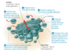
Eukaryotic cells
Have a double membrane-bound nucleaus that stores the cells DNA
Contain membrane-enclosed organelles
Cytoplasm is in the region between the plasma membrane and nucleus
Generally much larger than prokaryotic cells
Meaning -after the nucleaus
Prokaryotic cells
Lack a nucleus and membrane-enclosed organelles
DNA is concentrated in a non-membrane bound region called the nucleoid
Meaning -before the nucleus
Genomics
The large-scale analysis of the DNA sequence of a species- its genome
Comparatively studies genomes of different species
Hierarchy of life
Atoms < molecules < organelles < cells < tissues < organs < organ systems < organisms < populations < communities < ecosystems < biosphere
Linnaean system
Dear King Philip Came Over From Great Spain
- Domain
- Kingdom
- Phylum
- Class
- Order
- Family
- Genus
- Species
Domains of life
Domain Bacteria
Domain Archaea
Domain Eukarya
Which elements comprise the remaining 4% of essential elements?
Calcium
Phosphorus
Potassium
Sulfur
Emergent properties of water
- Cohesive behavior
- Ability to moderate temperature
- Expansion upon freezing
- Versatility as a solvent
Temperature at which water reaches its greatest density
4º C
What is an isomer?
A compound that has the same number of atoms but a different structure
Different types of isomers
Structural isomer- differ in the arrangement of atoms
Cis-trans isomers (formerly called geometric isomers)- carbons are bonded to the same atoms but differ in their spatial arrangements due to the rigidity of a double bond
Enantiomers- are mirror images that differ in shape due to an asymmetric carbon- one that is attached to four different atoms or groups of atoms
Hydroxyl group
Alcohol
Are polar due to electronegative oxygen
Compound names usually end in -ol
Carbonyl group
Ketone- carbonyl group within a carbon skeleton
Aldehyde- carbonyl group at the end of a carbon skeleton
Carboxyl group
Carboxylic acid or organic acid
Ionized form —COO– (carboxylate ion) is found in cells
Amino group
Amine
Acts as a base
Ionized form —NH3 is found in cells
Sulfhydryl group
Thiol
Two sulfhydryl groups can react to form a disulfide bond- help to stablize proteins
Phosphate group
Organic phosphate
Contributes a 1– charge when inside a chain and a 2– charge when at the end
Confers the ability of a molecule to react with water when attached
Methyl group
Methylated compound
Affects the espression of genes when on DNA or on proteins bound to DNA
Affects the shape and function of male and female sex hormones
Dehydration reaction
Formation of a bond by the removal of a water molecule
Hydrolysis reaction
The breaking of a bond by adding a water molecule
Glycosidic linkage
Covalent bond formed between two monosaccharides by a dehydration reaction
Polysaccharides
Polymers composed of monosaccharides joined by glycosidic linkages
Storage polysaccharides
Starch- primary form of energy storage in plant cells, form granules known as plastids which include chloroplasts, composed of a-glucose monomers
- Amylose- simplest form of starch, unbranched
- Amylopectin- a more complex starch, somewhat branched
Glycogen- primary form of energy storage in animal cells
- Stored mainly in liver and muscle cells, extensively branched
Structural polysaccharides
Cellulose- major component of cell walls in plant cells
- Composed of ß-glucose molecules making every glucose monomer “upside down” with respect to their neighbor
- Never branched, held together laterally by hydrogen bonds
Chitin- structural carbohydrate used by arthropods to build their exoskeletons
- Also provides structural support for the cell walls of many fungi
Lipids
Small hydrophobic molecules generally not big enough to be considered as macromolecules
Subcomponents of lipids
Glycerol- an alcohol whose three carbons each bear a hydroxyl group
Fatty acid- has a long carbon skeleton, usually 16 to 18 carbon atoms in length
- Relatively non-polar C—H bonds are the reason why fats are hydrophobic
Triglycerol
Three fatty acids linked to one glycerol molecule by a dehydration reaction
Hydrogenation
Process of converting unsaturated fats to saturated fats by adding hydrogen
Phospholipid
Two fatty acids and a phosphate group attached to glycerol
The oxygen of the phosphate opposite of the attachment site to the glycerol is connected to an additional small charged or polar molecule such as choline but can vary
Steroid
Lipids characterized by a carbon skeleton consisting of four fused rings
Different steroids are distinguished by the particular chemical groups attached to the rings
Cholesterol, a type of steroid, is a component in animal cell membranes and a precursor from which other steroids are synthesized
Amino acid
Is an organic molecule with both an amino group and a carboxyl group
At its center is an asymetric carbon called an alpha carbon whose four different partners are and amino group, a carboxyl group, a hydrogen atom, and a variable R-group called a side chain
Amino groups are usually ionized in a cell
Polypeptide bond
Peptide bond- covalent bond between the carboxyl group of one amino acid and the adjacent amino group of another, formed through a dehydration reaction
Forms the polypeptide backbone of an amino acid
Protein shapes
Globular- roughly spherical in shape
Fibrous- shaped like long fibers
Primary structure
Is the actual sequence of individual amino acids that make up a protein, dictates the secondary and tertiary structure
Secondary structure
Repeated coils and folds that form in a protein as the result of hydrogen bonds between repeating constituents of the polypeptide backbone, NOT the side chains
Two main structures:
- a helix- delicate coil held together by hydrogen bonding between every fourth amino acid
- ß pleated sheet- two or more segments of the polypeptide chain lying side by side (called ß strands) connected by hydrogen bonds
Tertiary structure
Overall shape of the polypeptide resulting from the interactions between the side chains of the constituent amino acids
Stabalized by three interactions:
- Hydrophobic interactions- hydrophobic side chains are clustered in the core of the protein, held together by van der Waals interactions
- Hydrogen bonds form between exterior polar side chains
- Disulfide bridges can reinforce the structure of the protein where two cysteine monomers are brought close together by folding
Quaternary structure
The ocerall protwin structure that results from the aggregation of seperate polypeptide subunits, not all proteins have multiple subunits
What are the protein molecules that assist in the folding of other proteins?
Chaperonins- keep the nascent polypeptide segregated from the disruptive chemical conditions in the cytoplasm while it folds spontaneously
Nucleic acids
Exist as polymers called polynucleotides made up of monomers calles nucleotides
Nucleotide components
Composed of three parts:
- Pentose- five carbon sugar
- Nitrogenous base- a nitrogen containing base
- Phosphate group
The portion of a nucleotide without any phosphate group is called a nucleoside
Pentoses
Deoxyribose- the sugar molecule in DNA, lacks an oxygen molecule on the second carbon in the ring
Ribose- the sugar molecule in RNA- has an oxygen molecule on the second carbon in the ring
Nitrogenous bases
Pyrimidines- has one six-membered ring of carbon and nitrogen atoms
- Cytosine (C)
- Thymine (T)- only in DNA
- Uracil (U)- only in RNA
Purines- are larger with a six-membered ring fused to a five-membered ring
- Adenine (A)
- Guanine (G)
Nucleotide bonding
Phosphodiester linkage- a phosphate group that links the sugars of two nucleotides, results in a repeating pattern of sugar-phosphate units called the sugar-phosphate backbone
- One end has a phosphate attached to a 5’ carbon and the other end has a hydroxyl group on the 3’ carbon
- Nucleotide has built-in directionality along the sugar-phosphate backbone from 5’to 3’
Key parameters of microscopy
- Magnification- ratio of an object’s image size to its real size
- Resolution- measure of clarity; minimum distance two points can be separated and still be distinguished as seperate points
- Contrast- the difference in brightness between the light and dark areas of an image
SEM
Scanning Electron Microscope
Used to look at the surface of a specimen by focusing a beam of electrons onto it
Provides images that look 3-D
TEM
Transmission Electron Microscope
Used mainly to study the internal structure of cells
Focuses a beam of electrons through a specimen
Cell fractionation
Takes cells apart and separates the major organelles from one another
Enables scientists to determine the functions of organelles
Basic features of all cells
- Plasma membrane
- Semifluid substance called cytosol
- Chromosomes (carry genes)
- Ribosomes (make proteins)
Nuclear envelope
A double membrane composed of two sepearte lipid bilayers with associated proteins called pore complexes
Nuclear lamina
A netlike array of intermediate protein filaments on the inside (nuclear side) of the envelope that maintains the shape of the nucleus my mechanically supporting the nuclear envelope
Nuclear matrix
A framework of protein fibers that extends throughout the nuclear interior
May help organize genetic material so that it functions efficiently
Nucleolus
A prominent structure within the nondividing nucleus where ribosomes are assembled
Ribosomal RNA is synthesized in the nucleolus and combined with proteins imported from the cytoplasm to form large and small ribosomal subunits
These subunits exit to the cytoplasm and a large and a small subunit combine into a ribosome
Ribosomes
Complexes made of ribosomal RNA and protein
Carry out protein synthesis in two locations:
- Free ribosomes are present in the cytosol
- Bound ribosomes are on the outside of the endoplasmic reticulum or the nuclear envelope
Endomembrane system components
- Nuclear envelope
- Endoplasmic reticulum
- Golgi apparatus
- Lysosomes
- Vacuoles
- Plasma membrane
These components are either continuous or connected via transfer by vesicles
Smooth ER
Synthesizes lipids
Metabolizes carbohydrates
Detoxifies drugs and poisons
Stores calcium ions
Rough ER
Has bound ribosomes, which secrete glycoproteins- proteins covalently bonded to carbohydrates
Distributes transport vesicles- secretory proteins surrounded by membranes
Is the membrane factory of the cell- as the ER expands, portions are transferred in the form of transport vesicles to other components of the endomembrane system
Golgi Apparatus
Functions of the Golgi apparatus:
- Modifies products of the ER
- Manufactures certain macromolecules
- Sorts and packages materials into transport vesicles
The shipping and receiving center of the cell
Consists of flattened membranous sacs called cisternae (cisterns)
Golgi directionality
Cis face- is usually located near the ER and accepts transport vesicles that bud from the ER
Trans face- is on the opposite side and gives rise to the vesicles that pinch off and travel elsewhere
As products of the ER migrate through the Golgi apparatus they are modified and refined
Lysosomes
A membranous sac of hydrolytic enzymes that can digest macromolecules
Lysosomal enzymes work best in the acidic environment inside the lysosome
Hydrolytic enzymes and lysosomal membranes are made by rough ER and then transferred to the Golgi apparatus for further processing
Phagocytosis
Process in which an organism engulfs and consumes a smaller organism or food particles
Extends pseudopodia around a particle which is then pinched off to form a food vacuole
Food vacuole fuses with a lysosome which digests it
Autophagy
Process by which a cell’s lysosomes recycle the cell’s own organelles and macromolecules
Vacuoles
Diverse maintenance compartments
Large vesicles derived from the ER and Golgi apparatus
Perform a variety of functions in different kinds of cells:
- Food vacuoles are formed by phagocytosis
- Contractile vacuoles are found in many freshwater protists- pump excess water out of cells
- Central vacuoles are found in many mature plant cells- hold organic compounds and water
Mitochondria
Have a smooth outer membrane and an inner membrane folded into cristae
Cristae present a large surface area for enzymes that synthesize ATP
Inner membrane creates two compartments, the intermembrane space and mitochondrial matrix
Chloroplast components
Thylakoids- flattened interconnected membranous sacs
Granum- stacks of thylakoids that resemble poker chips
Stroma- the fluid outside of the thylakoids which contains the chloroplast’s DNA, ribosomes, and enzymes
Plastids
A family of closely related plant organelles which include:
- Chloroplasts
- Amyloplast- organelle that stores starch
- Chromoplast- contains the pigments that give fruits and flowers their orange and yellow hues
Peroxisomes
Specialized metabolic compartments bounded by a single membrane
Produce hydrogen peroxide and convert it to water
Have many different functions:
- Use oxygen to break down fatty acids which can be used for cellular respiration
- Detoxify alcohol and othe poisons by transferring hydrogen from the poison to oxygen
- Specialized peroxisomes called glyoxysomes convert fatty acids to sugar in seeds as an energy source until a plant can initiate photosynthesis
Cytoskeleton
Network of fibers extending throughout the cytoplasm
Helps to support the cell and maintain its shape
Interacts with motor proteins to produce motility
Vesicles can travel along tracks provided by the cytoskeleton
Components of the cytoskeleton
Microtubules- thickest of the three components of the cytoskeleton
Microfilaments- also called actin filaments; thinnest components
Intermediate filaments- fibers with diameters in a middle range
Microtubules
Composed of tubulin-a dimer consisting of a-tubulin and ß-tubulin
25 nm with a 15 nm lumen
Functions of microtubules:
- Shaping the cell
- Guiding movement of organelles
- Separating chromosomes during cell division
- Control the beating of flagella and cilia
Centrosome
Region of the cell where the microtubules originate from
Often located near the nucleus
Location of the centrioles
Only present in animal cells
Centrioles
Organize microtubule assembly in animal cells
Aid in the seperation of chromosomes during mitosis
Located as a pair in the centrosome perpendicular to each other
Composed of nine sets of triplet microtubules arranged in a ring
Cilia and flagella
Differ in their beating patterns but share a common structure
Composed of a core of microtubules sheathed by the plasma membrane
- Nine doublets of microtublues arranged in a ring with two single microtubules in its center in a “9+2” pattern
- This pattern is found in nearly all eukaryotic flagella and motile cilia
Basal body- anchors the cilium or flagellum
- Structure similar to a centriole with nine triplets of microtubules in a “9+0” pattern
Dyneins- motor proteins that are responsible for movement that attach each outer tubule doublet
Movement of cilia and flagella
Microfilaments
Actin filaments
Structural role:
- Bear tension by resisting pulling forces within the cell
- Form a 3-D network called the cortex just inside the plasma membrane to help support the cell’s shape
Motile role:
- Function in cellular motility in conjunction with the protein myosin
- Thicker filaments composed of myosin interdigitate with the thinner actin fibers
- Involved in amoeboid (crawling) movement by extending pseudopodia
Cytoplasmic streaming- the induced circular flow of cytoplasm within a cell which speeds distribution of materials within a cell
Intermediate filaments
Most common in vertebrates
Specialized for bearing tension
Various types are constructed by particular molecular subunits; includes keratin
Some make up the nuclear lamina which lines the interior of the nuclear envelope
Cell wall
Extracellular strucutre posesed by:
- Plant cells
- Prokaryotes
- Fungi
- Some unicellular eukaryotes
Plant cell walls are made of cellulose fibers embedded in other polysaccharides and protein
Cell wall components
Primary cell wall- relatively thin and flexible outtermost wall
- First secreted by young plant cells
- When a plant cell is done growing it can secrete a hardening substance into the primary wall
Middle lamina- a thin layer between primary walls of adjacent plant cells
- Layer is rich in sticky polysaccharides called pectins
- Serves to glue cells together
Secondary cell wall- between the primary cell wall and the plasma membrane
- Strong and durable matrix that affords the cell protection and support
- Often deposited in several laminated layers
- Wood consists primarily of secondary cell walls
Plasmodesmata- channels that perforate the cell wall that join adjacent cells
Cell wall functions
Protects the cell
Maintains its shape
Prevents excessive uptake of water
Extracellular matrix of animal cells
Composed of glycoproteins and other carbohydrate-containing molecules:
- Collagen
- Proteoglycans
- Fibronectin
Bind to receptor proteins in the plasma membrane called integrin
The ECM around a cell can influence the activity of gene in the nucleus
Mechanical signaling through cytoskeletal changes trigger chemical signals in the cell
Collagen
Most abundant glycoprotein in animal cells
Form strong fibers outside of the cell embeded in a web of proteoglycan complexes
Accounts for 40% of total protein in the human body
Proteoglycan complex
Can consist of hundreds of proteoglycan molecules attached noncovalently to a single long polysaccharide molecule
Proteoglycan molecules consist of a small core protein with many carbohydrate chains covalently attached; about 95% carbohydrate
Fibronectin
Proteins that bind to cell surface receptor proteins called integrins which are built into the plasma membrane
Integrins
Span the cell membrane
Bind to microfilaments of the cytoskeleton on their cytoplasmic side and fibronectins of the ECM on their extracellular side
Based on the word “integrate”
Transmit signals between the ECM and the cytoskeleton to integrate changes occuring outside and inside the cell
Plasmodesmata
Perforations in plant cell walls that unify most plant cells
Channels allow cytosol to pass through
Join the internal chemical environments of adjacent cells
Tight junction
Plasma membranes of neighboringcells are tightly pressed against each other
Bound together by specific proteins
Forms a continuous seal around cells
Establish a barrier that prevents leakage of extracellular fluid between cells
Desmosomes
Function like rivets, fastening cells together into strong sheets
Intermediate filaments made of keratin anchor desmosomes in the cytoplasm
Attach muscle cells to each other
- Some muscle tears involve the rupture of desmosomes
Also called anchoring junctions
Gap junctions
Provide cytoplasmic channels from one cell to an adjacent cell; similar to plasmodesmata of plant cells
Consist of membrance proteins that surround a pore and allow ions, sugars, amino acids, and other small molecules to pass
Necessary for communication between cells in tissue such as the myocardium
Also called communicating junctions
Fluid mosaic model
Views the membrane as a mosaic of protein molecules bobbing in a fluid bilayer of phospholipids
Proteins are not randomly distributed in the membrane but are gropued in long-lasting specialized patches that carry out common functions
Types of membrane proteins
Peripheral proteins- bound to the surface of the membrane
Integral proteins- penetrate the hydrophobic core
- Integral proteins that span the membrane are called transmembrane proteins
- The hydrophobic regions of an integral protein consist of one or more stretches of nonpolar amino acids, often coiled into alpha helices
Plasma membrane protein functions
- Transport
- Enzymatic activity
- Signal transduction
- Cell-cell recognition
- Intercellular joining
- Structural integrity
Transport proteins
Channel proteins- provide a hydrophilic channel through the membrane that allow for the passage of certain molecules or ions
- Many function as gated ion channels that open or close in response to stimulus
- Aquaporins- speed the passage of water molecules; allow entry of up to 3 billion water molecules per second
Carrier proteins- hold onto their passengers and change shape to ashuttle them across the membrane
- Are specific to the substance that they are designed to transport
- Allow for directionality of membrane permissibility
- Some actively hydrolyze ATP to pump against a concentration gradient
Osmoregulation
The control of solute concentrations and water balance
Plasmolysis
When the membrane pulls away from the cell wall causing the plant to wilt; usually lethal
Sodium-potassium pump
Oscillates between two shapes that result in the removal of 3 Na+ from the cell for every 2 K+ brought back into the cell
Membrane potential
Voltage difference across a membrane
Ranges from about –50 to –200 millivolts (mV)
Electrochemical gradient
Combination of two forces that drive the diffusion of ions across a membrane
- Chemical force- the ion’s concentration gradient
- Electrical force- the effect of the membrane potential on the ion’s movement
Electrogenic pump
A transport protein that generates voltage across a membrane
Help store energy that can be used for cellular work
- The sodium-potassium pump is the major electrogenic pump of animal cells
- The proton pump is the main electrogenic pump of plants, fungi, and bacteria cells
- Actively transports protons out of the cell
Cotransport
Occurs when active transport of a solute indirectly drives transport of other substances
Couples the downhill diffusion of one solute to the uphill transport of another
Plants use hydrogen ion gradient to drive nutrients
Exocytosis
Secretion of certain molecules from the cell
Transport vesicles migrate to the membrane, fuse with it, and release their contents outside the cell
Many secretory cells use exocytosis to export their products
The loss of membrane by exocytosis seems to be offset the addition of membrane via endocytosis
Endocytosis
The cell takes in macromolecules by forming vesicles from the plasma membrane
Small area of the plasma membrane sinks inward to form a pocket which gets pinched-off
The addition of membrane by endocytosis seems to be offset by the loss of membrane via exocytosis
Three types of endocytosis
- Phagocytosis (cellular eating)
- Pinocytosis (cellular drinking)
- Receptor-mediated endocytosis
Pinocytosis
Continuous process in which the cell forms tiny vacuoles of extracellular fluid
Cell obtains molecules dissolved in the droplets
Nonspecific for the substances brought into the cell
Parts of the membrance that form vesicles are lined with proteins on their cytoplasmic side; resulting vesicles are said to be coated
Receptor-mediated endocytosis
Specialized type of pinocytosis that enables the cell to acquire bulk quantities of specific substances that may not be highly concentrated in the extracellular fluid
Proteins embedded in the membrane act as receptor cites exposed to the extracellular fluid and bind to specific solutes
The receptor proteins then cluster in coated pits which form a vesicle containing the bound solutes
After the vesicles are emptied, the receptors are recycled to the outter membrane by the same vesicle
Binding of ligands to receptors triggers vesicle formation
Ligand
Any molecule that binds specifically to a receptor site on another molecule
First law of thermodynamics
Energy cannot be created or destroyed, only transferred or transformed
Second law of thermodynamics
Every energy transfer or transformation increases the entropy of the universe
Bioenergetics
The study of how energy flows through living organisms
Free energy
The portion of a system’s energy that can do work when temperature and pressure are uniform throughout the system
Gibb’s free energy formula
For a reaction to be spontaneous the change in Gibb’s free energy must be negative
As a reaction proceeds towards equilibrium the free energy decreases
A system at equilibrium is at the lowest possible energy state for that system
Exergonic reaction
Proceeds with a net release of free energy and is spontaneous
Magnitude of energy released represents the maximum amount of work that the reaction can perform
Endergonic reaction
Absorbs free energy from its surroundings and is nonspontaneous
Magnitude of energy absorbed is the quantity of energy required to drive that reaction
Energy coupling
The use of an exergonic process to drive an endergonic one
Energetic mechanism of ATP
Phosphates are crowded together and their mutual repulsion contributes toward instability in that region; equivalent to a compressed spring
Energy is released by the hydrolysis of the third phosphate group
- Can drive endergonic reactions as long as the energy required to drive the reaction is less than the energy released by the hydrolysis of ATP
Usually involves the phosphorylation:
- Phosphate group covalently bonds to a recipient molecule called a phosphorylated intermediate
- This phosphorylated intermediate is more reactive than the original unphosphorylated molecule
Structure of ATP
Ribose- five-sided monosaccharide
Adenine- nitrogenous base
Triphosphate group- chain of three phosphate groups bonded together
The ATP cycle
The ATP cycle is a revolving door in which energy passes from catabolic to anabolic pathways
It couples energy yielding exergonic reactions to energy consuming endergonic ones
ATP is regenerated by a dehydration reaction between ADP and a phosphate group
Transition state
Intermediate chemical stage a reactant must reach prior to the formaion of a product
Has a higher free energy than that of the initial reactants
Substrate
The reactant that an enzyme acts on
Properties of a catalyst
- Increase reaction rates by lowering the EA requiredf
- Form transient, reversible complexes with substrate molecules
- Change the rate at which equilibrium is achieved, not the position of the equilibrium
Enzyme-substrate complex
An enzyme and substrate as a singular structure
Substrate activation mechanisms
Template- when there are two or more reactants the enzyme can provide a template on which the substrates can come together in the proper orientation for a reaction to occur
Bond distortion- the enzyme may stretch the substrate molecules toward their transition state by stressing and bending the chemical bonds, thus reducing the free energy required for the reaction
Proton transfer- increases reactivity of substrate
Microenvironment- the active site may provide a more conducive environment than the solution it is in for the reaction to take place
Electron transfer- a brief covalent bond may form between the substrate and a side chain of an amino acid of the enzyme which is returned to its original state after completion
Cofactors
Nonprotein helpers for catalytic activity, often by functioning as electron acceptors
May be permanently bound to the enzyme bind loosely and reversibly along with the substrate
Called prosthetic groups, there are two main types:
- Inorganic metals such as zinc, copper, or iron
- Coenzymes- organic molecules often derived from vitamins
Enzyme classes
Under the Enzyme Commission (EC), enzymes are divided into six major classes based on general function:
Over The HILL
Oxidoreductases
Transferases
Hydrolases
Isomerases
Lysases
Ligases
The induced-fit model
Substrate binding at the active site induces a conformational change in the shape of the enzyme
The active site recognizes and binds to the substrate and activates it by providing the right environment for catalysis
Called substrate activation which proceeds via several possible mechanisms
In opposition to the “lock and key model” which viewed this interation as more static
The catalytic event
- The random collision of a substrate molecule with the active site results in it binding there
- Substrate binding induces a conformational change that tightens the fit, facilitating the conversion of substrate into products
- The products are then released from the active site
- The enzyme molecule returns to the original conformation with the active site available for another molecule of substrate
Enzyme kinetics
Describes the quantitative aspects of enzyme catalysis and the rate of substrate conversion into products
Reaction rates are influenced by factors such as the concentrations of substrates, products, and inhibitors
The Michaelis–Menten equation
Michaelis–Menten kinetics
Initial reaction velocity (v)- rate of change in product concentration per unit time is dependent on: the substrate concentration [S]
- At low [S], doubling [S] will double v
- As [S] increases though, each additional increase in [S] results in a smaller increase in v
- When [S] becomes very large the value of v reaches a maximum
Michaelis–Menten kinetics:
Vmax and saturation
As [S] tends toward infinity, v approaches an upper limiting value; maximum velocity (Vmax)
Vmax is an upper limit determined by:
- The time required for the actual catalytic reaction
- How many enzyme molecules are present
The value of Vmax can be increased by adding more enzyme
Saturation- the state where increasingly higher substrate concentrations will no longer increase the reaction velocity beyond a finite upper value
Michaelis–Menten kinetics:
Very low substrate concentration ([S] << Km)
At very low [S], the initial velocity of the reaction is roughly proportional to [S]
Michaelis–Menten kinetics:
Very high substrate concentration ([S] >> Km)
At very high [S] the initial velocity of the reaction is independent of variation in [S]
Vmax is the velocity at saturating substrate concentrations
Michaelis–Menten kinetics:
Substrate concentration and Km are constant
Shows that Km is the specific substrate concentration at which the reaction proceeds at one half its maximum velocity
Michaelis–Menten kinetics:
Km and Vmax relavence to cell biology
The lower the Km value for a given enzyme and substrate, the lower the [S] range in which the enzyme is effective
Vmax is important, as a measure of the potential maximum rate of the reaction
By knowing Vmax, Km, and the in vivo [S] we can estimate the likely rate of the reaction under cellular conditions
Enzyme inhibition
Enzymes are influenced (mostly inhibited) by products, alternative substrates, substrate analogs, drugs, toxins, and allosteric effectors
The inhibition of enzyme activity plays a vital role as a control mechanism in cells
Drugs and poisons frequently exert their effects by inhibition of specific enzymes
Enzyme inhibition:
Reversible and irreversible inhibition
Reversible inhibitors- bind enzymes noncovalently and can dissociate from the enzyme
Irreversible inhibitors- bind to the enzyme covalently; cause permanent loss of catalytic activity and are generally toxic to cells
- Heavy metal ions, nerve gas poisons, some insecticides
- Nerve agents block acetylcholinesterase which breaks down acetylcholine
Enzyme inhibition:
Noncompetitive inhibition
Bind to the enzyme molecule outside of the active site
Inhibit activity indirectly by causing a conformation change in the enzyme either by:
- Inhibits substrate binding at the active site
- Reduces catalytic activity at the active site
Enzyme inhibition:
Competitive inhibition
Bind the active site of an enzyme competing with substrate
Enzyme activity is inhibited directly because active sites are bound to inhibitors which prevents the substrate from binding
Can only be overcome by increasing the substrate concentration
Enzyme regulation:
Allosteric regulation
Allosteric regulation- a protein’s function at one site is affected by the binding of a regulatory molecule at a seperate site
Allosteric enzymes have two conformations, one in which it has affinity for the substrate and one in which it does not
May be an activator or inhibitor:
- The binding of an activator to a regulatory site stabalizes the shape that has functional active sites
- The binding of an inhibitor to a regulatory site stabalizes the inactive form of the enzyme
Enzyme regulation:
Cooperativity
A substrate molecule binding to one active site in a multisubunit enzyme triggers a shape change in all subunits
Increases catalytic activity at other active sites
One substrate molecule thus primes an enzyme to act on additional substrate molecules more readily
A kind of allosteric activation
Enzyme regulation:
Covalent modification
Enzymes can be regulated by the addition or removal of chemical groups
Activity is regulated by addition or removal of groups such as phosphate, methyl, or acetyl groups, etc
The reversible addition of phosphate groups by protein kinases is a common covalent modification
Enzyme regulation:
Feedback inhibition
Metabolic pathway is halted by the inhibitory binding of its own end-product to an enzyme that acts early in the pathway
Prevents overproduction or overaccumulation of metabilic products
Enzyme regulation:
Phosphorylation and dephosphorylation
The reversible addition of phosphate groups by protein kinases is a common covalent modification
Phosphorylation- occurs most commonly by transfer of a phosphate group from ATP to the hydroxyl group of Ser, Thr, or Tyr residues in a protein
Dephosphorylation- the removal of phosphate groups from proteins; catalyzed by protein phosphatases
Enzyme regulation:
Regulation of glycogen phosphorylase
Glycogen phosphorylase exists as two inter-convertible forms
- Glycogen phosphorylase–a- the active, phosphorylated form
- Glycogen phosphorylase–b- the inactive non-phosphorylated form
The enzymes responsible:
- Phosphorylase kinase phosphorylates the enzyme
- Phosphorylase phosphatase removes the phosphate
Enzyme regulation:
Proteolytic cleavage
The activation of a protein by a one-time, irreversible removal of part of the polypeptide chain
Proteolytic enzymes of the pancreas: trypsin, chymotrypsin, and carboxypeptidase are examples of enzymes synthesized in inactive form (zymogens) and activated by cleavage as needed
Common disaccharides
Common monosaccharides
Good Men have Regular GirlFriends
Glucose
Mannose
Ribose
Galactose
Fructose
Redox reactions and covalent bonds
Not all redox reactions involve the complete transfer of electrons
Pure covalent bonds such as between C–H share electrons EQUALLY
Oxygen being much more elctronegative “hogs” electrons
Electrons are much closer to oxygen in CO2 so the oxygen has partially gained the electrons, oxidizing the carbon
Electrons thus lose energy and free energy is released
Substrate-level phosphorylation
Mode of ATP synthesis where an enzyme transfers a phosphate group from a substrate molecule to ADP
In contrast to oxidative phosphorylation which transfers an inorganic phosphate group
Substrate molecule refers to an organic molecule that is generate as an intermediate during the catabolism of glucose
Glycolysis phases and net products
Glycolysis occurs in the cytoplasm and has two major phases
- Energy investment phase- consumes two ATP
- Energy payoff phase- yields four ATP, four NADH, and two pyruvate
Glycolysis occurs whether or not O2 is present
Stages of cellular respiration
Harvesting of energy from glucose has three stages
- Glycolysis- breaks down glucose into two molecules of pyruvate
- Citric acid cycle- completes the breakdown of glucose
-
Oxidative phosphorylation- accounts for most of the ATP synthesis
- The process that generates most of the ATP because it is powered by redox reactions
- Almost 90% of the ATP generated by cellular respiration
Glycolysis:
Energy investment phase
Intermediates and enzymes
Intermediates
Girls Get Fine Food, Gentlemen Dine Girl(s)
- Glucose
- Glucose 6-phosphate
- Fructose 6-phosphate
- Fructose 1,6-bisphosphate
- Glyceraldehyde 3-phosphate (G3P)
- Dihydroxyacetone phosphate (DHAP)
- DHAP converts to second molecule of G3P
Enzymes
Helen Paints Pictures Along Isolated Trails
- Hexokinase
- Phosphoglucoisomerase
- Phosphofructokinase
- Aldolase
- Isomerase- converts DHAP to G3P
- Triose phosphate dehydrogenase- leads to energy payoff phase
Primary rate limiting enzymes highlighted in red
Glycolysis:
Energy payoff phase
Intermediates and enzymes
Intermediates
Boys Prefer to Pick-up Pepperoni Pizza
- 1,3-Bisphosphoglycerate (BPG)
- 3-Phosphoglycerate (PG)
- 2-Phosphoglycerate (PG)
- Phosphoenolpyruvate (PEP)
- Pyruvate
Enzymes
Pretty People Enjoy Parties
- Phosphoglycerokinase
- Phosphoglyceromutase
- Enolase
- Pyruvate kinase
Primary rate limiting enzymes are highlighted red
Pyruvate oxidation
Links glycolysis to the citric acid cycle
Pyruvate is a charged molecule and must enter the mitochondria via active transport
Pyruvate is converted to a compound called acetyl CoA in a series of three reactions catalyzed by several enzymes called the pyruvate dehydrogenase complex
- Pyruvate’s carboxyl group is already fully oxidized and is thus removed and given off as one molecule of CO2
- Remaining two-carbon group is oxidized forming acetate
- Extracted electrons are transferred to NAD+ forming one molecule of NADH
- Coenzyme a (CoA) is attached via its sulfur atom to the acetate forming acetyl CoA
Citric acid cycle phases and net products
Pyruvate oxidation- yields one NADH, one acetyl CoA, and one CO2 as waste
Citric acid cycle- yields one ATP, three NADH, one FADH2, and two CO2 as waste
Inputs and outputs shown are for each pyruvate molecule thus, each glucose molecule nets:
- 2 ATP
- 8 NADH
- 2 FADH2
Citric acid cycle
Also called the Krebs cycle, completes the break down of pyruvate to CO2
The citric acid cycle has eight steps, each catalyzed by a specific enzyme
- The acetyl group of acetyl CoA joins the cycle by combining with oxaloacetate, forming citrate
- The next seven steps decompose the citrate back to oxaloacetate, making the process a cycle
The NADH and FADH2 produced by the cycle relay electrons extracted from food to the electron transport chain
Electron transport chain
The ETC is in the cristae of the mitochondrion
- Most of the chain’s components are proteins which exist in multiprotein complexes
The carriers alternate reduced and oxidized states as they accept and donate electrons
Electrons drop in free energy as they go down the chain and are finally passed to O2, forming H2O
- The ETC generates no ATP directly
Citric acid cycle intermediates
Intermediates
Can I Keep Selling Sex For Money Officer?
- Citrate
- Isocitrate
- a-Ketoglutarate
- Succinyl CoA
- Succinate
- Fumarate
- Malate
- Oxaloacetate
Citric acid cycle enzymes
Enzymes
Covert Atropian Infidels Keep Stealing Super Funny Memes
- Citrate synthase
- Inhibited by citrate and ATP
- Aconitase
- Isocitrate dehydrogenase- rate limiting
- Inhibited by NADH
- Stimulated by ADP and Ca2+
-
a-ketoglutarate dehydrogenase
- Inhibited by NADH, succinyl CoA, and acetyl CoA
- Stimulated by Ca2+
- Succinyl CoA synthetase
- Succiniate dehydrogenase
- Fumarase
- Malate dehydrogenase
Pyruvate dehydrogenase complex inhibition
PDH is allosterically inhibited by:
- ATP
- NADH
- acetyl CoA
- High ATP:ADP ratio
PDH is activated by:
- AMP
- NAD+
- Free CoA
- Low ATP:ADP ratio
Electron transport chain carriers
- Flavoproteins- have a prosthetic group called flavin mononucleotide (FMN)
- Iron-sulfur proteins- family of proteins with both iron and sulfur tightly bound
-
Ubiquinone (coenzyme Q)- small hydrophobic molecule
- Only enzyme in ETC that is NOT a protein
- Is individually mobile within the membrane
- Occur as large assemblies of proteins called respiratory complexes
-
Cytochromes- electron carriers between ubiquinone and oxygen
- Prosthetic group called a heme group has an iron atom that accepts and donates electrons
- Each cytochrome has a slightly different heme group
- Last cytchrome Cyt a3 passes its electrons to oxygen which picks up a pair of hydrogen ions, forming water
All enzymes except ubiquinone are proteins with prosthetic groups capable of being reversibly oxidized and reduced
Proton gradients
Gold arrows trace the transport of electrons which are finally passed to a terminal acceptor
As the complexes shuttle electrons they pump protons from the mitochondrial matrix into the intermembrane space
NADH deposits its electrons in complex I
FADH2 deposits its electrons via complex II which is at a lower energy level than complex I
- Consequently, results in fewer protons pumped into the intermembrane space
Chemiosmosis
The use of the energy in a H+ gradient to drive cellular work
The H+ gradient results in a proton-motive force which drives H+ back across the membrane
During chemiosmosis protons flow back down their gradient via ATP sythase
In general terms chemiosmosis is an energy coupling mechanism that uses the proton-motive force to drive cellular work
ATP sythase
Protein complex that functions as a mill powered by the proton-motive force
Makes ATP from ADP and inorganic phosphates
Multiple ATP sythases reside in eukaryotic mitochondrial and chloroplast membranes
- Located in cellular membrane in prokaryotic cells
Dinitrophenol (DNP) is known to uncouple ATP synthesis from electron transport
- Allows protons to cross the membrane freely so that no proton gradient can be formed
Yields of each stage of cellular respiration
Per molecule of glucose
Glycolysis
- 2 ATP via substrate-level phosphorylation
- 2 NADH
- 2 Pyruvate
Pyruvate oxidation
- 2 NADH
- 2 Acetyl CoA
Citric acid cycle
- 2 ATP via substrate-level phosphorylation
- 6 NADH
- 2 FADH2
Oxidative phosphorylation
- Roughly 26-28 ATP
Total ATP
Maximum 30-32 ATP
Anaerobic respiration
Without oxygen glycolysis couples with anaerobic respiration or fermentation to produce ATP
Anaerobic respiration uses an ETC with a final electron acceptor other than oxygen such as sulfate
- Some sulfate-reducing marine bacteria use the sulfate ion at the end of their respiratory chain
Fermentation
Fermentation uses substrate-level phosphorylation instead of an ETC to generate ATP
Consists of glycolysis plus reactions that regenerate NAD+ by transferring a hydride from NADH to pyruvate or derivatives of it
Two common types:
-
Alcohol fermentation- pyruvate is converted to alcohol in two steps
- The first releases CO2 from the pyruvate which is converted to a two-carbon compound acetaldehyde
- Acetaldehyde is reduced by NADH to ethanol, forming NAD+
-
Lactic acid fermentation- method of fermentation used by human muscle cells when oxygen is scarce
- Pyruvate is reduced directly by NADH to form lactate as an end product with no release of CO2
Hydrophilic amino acids
Amino acids with polar side chains
Santa’s Team Crafts New Quilts Yearly
S- serine- Ser
T- threonine- Thr
C- cysteine- Cys
N- asparagine- Asn
Q- glutamine- Gln
Y- tyrosine- Tyr
Santa’s workshop IS near the North Pole
Hydrophobic amino acids
Amino acids with a non-polar side chain
Grandma Always Visits London In May For Winstons’s Party
G- glycine- Gly
A- alanine- Ala
V- valine- Val
L- leucine- Leu
I- isoleucine- Ile
M- methionine- Met
F- phenylalanine- Phe
W- tryptophan- Trp
P- proline- Pro
London is NOT near the North Pole
Amino acids with acidic side chains
Dragons Eat acid
D- asparate
E- glutamate
Aspiring Gluttons
Also, suffix -ate associated with dragons eating
Amino acids with basic side chains
Knights Ride Horses
K- lysine- Lys
R- arginine- Arg
H- histidine- His
Knights riding horses in a very BASIC literary motif
Obligate anaerobes
Only carry out fermentation of anaerobic respiration
Organisms cannot survive in the presence of oxygen
Faculative anaerobes
Species that can make enough ATP to survive by either fermentation or respiration
Human muscle cells behave in a similar way
- Under aerobic conditions pyruvate is converted into acetyl CoA and oxidation continues to the citric acid cycle
- Under anaerobic conditions lactic acid fermentation occurs and pyruvate is diverted from the citric acid cycle to serve as an electron acceptor to recycle NAD+
Protein catabolism
Begins with peptide bond hydrolysis or proteolysis
- Enzymes responsible for it are called proteases
- Products are small peptides and free amino acids
Free amino acids can be catabolized for energy
- Amino group must first be removed via process of deamination
- Nitrogenous refuse is excreted in the form of ammonia (NH3)
- Converted into intermediates of mainstream catabolism in as few steps as possible
Fat catabolism
Fats are highly reduced compounds that liberate more energy per gram upon oxidation than carbohydrates
Triacylglycerol catabolism begins with their hydrolysis to glycerol and free fatty acids
- The glycerol is channeled into the glycolytic pathway by oxidative conversion to dihydroxyacetone phosphate
- Fatty acids are linked to coenzyme A to form fatty acyl CoA which is then degraded by ß-oxidation
Steps of ß-oxidation
Each cycle involves
- Oxidation
- Hydration
- Reoxidation
- Thiolysis
The result is the production of one (1) FADH2, one (1) NADH, and one (1) acetyl CoA per cycle
Formation of fatty acyl CoA
Begins with an activation step in the cytosol (FA-1) that requires the energy of ATP hydrolysis
- FA-1⇢ drives the attachment of a CoA molecule to the fatty acid forming FA-1
The fatty acetyl CoA is then transported into the mitochondrion by a translocase in the inner membrane
Degradation of fatty acetyl CoA
FA-2⇢ An integral membrane dehydrogenase oxidizes the fatty acetyl CoA, forming a double bond between the α and ß-carbons
- The two electrons and protons removed are transferred to FAD, forming FADH2
FA-3⇢ water is added across the double bond by a hydratase
FA-4⇢ another dehydrogenase oxidizes the ß-carbon, converting the hydroxyl group to a keto group
FA-5⇢ the bond between the α and ß-carbons is broken by a thiolase and a two-carbon fragment is transferred to a second acetyl CoA
The steps FA-2 to FA-5 are repeated until the original fatty acid is completely degraded
- Most fatty acids have an even number of carbons and are completely degraded
- Unsaturated fatty acids require one or two additional enzymes
Versatility of catabolism
Catabolic pathways funnel electrons from many kinds of organic molecules into cellular respiration
Glycolysis accepts a wide range of carbohydrates
Proteins must be digested to amino acids; amino groups can feed glycolysis or the citric acid cycle
Fatty acids are linked to coenzyme A, to form fatty acetyl CoA which is then degraded by β-oxidation
Gluconeogenesis
Pyruvate and lactate are the most common starting materials
Simple reversal of glycolysis using the same enzyme in both directions
Gly-1, Gly-3, and Gly-10 are accomplished by other means
- These are the most exergonic reactions of glycolysis
Enzymes that catalyze the bypass reactions in gluconeogenisis
Gly-10
- Pyruvate carboxylase (PC)
- Phosphoenolpyruvate carboxykinase (PEPCK)
Gly-3
- Fructose 1,6-bisphosphatase (FBPase)
Gly-1
- Glucose 6-phosphatase
Regulation of cellular respiration via feedback mechanisms
Control of catabolism is based mainly on regulating the activity of enzymes at strategic points in the catabolic pathway
Feedback inhibition is the most common mechanism for metabolic control
- If ATP concentration begins to drop respiration speeds up
- When there is plenty of ATP respiration slows down