Embryology of the CVS 2 Flashcards

1
Q

What are the 2 methods that blood vessels develop by?

A

Vasculogenesis

Angiogenesis

2
Q

What is vasculogenesis?

A

New formation of a primitive vascular network

3
Q

What is the new formation of a primitive vascular network called?

A

Vasculogenesis

4
Q

What is angiogenesis?

A

The growth of new vessels from pre-existing blood vessels

5
Q

What is the growth of new vessels from pre-existing blood vessels called?

A

Angiogenesis

6
Q

What is the process of the formation of the aortic sac?

A

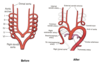
1) First arteries to appear in the embryo are the left and right primitive aortae
2) Each primitive aorta has a ventral part (ventral aorta) and a dorsal part (dorsal aorta)
3) After the fusion on the two endothelial tubes the two ventral aortae partially fuse to form aortic sac
4) Aortic branches arise from the aortic sac

7
Q

What are the first arteries to appear in the embryo?

A

Left and right primitive aorta

8
Q

What is the process of the development of the pharyngael arch arteries and aortic branches?

A

1) Pharyngeal arches (future neck) develop during 4th and 5th week
2) Each arch receives its own nerve and artery (pharyngael arteries)
3) Pharyngael arteries communicate with aortic branches (now called aortic arches)
4) 6 aortic arches are formed on each side, all in communication with the dorsal aortae

9
Q

When does the pharyngael arches develop?

A

During 4th and 5th week

10
Q

How many pairs are present in the aortic arches?

A

6 pairs

11
Q

What do the 6 pairs of the aortic arches develop from?

A

Aortic branches and pharyngael arteries

12
Q

What should be noted about the aortic arches and how long they are present for?

A

They are not all present at the same time

13
Q

Where do all of the aortic arches terminate?

A

Dorsal aorta

14
Q

What is the fate of aortic arch 1 and 2?

A

Disappear early, remnant of the 1st arch forms part of the maxilary artery

15
Q

What is the fate of aortic arch 3?

A

Common carotid and commencement of the internal carotid artery (so is named carotid arch)

16
Q

What is the fate of aortic arch 4?

A

4th right arch forms the right subclavian

4th left arch constitutes the distal part of aortic arch

17
Q

What is the fate of the 5th aortic arch?

A

Never forms or forms incompletely and then regresses

18
Q

What is the fate of the 6th aortic arch?

A

Proximal part of the 6th right arch persists as the proximal part of the right pulmonary artery

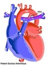
6th left arch gives of the left pulmonary artery and forms the ductus arteriosus, within 1-3 months the ductus is obliterated and becomes the ligamentum anteriosum

19
Q

What does the ductus arteriosus become once it is obliterated?

A

Ligamentum arteriosum

20
Q

What are the maxillary arteries derived from?

A

Aortic arch 1

21
Q

What is the common carotid and first part of internal carotid artery derived from?

A

Aortic arch 3

22
Q

What is the arch of aorta (distal portion) derived from?o

A

Aortic arch 4 left side

23
Q

What is the right subclavian artery (proximal portion) derived from?

A

Aortic arch 4 right side

24
Q

What is the left pulmonary artery and ductus arteriosus derived from?

A

Aortic arch 6 left side

25
What is the right pulmonary artery derived from?
Aortic arch 6 right side
26
What do most defects of the great arteries arise due to?
Persistance of aortic arches that normally should regress or regression of arches that normally shouldn't
27
What are examples of defects of the great arteries due to aortic arches not regressing or regressing when they shouldn't?
Aberrant subclavian artery Double aortic arch Patent ductus arteriosus (PDA) Coarctation of the aorta
28
What is aberrant subclavian artery?
With regression of the right aortic arch 4 and the right dorsal aorta the right subclavian artery has an abnormal origin on the left side, meaning to supply blood to the right arm this forces the right subclavian artery to cross the midline behind the trachea and oesophagus which may constrict these organs
29
What is double aortic arch?
Occurs with the development of an abnormal right aortic arch in addition to the left aortic arch, forming a ring around the trachea and oesophagus which usually causes difficulty swallowing and breathing
30
What is patent ductus arteriosus (PDA)?
Ductus arteriosus fails to close after birth Early symptoms are common but in the first year of life include increased work of breathing and poor wait gain Uncorrected PDA may lead to congestive heart failure with increasing age
31
What does PDA stand up for?
Patent ductus arteriosus
32
What are symptoms of patent ductus arteriosus (PDA) in the first year of life?
Increased work of breathing Poor weight gain
33
What could patent ductus arteriosus lead to in later life?
Congestive heart failure
34
What is coarctation of the aorta?
Aorta is narrow, usually in the area where the ductus arteriosus (ligamentum arteriosum after regression) inserts
35
What are the 2 kinds of coarctation of the aorta?
Preductal (proximal to ductus arteriosus) Post ductal (distal to ductus arteriosus)
36
What supplies the yolk sac with blood?
Vitelline vein and artery
37
What supplies the placenta with blood?
Umbilical artery and vein
38
What supplies the rest of the embryonic body with blood?
Cardinal artery and vein
39
What does the vitelline arteries and veins supply?
Yolk sac
40
What does the umbilical arteries and veins supply?
Placenta
41
What does the cardinal arteries and veins supply?
The rest of the body
42
What is the fate of the vitelline arteries after birth?
Represented by arteries to the foregut, midgut and hindgut
43
What is the fate is the fate of the umbilical arteries after birth?
Proximal portion persists as internal iliac and superior vesical branches to urinary bladder
44
What is the embryonic venous system composed of?
Vitelline veins Umbilical veins Cardinal veins
45
What do the vitelline veins do?
Carry blood from the yolk sac to the sinus venosus
46
What do the umbilical veins do?
Originate from chorionic villi of placenta carrying oxygenated blood to the embryo
47
What do the cardinal veins do?
Drain the body of the embryo
48
What veins drain the body of the embryo?
Cardinal veins
49
What supplies the embryo with oxygenated blood from the chorionic villi of the placenta?
Umbilical veins
50
What vein carries blood from the yolk sac to the sinus venosus?
Vitelline veins
51
What is the fate of the cardinal veins?
Form vena caval system by anastomosis amoung the veins
52
What is the main venous system of the embryo composed of?
Anterior cardinal veins Posterior cardinal veins Common cardinal vein all draining to sinus venosus
53
What is anastomosis?
Corss connections between adjacent channels
54
What are cross connections between adjacent channels called?
Anastomosis
55
What are examples of venous system abnormalities?
Double IVC Absence of IVC Left SVC Double SVC
56
What does IVC stand up for?
Inferior vena cava
57
What does SVC stand up for?
Superior vena cava
58
When does the lymphatic system develop?
End of the 6th week
59
What does the lymphatic system develop around?
Main veins
60
In terms of lymphatics, what develops at the end of the embryonic period?
Six primary lymph sacs
61
What are some of the lymph sacs that develop at the end of the embryonic period?
Jugular lymph sac Retroperitoneal lymph sac Cisterna chyli Iliac lymph sac
62
What is good respiration in neonate dependent upon?
Normal circulatory transition at birth (placental circulation to pulmonary circulation)
63
What circulation transition occurs at birth?
Placental circulation to pulmonary circulation
64
What are important vascular structures in the transitional circulation?
Ductus venosus (shunts left umbilical vein blood flow directly to inferior vena cava allowing oxygenated blood from the placenta to bypass the liver) Oval foramen (allows blood to enter the left atrium from the right atrium, allowing it to bypass the lungs) Ductus arteriosus (allows blood that still escapes to the right ventricle to bypass the lungs)
65
What does the ductus venosus allow?
Shunts left umbilical vein blood flow directly to inferior vena cava, allowing oxygenated blood from the placenta to bypass the liver
66
What allows blood to bypass the liver?
Ductus venosus
67
What does the oval foramen allow?
Blood to enter the left atrium from the right atrium, allowing it to bypass the lungs
68
What allows blood to bypass the lungs?
Oval foramen Ductus arteriosus
69
What does the ductus arteriosus allow?
Blood that still escapes to the right ventricle to bypass the lungs
70
What are the vessels that blood in fetal circulation goes through?
Aorta \> descending aorta \> abdominal aorta \> common iliac artery \> placenta \> umbilical vein
71
What does the ductus venosus become after birth?
Ligamentum venosum of the liver
72
What is the ligamentum venosum of the liver derived from?
Ductus venosus
73
What happens to the oval foramen after birth?
Closes by tissue proliferation and adhesion of septal structures, becoming the fossa ovalis
74
What is the fossa ovalis derived from?
Oval foramen
75
What happens to the ductus arteriosus after birth?
Obliterates to form ligamenum arteriosum
76
What is the ligamentum arteriosus derived from?
Ductus arteriosus
77
Is patent ductus arteriosus (PDA) more common in males or females?
Females
78
What is patent ductus arteriosus (PDA) commonly associated with?
Maternal rubella infection (early pregnancy)
79
What is the cause of patent ductus arteriosus (PDA)?
Failure of muscular wall to contract Respiratory distress syndrome Lack of surfactant in the lungs
80
Where does coarctation (constriction) of the aorta occur in 90% of cases?
Opposite the ductus arteriosus
81
What causes coarctation of the aorta?
Incorporation of muscle tissue of the ductus arteriosus into arch of aorta (during development), ductus arteriosus constricts after birth so part of the arch also does