Cutaneous histology Flashcards
What are the main characteristics of the epidermis?
- stratified squamous epithelium
- 4 layers (+ lucidum on acral skin)
- corneum
- granulosum
- spinosum
- basale
- mainly composed of keratinocytes
- typically 0.05-0.1 mm thick

Describe the stratum corneum
- cornified/keratin layer
- comprised of anucleated corneocytes
- primary barrier of the epidermis
- thicker at acral sites
- NO stratum corneum at mucosal sites

Describe the stratum lucidum
- “clear” layer; thin eosinophilic band beneath the stratum corneum
- ONLY on acral skin
- 3-5 cell layers thick
- may function to reduce friction

Describe the stratum granulosum
- flat cells filled with basophilic granules (keratohyaline and lamellar/odland bodies)
- barrier, cell cohesion, hydrolyltic enzymes

Describe the stratum spinosum
- “prickle”/spinous layer
- polygonal cells (progressively flatter towards surface)
- abundant eosinophilic cytoplasm
- oval vesicular nuclei
- conspicuous nucleoli
- 5-10 cell layers thick
- contains differentiating keratinocytes

Describe the stratum basale
- cuboidal or columnar cells (single layer)
- perpendicular to dermis
- more basophilic cytoplasm; dark large nuclei
- periapical cap of melanin
- connected by desmosomes (connected to basement membrane by hemidesmosomes)
- **Most mitotic activity

What are the major characteristics of melanocytes?
- found sparsely in the basal layer of the epidermis (1 melanocyte/10 keratinocytes… 1/4 in cheek… less with skin damage)
- neural crest origin
- no desmosomal attachments
- pale cytoplasm
- transfer pigment to keratinocytes

What are the major characteristics of langerhans cells?
- bone marrow derived
- dendritic antigen presenting cells
- normally in epidermis in concentration similar to melanocytes
**found at different layers (UNLIKE melanocytes which are only found in the basal layer unless there is pathology)

What are the major characteristics of merkel cells?
- Found in:
- basal layer of epidermis
- bulge of hair follicle
- oral mucosa
- not easily identified on H&E
- closely associated with sensory nerves
- tough receptors

What are the two layers of the dermis? What separates them?
- papillary dermis
- directly beneath epidermis (connects via dermal papillae containing capillaries)
- fine vertically oriented collagen
- reticular dermis
- coarse thicker collagen fibers parallel to surface epithelium
- contains sweat glands, lymph vessels, hair, and blood vessels
**separated by superficial vascular plexus

Describe meissner’s corpuscles
- at dermal papilla of palms, soles, and lips
- thick lamellated capsule surrounding core of cells and nerve fibers
- sensory light touch receptors

Describe fibroblasts
- thin, spindle shaped cells with elongated ovoid nuclei interspersed between collagen bundles
- synthesize collagen and elastin fibers and ground substance

Describe elastic fibers
- not easily visible without special stains
- horizontally oriented thicker fibers in reticular dermis
- vertically oriented and more fine fibrils in papillary dermis

Describe pacinian corpuscles
- at dermal-subcutaneous interface
- palms, soles, digits, genitalia, ligaments, joints
- ovoid ~1mm in length
- lamellated in cross section
- encapsulated sensory receptors for deep pressure and vibration

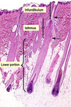
What are the three parts of a hair follicle?
- infundibulum
*follicular orfice to entrance of sebaceous gland
*normal keratinization - isthmus
*sebaceous duct to insertion of arrector pili muscle
*trichilemmal keratinization (no granular layer)
*no inner root sheath - lower portion
*dermal papillae
*matrix
Describe a hair follicle in cross section (parts outer to inner)
- fibrous sheath
- basemement membrane
- outer root sheath **continuous with the epidermis
- inner root sheath **supports hair fiber, degenerates at the level of sebaceous gland (aka no IRS higher than isthmus)
- henle’s layer
- huxley’s layer
- cuticle
- central cortex

Describe a sebaceous gland
- lobular; lined with thin outer layer of basophilic germinative cells
- central bubbly clear cells filled with lipid and “scalloped” nuclei
- duct lined with stratified squamous epithelium
- holocrine secretion (completely degenerates to secrete sebum)

Describe an apocrine gland
- single layer of columnar secretory cells with round nuclei
- coiled secretory portion (in lower reticular dermis or subcutaneous fat)
- straight duct opens into hair follicle above the level of the sebaceous gland (rarely opens to epidermal surface)
- found in axillae, anogenital area, external ear canal, eyelid, areola (inactive until puberty)
- decapitation secretion
**lumen may be larger than in eccrine tissue

Describe an eccrine gland
- present everywhere except vermillion of lips, glans, labia minora, nail beds, inner prepuce **greatest on palms, soles, axillae, forehead
- Three parts:
- spiraled intraepidermal portion (acrosyringium)
- intradermal duct (straight and coiled portions)
- coiled secretory portion

Describe the outermost portion of an eccrine gland
**the intraepidermal spiraled portion:
- also called acrosyringium or epidermal sweat duct unit
- empties directly onto epidermal surface

Describe the middle portion of an eccrine gland
**the intradermal straight portion
(two layers of small cuboidal cells)

Describe the innermost portion of an eccrine gland
**the coiled secretory portion
- one distinct layer of secretory cells surrounded by a layer of myoepithelial cells (similar to apocrine gland but smoother inner border and smaller lumen)
- lies in the lower reticular dermis
- surrounded by thick basement membrane

Describe how lymphatics look on histology
- thin walled vessels lined by attenuated epithelium
- have multiple valves
- usually collapsed and difficult to detect in the dermis… see with obstruction

Describe how subcutaneous fat looks on histology
“Hypodermis”:
- arranged in lobules, separated by vascular fibrous septa
- fat is dissolved by routine processing
- large single globule of lipid that displaces nucleus and cytoplasm


