Cranial Nerve VII, VIII, IX Flashcards
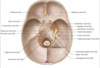
Identify the cranial nerves indicated via transvers cut of the skull

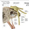
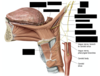
Identify the course of the Facial Nerve

- branches from the brainstem (pons) to the stylomastoid foramen
- It enters the petrous temporal bone via the internal acoustic meatus
- it bends as it passes fromthe internal coustic meatus to the facial canal
- as it bends, the geniculate ganglion is formed (sensory cell bodies and greater petrosal n.)
- while in facial canal, 2 major branches ariseto pass to middle ear cavity
- nerve to stapedius muscle
- chorda tympani, which continues through the petrotympanic fissue to enter the infratemporal fossal to merge with the lingual nerve
- CN VII exits the skull via the stylomastoid foramen
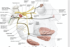
Label motor the branches of the Facial Nerve

- nerve to stapedius
- stylohyoid and posterior belly of digastri
- dives into parotid gland and divides into 5 branches that innervate facial muscles

Describe th pathway of taste from the anterior 2/3 on your tongue
The branches from the sensors on the anterior 2/3 of the tongue follow the lingual nerve, then branch off wiht chorda typani to synapse in the Geniculate ganglion (cell bodies). The post ganglionic fibers continue as the facial nerve.
Describe the pathway of innervation for the submandibular and sublingual galnds
- Pre-anglionic parasympathetic cell bodies in their own nucleus in the brainstem (pons).
- They pass through the geniculate ganglion without synapsing.
- They follow along the chorda tympani, then connect with the lingual nerve to reach the submandibular ganglion.
- They synapse on the submandibular ganglion, and post-synaptic fibers innervate both the sublingual and submandibular glands to stimulate salivaiton
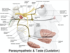
Describe the pathway of innervation for the nasal mucosa glands and lacrimal glands.
- Fibers from the facial nerve reach the genicular ganglion without synapsing, and diverge as the greater petrosal nerve which goes through the nerve to the pterygoid canal and the pterygopalatine fossa to the pterygopalatine ganglion where it synapses.
- Post-ganglionic fibers are sent with derivative V2 (maxilary) fibers into the glands in the nasal cavity
- Once the fibers reach the pterygopalatine ganglion to synapse, they can take ganglionic fibers up to use zygomatic n (V2) communicating branch of the lacrimal nerve V1 to reach the lacrimal gland.

Where are the cell bodies found for nerves that provide sensation from the posterior auricular nerve?
What sensory information is being transferred?
Describe its pathway.
geniculate gangloin
from auricle, external acoustic meatus, and oropharynx
the peripheral process seem to follow the posterior auricular nerve to mingle with the auricular branch of hte vagus, the posterior branch of the great auricular and lesser occipital to distribute to the auricle and external auditory canald
How does sympathetic innervation control glands?
Throgough controling the vasculature
contraction will decrease fluid in the gland and therefore fluid excretion
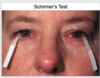
What does Schirmer’s Test look for?
It is a test of facial nerve dysfunction by looking at tear production bilaterally. A piece is paper is placed in the eye and length of wetted paper is measured after 5 minutes for each eye.
What types of problems with relation to the Facial nerve’s route can impact its functionality?
infection middle ear
infection mastoid air cells
major vasculature
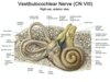
What are the three speial sensory fibers that are present in the vestibulocochlear nerve?
audition, equilibrium, and motion (mosty to the pons)
Why is the vestibulocochlear nerve (CNVIII) considered a composite nerve?
What are the difference between the components?
- Cochlear portion (audition)
- intefaces with the chochlea, very short peripheral processes
- cell bodies found in nmerous spiral ganglio within the chochlea
- central processes form cochlear nerve (roon) of CNVIII which passes through the internal acoustic meatus to enter the brainstem (at pontomedullary junction)
- Vestibular comoponent (equilibrium and motion)
- also within temporal labyrinth, short peripheral processes synapse with sensory receptors within th 5 organs of the vestibular apparatus
- form individual nerves that lead to cell bodies that are found within the two vestibular ganglia
- central process form the vestibular nerve (root) of CNVIII, which passes through the internal acoustic meatus to enter the brainstem (at pontomedullary junction)
Which cranial nerves could be impacted by enlarging acoustic neuroma?
CNIII and CNII because they have portions in the internal acoustic meatus
What cranial nerve is tested by the Rinne test?
Cranial Nerve VIII
conduction vs. mechanical characteristics
Can also test vestibular characteristics by having people stand up straight with their eyes closed
Identify the branches of the glossopharyngeal nerve (CNIX)

Describe the path of the visceral efferent fibers of the glossopharyngeal nerve
Emerges from teh medulla oblongata; leaves cranial cavity through jugular foramen
The nuclei is hte inferior salivatory nucleus
Parasympathetic fibers are sent to the otic ganglion; psot synaptic fibers are distributed to (1) parotid gland (2) Buccal gland (3) Labial gland
Describe the path of the special visceral efferent (somatic motor) fibers of the glossopharyngeal nerve
Emerges form medulla oblongata; leaves crainial cavity through jugular foramen
nucleus ambiguus
innervates the styloparyngeus muscle
Describe the path of the visceral afferent fibers of the glossopharyngeal nerve
Emerges from the medulla oblongata; leaves cranial cavity through the jugular foramen
nucleus of the solitary tract (inferior part)
receives sensory information from (1) chemoreceptors in carotid body (2) pressure receptors in the carotid sinus
Describe the path of the special visceral afferent fibers of the glossopharyngeal nerve
emerges from medulla oblongata; leaves crainial cavity through the jugular foramen
nucleus of the solitary tract (superior part)
receives sensory information from the posterior third of the tongue (via the inferior ganglia)
Describe the path of the somatic afferent fibers of the glossopharyngeal nerve
emerges from the medulla oblongata; leaves cranial cavity through the jugular foramen
spinal nucleus of hte trigeminal nerve
peripheral process of the intracranial superior ganglion or the extracranial inferior ganglion arise from
- (1) tongue, soft palate, pharyngeal mucosa and tonsils
- (2) mucosa of the tympanic cavity, internal surface of the tympanic membrane, pharyngotympanic tube (tympanic plexus)
- (3) skin of the external ear and auditory canal (blends with vagus nerve
Describe pathway for glossopharyngeal innervation of the parotid gland.
Glosopharyngeal nerve comes out of base of the skull, immediately gives of a tympanic nerve which pierces back up to the middle ear cavity and forming a plexus, which gives off a lesser petrosal nerve, paralleling the greater petrosal nerve (CNVII)
The lesser petrosal nerve will dive down with the mandibular nerve toward the otic ganglion, which will give branches that merge with the auricular temporal to innervate the parotid gland
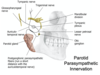
Identify the components of the pathway for innevation of the parotid gland.


