Clinical Syndromes Flashcards
Defn: Avascular Necrosis of the Femoral Head
Progressive eschemia and death of bone cells of the femoral head
Etiology: Avascular Necrosis of the Femoral Head
Disruption of arterial circulation due to trauma
MOI: Avascular Necrosis of the Femoral Head (2)
- Trauma - falls causing fx and dislocation causing damage to vessels
- Non-traumatic - long term corticosteroids, excessive alcohol causing occlusion of vessels
Symptoms: Avascular Necrosis of the Femoral Head (4)
- Pain: groin, prox thigh, glutes, increases with WB
- Limited ROM
- Axial loading increases symptoms
- Limb/Antalgic Gait
Modalities: Avascular Necrosis of the Femoral Head (2)
- Pulsed electromagnetic therapy
- Extracoporeal Shock Wave Therapy
TherEx: Avascular Necrosis of the Femoral Head (4)
- Stretch/ROM
- Strengthen
- Balance
- Gait training
Manual Therapy: Avascular Necrosis of the Femoral Head
Possible glides to facilitate ROM (depending on exam)
Education: Avascular Necrosis of the Femoral Head (3)
- Rest
- Limit smoking/drinking/steroid use
- Watch cholesterol levels
AD: Avascular Necrosis of the Femoral Head
Any device to offload the involved bone (femur)
HEP: Avascular Necrosis of the Femoral Head
Emphasis on gait and ROM
Defn: Piriformis Syndrome
Irritation or compression of the sciatic nerve caused by spasm or contracture of the piriformis muscle
MOI: Piriformis Syndrome (5)
- Overuse of glutes
- Inadequate stretching before/after activity
- Poor posture
- Prolonged sitting
- Trauma
Q: Who is Piriformis Syndrome more common in?
Women
Symptoms: Piriformis Syndrome (3)
- Pain, Numbness, and tingling over buttocks and down back of thigh
- Difficulty sitting
- Feeling of soreness
Manual Therapy: Piriformis Syndrome (3)
- Muscle Energy Techniques
- ST massage
- Myofasical release
TherEx: Piriformis Syndrome
Stretching - Figure 4 stretch
Modalities: Piriformis Syndrome (4)
- Moist Heat
- Ultrasound (+ stretching)
- Cold pack
- E-stim (after exercise/MT)
Education: Piriformis Syndrome (3)
- Rest
- Light and gradual stretching
- Posture
Q: What is the bimodal distribution of Femoral Neck Stress Fx?
- Young and active
- Elderly and osteoporotic
T/F: Men are more affected than women by femoral neck stress fx.
False, flip it
Defn: Femoral Neck Stress Fx
A Fx of the femoral neck that can be classified as either a compression or tension fx and puts the femoral head at a high risk of avascular necrosis
Classifications: Femoral Neck Stress Fx (2)
- Compression: inferior aspect of femoral neck
- Tension: superior aspect of femoral neck
MOI: Femoral Neck Stress Fx
Young = trauma
Older = falls/twisting
Typically fx 1-2 inches from the hip joint
Symptoms: Femoral Neck Stress Fx (4)
- Groin pain with activities
- Deep thigh pain
- May limp
- Pain eases with rest
TherEx: Femoral Neck Stress Fx (3)
Progressive, always with an emphasis on PAIN FREE movement
Acute (4-6 wk): NWBing to PWBing
Rehab (8-12 wk): FWB, progress from walk to run
Maintenance (12+wk): Monitor activities/form, increase distance
Manual Therapy: Femoral Neck Stress Fx
Joint mobilization once fx is healed
Modalities: Femoral Neck Stress Fx
Ice
Education: Femoral Neck Stress Fx
WB restrictions
AD: Femoral Neck Stress Fx
Crutches
Defn: Pubalgia
Groin pain in athletic individuals withouth inguinal hernia,
Pain from pubic symphsis to ASIS, can involves abdominal muscles/tendons/sheaths, inguinal ligament, adductor muscles, gracilis, pectineus, and iliopsoas
May also be known as a “sports hernia” (not an actual hernia)
Grades of Pubalgia (3)
1 = single/mutliple tears of rectus abdominis or adductor muscles
2 = partial avulsion from pubic symphsis
- = comples avulsion with micro tears
MOI: Pubalgia
Muscular imbalances between abdominals and adductors
Symptoms: Pubalgia (3)
- Insidious onset groin pain
- Hx of sudden tearing sensation
- Pulling sensation in groin with activity
Education: Pubalgia (3)
- Warm Up
- Rest
- NSAIDS
Modalities: Pubalgia (4)
- Ultrasound
- E-stim
- Hot pack
- Cold pack
TherEx: Pubalgia (3)
- Stretching as tolerated
- Strengthening (adductors, hip flexors/IR, abs, glutes)
- Proprioceptive training
Manual Therapy: Pubalgia
Transverse friction massage
Term: used to describe chronic, intermittent pain accompanied by tenderness to palpation overlying the lateral aspect of the hip
Trochanteric bursitis (TB)
Q: What is another name for trochanteric bursitis?
Greater trochanteric pain syndrome (GTPS)
Q: What can GTPS associted with? (4)
- Tendinitis
- Muscles tears
- Trigger points
- IT band disorders
MOI: GTPS (6)
- Chronic microtrauma
- Regional muscle dysfunction
- Overuse
- Acute injury
- Obesity
- Muscle fatigue
Q: What’s the profile for GTPS? (2)
- Femal:Male = 4:1
- 40-60 yo
Symptoms: GTPS (3)
- Persisent pain inthe lateral hip/buttocks
- Lying on affected side or prolonged standing provokes pain
- Sit>stand, stair climbing, high impact probokes pain
T/F: PT alone will cure GTPS.
False: need to eliminate the cause (prolonged standing)
Education: GTPS
Avoid MOI, side laying, hard surfaces and lose wieght
Modalities: GTPS (2)
- TENS
- US
AD: GTPS
Cusion/pads for protection, insoles if leng length discrepancy
TherEx: GTPS (3)
- Stretching
- Strengthening
- Functional exercises
MT: GTPS
Manipulations for mobility if needed
Symptoms: Hip Muscular Strain (Pull or Tear) (4)
- Pain over injured muscle
- Increased pain with contraction
- Swelling
- Loss of strength
Q: What muscles are commonly affected in Hip Muscular Strain (Pull or Tear) (3)
- Hamstrings (high speed movement)
- Quadriceps
- Adductors (socer/ice hockey)
MOI: Hip Muscular Strain (Pull or Tear) (6)
- Stretched muscled forced to suddenly contract
- Overstretching/Overuse
- Fall/direct blow
- Inadequate warm up
- Lack of flexibilty
- Poor posture
Grade 1 Hip Muscular Strain (Pull or Tear) (2)
- Small tears in fibers
- Pain but minimal strength and ROM loss
Grade 2 Hip Muscular Strain (Pull or Tear) (3)
- More fibers torn, but lesion not complete
- Pain, swelling, and bruisin may occur
- Compromised strength, but still within NFL
Grade 3 Hip Muscular Strain (Pull or Tear) (2)
- Most fibers torn, in some cases complete ruptured
- Movement is difficult, not impossible, but loss of function
Education: Hip Muscular Strain (Pull or Tear)
RICE 48 hours after injury
Modalities: Hip Muscular Strain (Pull or Tear) (2)
- TENS
- US
MT: Hip Muscular Strain (Pull or Tear)
Gentle massage
TherEx: Hip Muscular Strain (Pull or Tear) (4)
- Isometric > isotonic > functional
- Subacute: cycling, treadmill
- Plyometric training
- Improve flexibility/posture
Defn: Trochanteric Bursitis
Inflammation of the bursa located on the superior lateral part of the thigh bone
Q: Apart from the greater trochanteric bursa, what is the other major bursa in the hip?
Iliopectineal bursa - front of the hip joint
MOI: Trochanteric Bursitis (5)
- Prolonged pressure
- Overuse
- Arthritis
- Injury
- Infection
Symptoms: Trochanteric Bursitis (3)
- Pain/tenderness with motion and at rest
- Pain over outer thigh
- Difficulty walking
Education: Trochanteric Bursitis (2)
- Explain MOI
- Identify and change aggravting factors
Modalities: Trochanteric Bursitis (4)
- Ice (Massage)
- Heat
- Ultrasound
- TENS
TherEx: Trochanteric Bursitis
Emphasize stretching
MT: Trochanteric Bursitis
Mobs to improve motion
AD: Trochanteric Bursitis
If a leg discrepancy exists gradually increase the height of the insoles
Defn: Baker’s Cyst
a fluid filled cyst that causes a bulge and feeling of tightness behind your knee, typically develops in the gastrocnemius-semimembranosus bursa
Q: Who is a Baker’s Cyst more common in?
Typically unilateral and medial, twice as common in men
MOI: Baker’s Cyst
Inflammation of the joint can cause an excess of synovial fluid
Q: What is the difference between primary and secondary Baker’s Cyst?
Primary = no knee pathology
Secondary = underlying knee problem
Symptoms: Baker’s Cyst (4)
- Swelling
- Pain with flexion and extension
- Stiffness
- Clicking, locking, buckling
Modalities: Baker’s Cyst
Ice and Compression (RICE)
TherEx: Baker’s Cyst (2)
- Strengthening
- ROM
Education: Baker’s Cyst (3)
- RICE
- Educate about Cause
- Prognosis
Defn: Osgood-Schlatter’s Disease
A benign traction apophysitis (inflammation of an apophysis) that occurs in the tibial tubercle
MOI: Osgood-Schlatter’s Disease
During periods of rapid growth, stress from repetitive quad contractions is transmitted through the patellar tendon onto the partially developed apophysis
Can result in avulsion fx, inflammation of the tendon, and heterotrophic bone formation
Symptoms: Osgood-Schlatter’s Disease (3)
- Pain with activities
- Viisible lump over site
- Pain with knee extension
Q: What is the typical population of Osgood-Schlatter’s Disease?
11-18 yo, boys > girls, typically unilateral
Modalities: Osgood-Schlatter’s Disease
Ice
Education: Osgood-Schlatter’s Disease
- Avoid aggravating factors
- Rest
- Length of recovery (can be 1-2 years)
AD: Osgood-Schlatter’s Disease
Infrapatellar strap
MT: Osgood-Schlatter’s Disease
Patellar glides
TherEx: Osgood-Schlatter’s Disease (3)
- SLR
- Stretching
- Knee stabilization
Defn: Femoral Condyle Injury
Focal articular cartilage defect - osteochondritis dissecans lesion
Typical Profile: Femoral Condyle Injury (3)
- Sports trauma (most common cause)
- 12-35 yo
- Males > females
Symptoms: Femoral Condyle Injury (5)
- Focal tenderness
- Swelling/Joint effusion
- Catching
- Limited ROM
- Pain with WB
Conservative Treatment: Femoral Condyle Injury (4)
- Bracing
- ROM/Strength
- Pt Education
- Corticosteroid injections
T/F: Lateral meniscus injuries happen more often then medial meniscus injuries.
False: Flip It
MOI: Meniscus Injury (2)
- Trauma/Sports (Non-Contact)
- Degenerative
Q: What type of forces can cause traumatic meniscus injury? (3)
- Compression + rotation
- Flexion OR extension + rotation during WB
- Sudden acceleration/deceleration with direction change
T/F: Meniscal injuries are associated with cruciate ligament injuries.
True
Q: Who typically has degenerative meniscal injuries? (3)
- > 40 yo
- Menisci are stiff and less compliant
- Minimum to no healing potential
Diagram: Identify the types of meniscal injury
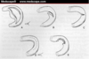
A = Bucket handle
B = Oblique
C = Radial
D = Longitudinal
E = Degenerative
Symptoms: Mensicus Injury (4)
- Pain
- Limited extension
- Hard end feel with extension
- Locking, giving out
Q: What is the healing response of meniscal injuries influenced by? (2)
- Location
- Extent of injury/protection
AD: Mensicus Injury
Progressive PWB > WBAT with brace and crutches
Modalities: Mensicus Injury
Ice and E-stim
MT: Mensicus Injury
Patellar mob
Education: Mensicus Injury
WB compliance
TherEx: Mensicus Injury
Strengthen quadriceps and hamstrings and ROM
MOI: ACL injury
Non-contact:
- Hyperextension with anterior translation (anterior tilt of femur stretches posterior part of ACL)
- Sudden deceleration (most commont)
Contact:
- Excessive twisting of the knee or valgus stress on the knee (usually = ACL + other structures)
MOI: PCL Injury
Hyperflexion with posterior translation
MOI: MCL Injury
Valgus stress
MOI: LCL Injury
Varus stress
MOI: ACL + medial meniscus
Hyperextension with rotation
MOI: ACL + medial meniscus + MCL
Extension, valgus with rotation
Symptoms: ACL Injury (6)
- Knee giving out (#1 complaint of complete or partial tear)
- Pain, edema, joint stiffness
- Lack of quad control
- Gait deviation
- Pop with MOI?
- Ability to walk with extended knee (extend of injury)
Conservative Treatment: ACL Injury (4)
- Full extension, quad control
- Proprioceptive training to max dynamic stability (ligaments major source of proprioception)
- Sports restriction
- Risk of developing joint degeneration
Presurgical Requirements: ACL Injury (4)
- Edema control
- Gain full extension and quad tone
- Pt. education
- Quad and Hamstring strengthening
Q: What are the options for ACL reconstruction? (2)
- Bone, Patellar tendon, Bone
- Hamstring Tendon
Rehab: ACL Injury (3)
- Usually spancs 3-6 mo
- Protective, rehab, functional phases
- Return to sport 6-12 mo
Modalities: ACL Injury (2)
- Ice
- IFC
MT: ACL Injury
Patellar mobilization
Education: ACL Injury
Protection, importance of exercises, expectation
MOI: PCL Injury (2)
- Forceful posterior translation of tibia (“Dashboard Injury”)
- Falling onto flexed knee
MOI: MCL/LCL Injury
Excessive valgus/varus force with planted foot
AD: MCL/LCL Injury
Bracing to limit extension and minimize valgus/varus forces
TherEx: MCL/LCL Injury
Strengthening, proprioception, functional/agility training
Defn: Patellofemoral (PF) pain syndrome
Patellar tracking problem (dislocation)
Contributing factors: PF syndrome (4)
- Anatomical abnormalities: increased Q angle, patella alta, pes planus
- Muscle weakness: hip ABD/ER, quads (VMO)
- Flexibility: tight ITB
- Poor motor control
Symtpoms: PF syndrome (4)
- Anterior/lateral/retro-patellar pain
- Dull ache
- Clicking/popping
- Knee giving out
Aggravating Factors: PF syndrome (4)
- Walking
- Stair climbing
- Kneeling
- Squatting/sit to stand
Q: What muscle strength is important to assess with PF syndrome?
Hip
AD: PF Syndrome
Patellar taping (for pain control)
Bracing (provides stability)
Education: PF Syndrome
Activity modification
TherEx: PF Syndrome (3)
- Quad, Hip ABD, Hip ER strengthening
- Flexibility: ITB, HS, quads
- Motor control: squatting, landing, running
Q: What are other names for patellar tendinopathy?
Jumper’s knee
Defn: Patellar Tendinopathy
Chronic degeneration of patellar tendon due to overuse and microtrauma
Not to be confused with patellar tendinitis (inflammation of the tendon)
Q: Where is patellar tendinopathy most common?
Posterior proximal portion of tendon - tender and thickened
Contributing factors: Patellar Tendinopathy (4)
- Lack of flexibility/strangth can resist ROM and increase load on anterior knee
- High patella/patella alta
- Overuse
- Postural alignment, reduced patellar glide, foot structure
Symptoms: Patellar Tendinopathy (4)
- Pain over posterior tendon
- Mild stiffness after prolonged sitting
- Pain worse with activity
- Palpable tenderness
Aggravating Factors: Patellar Tendinopathy (2)
- Jumping
- Landing
Modalities: Patellar Tendinopathy
Ice
MT: Patellar Tendinopathy
Friction massage
AD: Patellar Tendinopathy
Patellar tendon strap or taping
TherEx: Patellar Tendinopathy
Stretching and eccentric strengthening
Goal of Treatment: Patellar Tendinopathy
Initially reduce symptoms, then progress strengthening of muscles and quad tendon
Defn: IT Band Syndrome
Overuse of the TFL results in pain and inflammation at the outer thigh/knee
MOI: IT Band Syndrome (5)
- Overuse
- Poor flexibility
- Muscle imbalances
- Leg length discrepancy
- Bowed legs
Symptoms: IT Band Syndrome (3)
- Pain on outer knee/greater trochanter
- Snapping or popping
- Swelling
Modalities: IT Band Syndrome (3)
- Ultrasound
- Iontophoresis
- Ice
MT: IT Band Syndrome
Myofascial release
TherEx: IT Band Syndrome
Stretching and strengthening
AD: IT Band Syndrome
Shoe orthotic to control gait problem/pelvic tilt/leg length
Defn: Osteoarthritis
Worn down cartilage of the end of bones
MOI/Risk Factors: Osteoarthritis
MOI: Occurs gradually over time
Risk factors: overweight, age, joint injury, joint stress
Symptoms: Osteoarthritis (4)
- Pain and Tenderness
- Stiffness
- Loss of flexibility
- Grating sensation
T/F: There is a cure for Osteoarthritis.
False, no cure
TherEx: Osteoarthritis
LOW IMPACT stretching and strengthening
AD: Osteoarthritis
Orthotics, braces, canes, crutches, walker
MT: Osteoarthritis (2)
- Joint distraction to decrease pain and stiffess
- Glides to improve ROM
Modalities: Osteoarthritis
TENS
Q: For tendon issues what type of exercise should you focus on?
Eccentric exercises
- decline board more specfically targets patellar tendon with squats
progression: bilat > unilat > eccentric > concentric > PWB > FWB > resistance/increased speed
Defn: Heel Spur
Bony formation on the medial plantar aspect of the calcaneal tubercle
MOI: Heel Spur (3)
- Stress
- Inadequate footwear/Poor gait mechanics
- Prolonged standing
Symptoms: Heel Spur (3)
- Presents in similar fashioin as plantar fasciitis - pain in morning
- Pain with WB, heel strike, palpation
- Inflammation of Achilles Tendon
Modalities: Heel Spur (2)
- Ionto
- Ice massage/moist heat
MT: Heel Spur
Deep friction massage
Soft tissue mob
Manipulation
AD: Heel Spur
Shoe modifications: orthotic, heel cup, taping
TherEx: Heel Spur (3)
- Foot stabilization exercises for motor control
- Strengthening
- Achilles stretching
Defn: Achilles Tendinopathy
Tendinitis or Tendinosis of the Achilles tendon
Can be inserstional or non-insertional
MOI: Achilles Tendinopathy
Quick repetitive pronation/supination cuasing whipping/twisting effect of Achilles tendon
Symptoms: Achilles Tendinopathy (3)
- Pain stiffness along tendon
- Increasing pain with activity
- Thickening of tendon
Education: Achilles Tendinopathy
Rest and activity modification
Modalities: Achilles Tendinopathy (3)
- Ionto
- Ultrasound
- Ice
TherEx: Achilles Tendinopathy
Eccentric strenghtening and ROM
MT: Achilles Tendinopathy
PNF stretching
Defn: Ankle Sprain
Stretching or tearing of latera/medial ligaments of the ankle joint
MOI: Ankle Sprain (2)
- IR, PF, inverted (rolling foot inward) - 85%
- ER, DF, everted (rolling foot outward)
Education: Ankle Sprain
Resting and NWB
Modalities: Ankle Sprain
RICE
MT: Ankle Sprain
Acute phase = gentle massage, grade 1-2 mob
Grade 3-4 when pain decreases
TherEx: Ankle Sprain
Early - limit inv/ever and WB, DF/PF ROM, stretching
Late - Isotonic, isokinetic, WB, total ROM including inv/ever, balance
Functional - running, jumping, stairs, balance
Defn: Functional Ankle Instability
A condition in which pts. experience recurrent sprains and/or a feeling of their ankle “giving way”
Symptoms: Functional Ankle Instability
Recurrent ankle sprain and sensation of ankle instability
MOI: Functional ankle instability (4)
- Increased joint flexibility/stiffness
- Muscular weakness
- Proprioceptive/balance impairments
- Delayed peroneal activation time
MT: Functional Ankle Instability
Grad 3-4 mob when hypomobile
TherEx: Functional Ankle Instability
Balance, proprioception, strengthening exercises
Term: a slowly progressive joint disease typically seen in middle-aged to elderly people.
Osteoarthritis
MOI: Osteoarthritis (Hip) (5)
- Aging process
- Joint trauma
- Repetitive abnormal stress
- Obesity
- Systemic diseases (RA)
Symptoms: Osteoarthritis (Hip) (2)
- Insidious onset of pain anterolateral hip and groin
- Decreased ROM
Aggravating factors: Osteoarthritis (Hip) (5)
- Standing, walking, or sitting for a long time
- Squatting
- Active hip flexion causing lateral hip pain
- Scour test with adduction causing lateral hip/groin pain
- Active hip extension causing pain
Education: Osetoarthritis (Hip)
Lose weight, yoga, tai chi classes
Modalities: Osetoarthritis (Hip)
Thermal agents or ice
MT: Osetoarthritis (Hip)
Maneuvers for general mobility and traction manipulation
TherEx: Osetoarthritis (Hip)
Stretching, strengthening, endurance, aerobic exercise
AD: Osetoarthritis (Hip)
Walking aids and cane
Defn: Labral Tear
Tear of the acetabular labrum resulted from excessive forces at the hip joint
Symptoms: Hip Labral Tear (5)
- Pain is usually anterior/groin (90%)
- Clicking
- Catching
- Giving way
- Stiffness
MOI: Labral Tear of the Hip (5)
- Motor vehicle accidents/slipping/falling (with or without hip dislocation)
- Sporting activities (that require frequent ER)
- Forces movements (torsion/twisting, hyperABD, hyperEXT, hyperEXT with ER)
- Repetitive microtrauma
- Hip dysplasia (lead to bone abnormalities)
Education: Hip labral tear
Limited WB, avoid pivoting (under load with extension)
Modalities: Hip labral tear
Ice
MT: Hip labral tear
Depending on the PAM examination
TherEx: Hip labral tear (5)
- Optimize control of hip ABD, deep ER, Glute Max and Iliopsoas
- Correction of dominant participation of quads and hams
- Correct gait (hyperEXT)
- Avoid exercises causing hip hyperEXT
- Avoid weight training of quads and hams
AD: Hip labral tear
Crutches and cane in acute phase
Defn: What is Legg-Calve-Perthes
Disease casuing decreased blood supply to the femoral head
Education: Foot Deformities
Rest, activity modification, shoe wear
TherEx: Foot Deformities
Stretching, strengthenin, proprioception
MT: Foot Deformities
Massage, mobilization, manipulation
AD: Foot Deformities
Orthotics!!
Defn: LE Compartment Syndrome
Increase pressure in small fascia compartment due to edema or hypertrophy of muscle
MOI: LE Compartment Syndrome
Acute - vascular impairment, fx, soft tissue injury
Chronic - bilateral, hypertrophy/overuse
Symptoms: LE Compartment Syndrome
Early = pain, swelling
Late = paraesthesia, reduced pulse, paralysis
Education: LE Compartment Syndrome
Stop painful activity
Modalities: LE Compartment Syndrome
Acute - DO NOT ice/elevate
MT: LE Compartment Syndrome
Rare, maybe massage or mob
TherEx: LE Compartment Syndrome
Stretching, typical post-op recovery
AD: LE Compartment Syndrome
Maybe orthotics
Defn: Calcaneal Fx
Fx of calcaneous or tarsal
MOI: Calcaneal Fx
Most often due to high impact/traumatic events
Symptoms: Calcaneal Fx (4)
- Sudden onset heel pain
- Swelling
- Ankle bruising
- Pain with palpation
TherEx: Calcaneal Fx (4)
- Early ROM
- Progressive WB
- Strengthening
- Gait/Balance training
Education: Calcaneal Fx
REST is key
MT: Calcaneal Fx
Joint mob, soft tissue massage
Modalities: Calcaneal Fx
Ice/Heat, E-stim, Ultrasound
Defn: Plantar Fasciitis
Inflammation of the plantar fascia (shock absorber and arch support)
MOI: Plantar Fasciitis (4)
- Overstretch/strain which produces microtears
- Prolonged standing/walking
- High arches or flat feet
- Poor shoe support
Symptoms: Plantar Fasciitis (2)
- Pain and stiffness on the bottom of the foot, slightly anterior to the heel
- Pain with first steps in the morning
Modalities: Plantar Fasciitis
Ice, Ionto, Corticosteriod injections, Ultrasound
MT: Plantar Fasciitis
Soft tissue mobilization (myofasical release, massage, etc)
TherEx: Plantar Fasciitis
Stretching, strengthening, rolling massage
AD: Plantar Fasciitis
Foot orthotics or heel cup, strap sock, night splint/brace
Education: Plantar Fasciitis
Proper foot wear, avoid aggravating factors
Defn: Cuboid Syndrome
Distruption of the arthrokinematics of structural congruity of the calcaneocuboid joint (due to tearing of supporting soft tissue)
MOI: Cuboid Syndrome
Trauma (sprain) or repetitive use
Symptoms: Cuboid Syndrome (4)
- Pain on lateral foot
- Restricted ROM
- Inflammation
- Antalgic gait
Education: Cuboid Syndrome
Rest
Modalities: Cuboid Syndrome
Taping, Ice, Ultrasound
TherEx: Cuboid Syndrome
Stretching, Strengthening, Proprioception
MT: Cuboid Syndrome
Cuboid whip and cuboid squeeze
AD: Cuboid Syndrome
Orthosis, Cuboid padding
