Ch. 6-5 Bone Growth Flashcards
The deposition of calcium salts - occurs during ossification, but also in other tissues
calcification
- Bone replaces existing cartilage
- Most originate as hyaline cartilage
Endochondral ossification
bone develops directly from mesenchyme or fibrous connective tissue
intramemranous ossification
- Cartilage enlarges (outer appositional and inner interstitial growth)
- chondrocytes near the center of the shaft increase in size
- matrix is reduced to a series of small struts that begin to calcify
- enlarged chondrocytes die and disintegrate

First step of endochondral ossification
- Blood vessels grow around edges of cartilage
- cells of perichondrium convert to osteoblasts
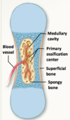
- shaft of cartilage ensheathed in superficial bone layer

2nd step of endochondral ossification
- Blood vessels penetrate the cartilage and invade the central region
- Fibroblasts migrating with the blood vessels differentiate into osteoblasts and produce spongy bone at the primary ossification center
- Bone formation spreads towards both ends

3rd step of endochondral ossification
- Remodeling occurs as growth continues, creating medullary cavity
- Osseous tissue of shaft becomes thicker
- Cartilage near each epiphyses replaced with bone
- Growth to increase length & diameter (2 distinct processes)

4th step of endochondral ossification
Capillaries and osteoblasts migrate into epiphyses, creating secondary ossification centers

5th step of endochondral ossification
- Epiphyses filled with spongy bone
- A thin cap of articular cartilage remains exposed to the joint cavity
- epiphyseal cartilage (epiphyseal plate) separates epiphysis from diaphysis

When osteoblasts ‘catch up’ with chondrocyte production of epiphyseal cartilage, which ultimately disappears. The former location of this cartilage forms a ____ ___ detectable in x-rays
epiphyseal line
Appositional Growth
(Endochondral ossification)
- Cells of the inner layer of periosteum differentiate into osteoblasts and deposit superficial layers of bone matrix
- Osteoblasts become surrounded by matrix and differentiate into osteocytes
- Appositional growth adds a series of layers that form circumferential lamellae.
osteoblasts differentiate within a mesenchymal or fibrous connective tissue.
Intramembraneous ossification
(dermal ossification)
Flat bones of the skull
Mandible (lower jaw)
Clavicle
Dermal bones that result from ossification that occurs in the deeper layers of the dermis (dermal ossification)
- Mesenchymal cells cluster (ossification center) and start to secrete organic components of matrix.
- As calcification occurs, mesenchymal cells differentiate into osteoblasts
- Developing bone grows outward in smalls truts (spicules)
- Trapped osteoblasts differentiate into osteocytes
First step of intramembranous ossification
Blood vessels begin to grown into ossification areas; spicules meet and fuse toegher.
2nd step of intramembraneous ossification


