Ch 38. Squamous neoplasms of the conjunctiva Flashcards
(16 cards)
from which region of the eye, does the squamous neoplasms most frequently arise?
The lymbus
The biopsy of this patient reports dyskeratosis, parakeratosis and acanthosis, with no atypia or dysplastic potential. The lesions are bilateral.
What is the diagnosis?

Benign hereditary intraepithelial dyskeratosis
<em>An autosomal dominant, bilateral, and highly penetrant disease mostly present in native Americans on the Haliwa tribe, north Carolina. </em>
Diagnosis.

Pterygium with pseudoepitheliomatous hyperplasia.
<em>PEH has a more rapid onset and lacks the capillary fronds seen as regular red dots in cases of squamous papilomas and carcinomas</em>

Management of these lesions?
These are both Conjunctival squamous papillomas.
The most effective treatment in preventing recurrences is excision and cryotherapy to the base of the lesion. Oral Cimetidine act as immunomodulatory.
It can also be treated with courses of MMC or alfa-interferon.
Most papillomas behave in a benign fashion, with little tendency to undergo malignant transformation. Signs of dysplasia include keratinization, symblepharon formation, inflammation, and palpebral conjunctival involvement.
Given the histology and clinical appearances of the lesion shown in the pictures, what is the most likely diagnosis?

Dacryoadenoma of the conjunctiva.
Which HPV serotypes are associated with benign/malignant conjunctival tumors?
6,8,11: Benign.
16,18: Malignant Transformation.
How can CIN be distinguished from flat conjunctival papillomas and pseudo epitheliomatous hyperplasia?
the latter have a low nuclear-cytoplasmic ratio and an imperceptible blending of the hyperplastic epithelium to normal epithelium.
What is the most appropriate treatment for CIN
full excision with 2mm margin (conjunctival, limbal, and corneal) + double or triple freeze.
mmc can be used as a treatment for small primary lesions or as adjunctive treatment of recurrent or large lesions.
up to 50% of lesions recur, therefore long-term follow-up is mandatory.
What differentiates invasive conjunctival carcinoma associated with HPV from the classical forms
they are more frequently multifocal and/or bilateral, bigger lesions, and have a more verrucous/papillary morphology.
Name the most likely diagnosis.

Squamous cell carcinoma in a pigmented individual.
these lesions are often confused with malignant melanomas, but the frosted-looking epithelium and leukoplakia components are most likely to be associated with SCC.
Name predictors for recurrence of squamous cell neoplasia of the conjunctiva.
- Size
- positive margins
- age
- elevated proliferation index (by Ki-67 index)
- scleral involvement
What is the index of intraocular invasion of SCC?
3-13%
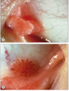
Top and bottom are two very similar clinical entities, how would you manage them?

Top: Corneal Epithelial Dysmaturation
Bottom: Primary corneal epithelial dysplasia.
The former is benign, with no atypia present. The latter does have atypia present, but both are usually cured by corneal scraping and, if present, excision of the limbal component.
A patient presents with a recurrence of a fast-growing lesion a few months after SCC excision.
What other Diagnosis must be considered and what precautions this requires.
Mucoepidermoid carcinoma.
These lesions are clinically indistinguishable from SCC (they can be suspected by lesions growing anywhere else outside the limbus or by lesions recurring after what was thought to be SCC)
This is an early locally invasive lesion and metastatic to lymph nodes.
Therefore invasion studies like UBM or AS-OCT must be performed. Staging through Centinel lymph node biopsy must also be performed.
Given the clinical appearance and the spindle cells reported from the biopsy, what is your differential diagnosis?

the differential diagnosis of this pathologic appearance includes spindle cell carcinoma, fibrosarcoma, leiomyoma, rhabdomyosarcoma, sebaceous cell carcinoma, and malignant melanoma.
Immunohistochemistry is often needed to make the distinction.
Biopsies taken from a sebaceous cell carcinoma with pagetoid spread can miss the correct dx in 50% of cases.
This added to the fact that this presentation may not have thickened eyelid or madarosis, how could you suspect it from the biopsy report?
These lesions spread superficially in the epithelium causing general denudation (pre, during, or after the biopsy procedure), which is in part responsible for the lack of accuracy of the biopsy, since the affected cells can be partially or totally lost, leaving confusing changes suggestive of SCC.
Suggest this diagnosis if Biopsy shows epithelial denudation, or if changes are mostly superficial in the epithelium since the SCC arrises from the deeper layer and progress outwards.
