CBL_acute abdo Flashcards
What’s meant by ‘an acute abdomen’?
Pain that:
- lasts <1 week
- requires hospital admission
- hasn’t been treated/investigated previously
Apart from SOCRATES, what other questions would you ask in Hx of acute abdo pain?
- Fevers
- Weight change
- Appetite disturbance
- Vomiting
- Gynaecological symptoms – is there a coil?
- Urinary symptoms
- Alcohol intake, smoking
- Family history
- Medication history
What is ‘swinging pyrexia’?
What is it typical of?
- An intermittent fever in which the daily oscillations are very large, often associated with severe chills and sweats
- typical of abscess
What’s Murphy’s sign?

What’s Mc Burney’s sign?
Associated condition
- It is tenderness at Mc Burney’s point

the point over the right side of the abdomen that is one-third of the distance from the anterior superior iliac spine to the umbilicus (navel). This point roughly corresponds to the most common location of the base of the appendix where it is attached to the cecum
- Typical of acute appendicitis
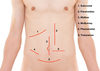
Name these incisions (1-5)
*picture

What’s the psoas sign?
What does it indicate?
How to perform?
- The psoas sign, also known as Cope’s psoas test or Obraztsova’s sign
1. is a medical sign that indicates irritation to the iliopsoas group of hip flexors in the abdomen
2. indicates that the inflamed appendix is retrocaecal in orientation (as the iliopsoas muscle is retroperitoneal)
3. How to carry out:
carried out on the patient’s right leg. The patient lies on his/her left side with the knees extended. The examiner holds the patient’s right thigh and passively extends the hip. Alternatively, the patient lies on their back, and the examiner asks the patient to actively flex the right hip against the examiner’s hand.
If abdominal pain results, it is a “positive psoas sign”

Look at the picture

Rovsing’s sign
If palpation of the left lower quadrant of a person’s abdomen increases the pain felt in the right lower quadrant -> a positive Rovsing’s sign and may have appendicitis

Carvical excitation as a sign
- It is tenderness on a cervical motion
It is also known by the colloquial name “chandelier sign” due to the pain being so excruciating upon bimanual pelvic exam
- Associated with: PID or ectopic pregnancy
*some use to help differentiate PID from appendicitis
SIRS criteria
Indicative of SIRS:
- Temp <36°C or >38°C
- P >90 BPM
- RR >20/min
- WCC <4 or >12 x10^9 cells/L
- Hyperglycaemia in non-diabetic
- New onset altered mental state
*2 or more consistent with SIRS
*If proven infective cause -> sepsis
What does sepsis commonly causes? (in terms of clinical picture)
Loss of glycaemic control and deranged clotting
Investigations for SIRS and sepsis with acute abdomen
- urinealysis
- betaHCG
- FBC
- U&Es
- LFTs
- Amylase
- Coagulation
- ABGs
- Venous blood cultures
- Erect CXR
- AXR
- ECG
- USS (biliary, KUB, pelvis)
- CT scan
- Endoscopy
- MRCP
Contraindications to CT with contrast
- pregnancy (CT contraindicated in general)-> amount of radiation is harmful to foetus particularly in 1st-trimester
- allergy to IV contrast media
- renal impairment -> creatinine and eGFR checked before the referral -> if eGFR is less than 30ml/min then the risk of AKI is increased
- Hyperthyroidism or Goitre -> as contrast media may induce thyrotoxic crisis
- Phaeochromocytoma -> hypertensive crisis may be induced
- Myasthenia gravis -> a small risk of worsening of the condition (may induce respiratory muscle depression - so administer with caution and monitor patient afterwards)
*weight and size limitations -> some scanners can accommodate up to 220kg person and 70 cm in diameter
*special precautions in a patient taking metformin
(as IV contrast media + metformin -> lactic acidosis possible when a renal failure is caused)
Metformin and contrast media
- what’s the danger? (pathophysiology)
- what to do to avoid this?
Danger: When a patient is on metformin and renal failure occurs following IV contrast media -> lactic acidosis will be caused
Pathophysiology: metformin is excreted primarily by the kidneys -> continued intake of metformin after the onset of renal failure -> toxic accumulation of this drug and subsequent lactic acidosis.
To avoid this complication:
Metformin must be withheld after the administration of the contrast agent for 48 hours (during which the contrast-induced renal failure becomes clinically apparent) -> If renal function is normal at 48 hours, the metformin can be restarted
General treatment options for acute abdomen (considering the variety of conditions)
- Fluids ± antibiotics, observation
- Correct electrolyte and clotting abnormalities
- Endoscopy, including intervention
- ERCP
- Cholecystostomy (rarely)
- Laparoscopy
–Appendicectomy, Cholecystectomy, Gynae
•Laparotomy
What fluid and how much should be given if suspected SIRS and shock? (Fluid Challenge)
20-30ml/Kg of crystalloid
What’s the aim of urine output in a septic patient?
>0.5/mls/kg/hr
Causes of post-operative fever
5 W’s (Wind, Water, Walking, Wound, Wonder Drugs)

What is the temperature range for post-op fever
- >38.0 C for 2 consecutive days post-op
- temperature >39.0 C for one day post - op
Is an infection a common cause of post-op fever in 48 hours after surgery? Why?
No. This is because there is not enough time for the bacteria to multiply
The common cause of post-op fever in 48 hours after surgery
Pyrectic stress response induced by the surgery (activated SNS response)
5 Ws in differentials for a post-op fever
W - wind
- What does it refer to?
- How the risk for that are increased?
W - wind
Chest infections
- Increased risk due to GA and artificial airway ventilation -> as widening of air spaces will # their natural structure and increase risk of infection
- patient may not take adequate breaths due to pain in the chest (e.g. from post-op wound)
5 Ws in differentials for a post-op fever
W - water
- What does it refer to?
- when does it occur
- How the risk for that are increased?
W - water
UTI
- post op day 3-5
*consider especially if a patient has urinary catheter in
5 Ws in differentials for a post-op fever
W - walking
- What does it refer to?
- when does it occur
- How the risk for that are increased?
W- walking
Venous Thromboembolism
- day 5-7 post-op
- reason: decreased mobility, the stress response to the surgery
5 Ws in differentials for a post-op fever
W - wound
- What does it refer to?
- when does it occur
- How the risk for that are increased?
W - wound
Surgical scar infection
- post-op day 10
- check the wound for the signs of infection
5 Ws in differentials for a post-op fever
W - wonder about drugs/ what have we done to a patient
W- Wonder about drugs/ what have we done to the patient
- check IV/cannula sites
- is the fever a blood transfusion reaction?
Define (in terms of SIRS)
- sepsis
- sepsis: SIRS criteria + identified source of infection
Define severe sepsis
Severe sepsis
Sepsis (SIRS + infection) + organ damage
Criteria (signs) for severe sepsis
- systolic BP <90 mmHg or MAP <65 mmHg
- drop in patient’s baseline BP of 40 mmHg or more
- lactate >2 -> indicates organ underperfusion
- urine output <0.5 ml/kg/hr for 2 consecutive hours
- drop in patient’s GCS or altered mental state
What’s septic shock?
Septic shock
- when a patient with a diagnosis of severe sepsis does not respond to fluid resuscitation (resistant hypotension)
Assessment of a patient with post-op fever
- components (steps of approach) (7 points)
- what questions to ask
- Initial assessment - vital signs, is a patient alert?
- ABCDE
- Patient’s notes - comorbidities, operative (any complication?), last word round
- System based assessment -> examine and ask the patient (in another flashcard)
- Vascular access sites -> look at every cannula/ line
- Sites of infection: cellulitis, pressure sores, DVT (calves)
- Look at the drug charts (another flashcard)
Assessment of post-op fever
System based approach
- what to ask the patient/ examination
System based assessment -> examine and ask the patient
- respiratory: sats, SOB, pleuritic pain
- UTI: catheter, urinary symptoms
- abdomen (id abdominal surgery): has a pain changed? wound appearance (pus, cellulitis, dry and healthy)
Assessment of post-op fever
System based approach
- what to look for in patient’s charts
Look at the chart:
- drug chart-> are they on antibiotics? are they treated for something? (e.g. change in antibiotic may be required)
- drug chart -> is a patient getting DVT prophylaxis?
- stool chart -> type 7 -> possible C. Diff if a patient is treated with antibiotic
What is an essential test if a post-op patient develops hypotension?
What does the test inform us about?
We test for lactate
Lactate is a product of anaerobic respiration -> increased if there is a tissue hypoperfusion
If a post-op patient is tachycardic what important investigation we need to do and why?
We do bedside ECG -> to exclude a new onset of AF ( possible as a stress response to the surgery) + to role out MI
What bedside test to perform in a post-op patient with fever
- vital signs
- ECG -> to role out AF, MI
- check lactate (venous gas) -> as lactate increased in organ underperfusion (anaerobic respiration)
- urinalysis -> to check for UTI
- sats -> to check for chest infection
- BM -> hyperglycaemia may be part of a post-op stress response
What blood tests to perform in a post-op patient with fever and why?
- Venous gas -> lactate
- FBC* -> to see if a picture of inflammation; haemoglobin - as post-op may be anaemic due to blood loss during surgery (maybe we need to do blood transfusion); look at platelets as in sepsis there are haemostatic abnormalities
- CRP -> together with FBC -> to confirm inflammation and sepsis
- U&E -> AKI? renal failure? electrolyte abnormalities?
- LFTs -> is there a liver injury? source of sepsis in hepato- biliary system
- clotting* -> may be deranged in sepsis
*sepsis -> inflammatory mediators-> induce hypercoagulation -> thrombosis is possible / DIC (microvascular thrombi consuming platelets and clotting factors -> haemorrhage)
Why blood cultures and microbiology is super important in sepsis?
Get the cultures before starting antibiotic -> to identify the bug and therefore to get the right antibiotic (when it is narrowed down) -> but start with broad-spectrum
What should be sent off to microbiology in any post-op patient with fever?
- minimum (3)
- extra, when specific sites of infectionss are suspected
Minimum of:
- urine
- cultures
- wound swabs
* sputum sample (if produced and if chest infection is suspected)
* if the infection of the line is suspected -> send off the blood from that line -> remove line -> send off the tip of that line
What imaging needs to be done as minimum for a post-op with the patient with suspected sepsis
- CXR -> to role out infection there
- abdominal US -> if the operation was there if symptoms point out at that location
- possibly CT -> to look for intraabdominal collection
Initial management of post-op patient with fever/sepsis
- Oxygen
- IV fluids
- Catheter
- Drugs: antibiotics, analgesia, anti-emetic
- DVT prophylaxis
- Escalation -> inform the seniors and ask about further management
What age group is the most common to develop acute appendicitis?
20 - 30 years old
Symptoms of appendicitis
(not signs)
- abdominal pain -> often starts centrally (peri-umbilical area) and then moves to RIF
- vomiting and nausea (but after pain has started, rarely proceeding pain)
- anorexia
- diarrhoea or constipation
Signs of appendicitis
(no symptoms)
- tachycardia
- tachypnoea
- pyrexia
- rebound tenderness
- percussion pain over McBurney’s point
- guarding (if perforated)
- RIF mass -> if the appendicular abscess
- Rovsing’s sign -> pain in RIF upon palpation of LIF
- Psoas sign -> pain in RIF upon extension of the hip (suggest retro-peritoneal position)
What else to look for during an appendectomy surgery when a normal appendix is found?
Look for an inflamed Meckel’s diverticulum

Investigations and rationale in patient with suspected appendicitis
(bloods and tests/ not imaging)
Tests:
- urinalysis -> to exclude UTI
- bHCG in females of reproductive age -> to check for the possibility of ectopic pregnancy
Bloods:
- FBCs, CRP -> markers of inflammation and as part of the pre-op assessment
- serum bHCG -> if an ectopic pregnancy is still not excluded
Imaging investigation and rationale in patient with suspected appendicitis
Appendicitis should be mostly a clinical diagnosis - use imaging if clinical diagnosis is inconclusive or if an alternative diagnosis is considered
- Trans-abdominal US – good sensitivity and most useful in children, who have less abdominal fat and should not be exposed to radiation
- CT scan – More commonly used in older patients, especially to identify any potential malignancy masquerading as or causing an appendicitis
What (2) scores are used in order to allow the clinician to place a patient in a risk category for having appendicitis?
Alvarado Score
Appendicitis Inflammatory Response (AIR)
These are used only to ASSIST in decision making, they do not replace a clinical judgement

What is a surgical procedure commonly used for the treatment of appendicitis?
Laparoscopic appendectomy
What else should be done during a laparoscopic appendectomy?
- the removed appendix should be sent for histopathology -> to exclude cancer
- other parts of the abdominal cavity are looked at -> to exclude pathology there
- check if any Meckel’s diverticulum is present
What type of the appendectomy surgery would you use in the pregnant patient?
Open approach
Possible complications of appendectomy
- Perforation -> appendix can perforate and cause peritoneal contamination
- Surgical site infection
- Appendix mass -> omentum and small bowel adhere to the appendix
Traditional management involves conservative approach with antibiotics, yet much debate remains surrounding the role of surgical intervention
-
Pelvic abscess
- Presents as fever with a palpable RIF mass, yet typically requires US scan or CT scan for confirmation
- Management is usually with antibiotics and percutaneous drainage of abscess.
- Follow-up with CT scan after conservative treatment is recommended in patients >40yrs, due to around 2% prevalence of concurrent malignancy.
Which pain is more localised: visceral peritoneal or parietal peritoneal?
parietal pain is more localised
What’s the aim of initial assessment in a patient presenting with ‘acute abdomen’?
To determine if patient requires:
A. urgent surgical intervention
B. urgent medical therapy
What to do in first 10 seconds of assessment of a patient with acute abdomen?
Look - end of the bed
If the patient looks critically unwell -> give oxygen, start suitable initial steps and call for help (all before taking history or looking into patient’s notes)
What’s the most serious cause of abdominal bleeding?
What to do?
Ruptured abdominal aortic aneurysm
Refer to the vascular team -> urgent surgical intervention is required
What are the other than AAA common causes of abdominal bleeding that require prompt surgical intervention?
a slower rate of bleeding (than in AAA) but still a prompt surgical intervention is required
- ruptured ectopic pregnancy
- bleeding gastric ulcer
- trauma
What happens to the patient with bleeding in the abdomen? What are the signs of it?
Typically go into hypovolemic shock.
Clinical features include:
- tachycardia
- hypotension,
- pale and clammy on inspection
- cool to touch
- thread pulse
What’s the pathophysiology of peritonitis?
What are the common causes of it?
Peritonitis is when an organ/viscus will perforate causing an inflammation of the peritoneum
Common causes:
perforated ulcer, IBD, diverticular disease, small or large bowel obstruction
Clinical picture of a patient with peritonitis (characteristic features)
- Patients often lay completely still, not to move their abdomen, and look unwell
- This is especially important when compared to a renal colic, whereby patients are constantly moving and cannot get comfortable
- Tachycardia and potential hypotension
- A completely rigid abdomen with percussion tenderness
- Involuntary guarding – the patient involuntarily tenses their abdominal muscles when you palpate the abdomen
- Reduced or absent bowel sounds, suggesting the presence of a paralytic ileus
What happens in an ischaemic bowel?
Acute mesenteric ischaemia is the sudden decrease in blood supply to the bowel, resulting in bowel ischaemia and, if not promptly treated, rapid gangrene.
What diagnosis do we need to consider in a patient with a severe abdo pain that is out of the proportion for clinical signs?
Every patient with that characteristic has an ischaemic bowel unless proven otherwise
Clinical picture of a patient with ischaemic bowel (4 features)
- severe pain/out of proportion for clinical signs
- acidosis
- raised lactate
- pain is diffuse and constant
*clinical examination may still be normal
What’s a mode for a definitive diagnosis of ischaemic bowel?
CT with IV contrast

Characteristics of a colic
How different is biliary colic to it?
Colic -> abdominal pain that crescendos to become very severe and then goes away completely
*This is most typically seen in either ureteric obstruction or bowel obstruction
Biliary ‘colic’ is not a true colic as the pain does not go away completely, instead getting periodically better and worse (colloquially termed ‘waxes and wanes’).
What’s peritonism?
What happens to the pain in it?
Peritonism (not peritonitis) refers to the localised inflammation of the peritoneum, usually due to inflammation of a viscus that then irritates the visceral (and subsequently, parietal) peritoneum.
This leads to patients stating that their abdominal pain starts in one place (irritation of the visceral peritoneum) before localising to another area* (irritation of the parietal peritoneum) or becoming generalised.
*The classic example of this is acute appendicitis, with the pain migrating from the umbilical region to the right iliac fossa
The differential diagnosis for pain in the different regions of the abdomen
(four quadrants, epigastric, peri-umbilical)

What levels of amylase would be suggestive of pancreatitis?
What if the levels are not raised that much?
- Any amylase 3x greater than the upper limit is diagnostic of pancreatitis
- Any raised value lower than this may also be due to another pathology, such as perforated bowel, ectopic pregnancy, or diabetic ketoacidosis (DKA)
Initial management of a patient with acute abdomen -> regardless of cause
- admission
- IV access
-NBM
analgesia +/- antiemetics
- imaging
- VTE prophylaxis
-urine dip
- bloods
Consider a urinary catheter and/or nasogastric tube if necessary. Start IV fluids and monitor fluid balance.
Modes of imaging for acute abdomen and rationale
In the emergency setting, every patient with abdominal pain should have an ECG to exclude myocardial infarction.
Other imaging modalities that may be initially requested include:
-
Ultrasound:
- Kidneys, ureters and bladder (‘KUB’) – for suspected renal tract pathology
- Biliary tree and liver – for suspected gallstone disease
- Ovaries, fallopian tubes and uterus – for suspected tubo-ovarian pathology
-
Radiological:
- An erect chest X-ray (eCXR) – for evidence of bowel perforation
- CT imaging, often best discussed with a senior depending on the suspected underlying diagnosis
How would arterial bowel ischaemia show on the CT scan with contrast?
Stages of progression
Arterial bowel ischaemia will initially show on CT imaging as oedematous bowel, secondary to the ischaemia and vasodilatation -> progressing to a loss of bowel wall enhancement -> pneumatosis

Initial management of a bowel ischaemia
Acute mesenteric ischaemia = surgical emergency
Initial management
- IV fluids
- catheter inserted
- fluid balance chart
- broad-spectrum antibiotics -> risk of faecal contamination in case of perforation of the ischaemic (and potentially necrotic) bowel
The patient will have a significant acidosis and is at a high risk of developing multiorgan failure, therefore early ITU input to optimise the patient is necessary.
Taking a patient to theatre for potential bowel resection without the support of ITU is likely to be futile.
Definitive management of an ischaemic bowel
- Excision of necrotic or non-viable bowel, if not suitable for (or able to access) revascularisation
- Revascularisation of the bowel, involving removal of any thrombus or embolism via radiological intervention; the decision for revascularisation is made depending upon the state of the patient, the bowel, and the angiographic appearance of the mesenteric vessels
What happens to the patient after emergency excision of necrotic bowel tissue?
Prognosis
- Post-operatively the patient should be on the intensive care unit under sedation
- planned for potential relook laparotomy in 24-48 hours;
Prognosis: the majority of patients will end up with a (either covering loop or end stoma) and there is a high chance of short gut syndrome
What’s the prognosis for ischaemic bowel?
Mortality is around 50-80%, even if the diagnosis is made and treatment performed; those that survive may have short gut syndrome
