CBL4 Bronchiectasis and Pneumonia Flashcards
What’s bronchiectasis?
(in terms of simple pathology)
Bronchiectasis describes a permanent dilatation of the airways secondary to chronic infection or inflammation.

Causes of bronchiectasis
Causes
- post-infective: tuberculosis, measles, pertussis, pneumonia
- cystic fibrosis
- bronchial obstruction e.g. lung cancer/foreign body
- immune deficiency: selective IgA, hypogammaglobulinaemia
- allergic bronchopulmonary aspergillosis (ABPA)
- ciliary dyskinetic syndromes: Kartagener’s syndrome, Young’s syndrome
- yellow nail syndrome
What characteristic features can be seen on that X-ray?
What’s the diagnosis?

- Chest x-ray showing tramlines, most prominent in the left lower zone
- Diagnosis: bronchiectasis
- What can be seen on that X ray? (characteristic feature) (2)
- What’s the diagnosis?
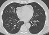
- CT chest showing widespread tram-track and signet ring signs
- diagnosis: bronchiectasis

Differential diagnosis for bronchiectasis

Symptoms of bronchiectasis

Characteristics of the cough in bronchiectasis
- productive
- worse in the morning
- large volume
- daily purulent sputum
Investigations in bronchiectasis (just in general)
- spirometry
- CXR
- CT
- sputum culture
What pattern of spirometry may be seen in bronchiectasis?
Obstructive or normal
Characteristic features of CXR in bronchiectasis
- May be normal
- Ring opacities, tram-tracks
- Fluid-filled cysts or bronchocoeles

Characteristic features of CT in bronchiectasis
- Signet ring sign and tram-tracks
- Lack of tapering of airways - thickness is NOT reduced towards the end
- Mucus impaction
- Mosaicism (vessels of different size in different regions of the lungs - smaller where less perfused)

Management of bronchiectasis
- physiotherapy (e.g. inspiratory muscle training) - has a good evidence base for patients with non-cystic fibrosis bronchiectasis
- postural drainage (airway clearance)
- antibiotics for exacerbations + long-term rotating antibiotics in severe cases
- bronchodilators in selected cases
- immunisations (influenza and bronchodilators)
- surgery in selected cases (e.g. Localised disease)
Most common organisms isolated from patients with bronchiectasis (4)
Most common organisms isolated from patients with bronchiectasis:
- Haemophilus influenzae (most common)
- Pseudomonas aeruginosa
- Klebsiella spp.
- Streptococcus pneumoniae
Management of infective exacerbations in bronchiectasis (what to do in general)
- Review previous sputum culture results & most recent course of antibiotics given
- Send more sputum for culture
- Choose antibiotic (in line with local guidance):
–amoxicillin/clarithromycin/doxycycline oral
–ciprofloxacin if Pseudomonas aeruginosa
–Tazocin/3rd generation cephalosporin IV
*Total course 10-14 days
*May need to consider outpatient IV antibiotics
•Chest physiotherapy
Antibiotics used in infective exacerbations of bronchiectasis
- what oral antibiotics
- what for Pseudomona aeurginosa
- what IV antibiotic
- how long for
* Choose antibiotic (in line with local guidance):
–amoxicillin/clarithromycin/doxycycline oral
–ciprofloxacin if Pseudomonas aeruginosa
–Tazocin/3rd generation cephalosporin IV
- Total course 10-14 days
- May need to consider outpatient IV antibiotics
Management of Pseudomonas Aeurginosa infection
- oral
- IV
- nebulised
•can colonise abnormal lungs
- also be associated with active infection
- Only orally active antimicrobial is Ciprofloxacin (fluoroquinolone antibiotic)
- IV: Tazocin (Piperacillin and Tazobactam), Ceftazidime (cephalosporin)
- Nebulised: colomycin (polymyxin antibiotic) can be used to suppress Pseudomonas in the case of colonisation with frequent exacerbations
Azithromycin
- class
- use
- usual dose
Azithromycin
Class: macrolide antibiotic
Antimicrobial and immuno-modulatory actions
Dose (usual):250mg three times a week
Use: aim of reducing exacerbation frequency
Side effects of Azithromycin
Key Side Effects:
–Prolonged QT and cardiac dysrhythmia
–Hearing loss (usually reversible)
–Hepatic dysfunction
Presenting symptoms of pneumonia
- cough
- sputum
- dyspnoea
- chest pain: may be pleuritic
- fever

What is pneumonia? (in general)
What is the most common cause?
Any inflammatory condition affecting the alveoli of the lungs, but in the vast majority of patients this is secondary to a bacterial infection
What is the likely organism causing pneumonia in the following presentation?
- Accounts for 80% of cases
Particularly associated with high fever, rapid onset
- A vaccine to pneumococcus is available
Streptococcus pneumoniae (pneumococcus)
What is the likely organism causing pneumonia in the following presentation?
Particularly common in patients with COPD
Haemophilus Influenzae
What is the likely organism causing pneumonia in the following presentation?
Often occurs in patient following influenza infection
Staphylococcus aureus
What is the likely organism causing pneumonia in the following presentation?
- One of the atypical pneumonias
- often present a dry cough
- atypical chest signs/x-ray findings
- Autoimmune haemolytic anaemia and erythema multiforme may be seen
Mycoplasma pneumoniae
What is the likely organism causing pneumonia in the following presentation?
- Another one of the atypical pneumonias
- Hyponatraemia and lymphopenia common
Legionella pneumophilia
What is the likely organism causing pneumonia in the following presentation?
Classically seen in alcoholics
Klebsiella pneumoniae
What is the likely organism causing pneumonia in the following presentation?
- Typically seen in patients with HIV
- Presents with a dry cough, exercise-induced desaturations and the absence of chest signs
Pneumocystis jiroveci
Signs of pneumonia
- signs of systemic inflammatory response: fever, tachycardia
- reduced oxygen saturations
- ausculatation: reduced breath sounds, bronchial breathing
What does it show?

- a classical signs of right upper lobe consolidation - abnormal opacity within the right upper lobe abutting the horizontal fissure.
- note how the ‘position’ of the consolidation on the film (i.e. in the ‘middle’ of the lung) doesn’t necessarily correlate with the lobe affected
What does it show?

- consolidation is harder to spot
- Look at the left heart border -> it is normally well dermaracted (borders are visible) with the lung
- Here -> it is fuzzy = this is a classic sign of left lingula consolidation
What types of investigations (in general) would you perform in pt presenting with Sx of pneumonia?
- CXR
- blood
- blood cultures and sputum samples
- ABG
Differential diagnosis of pneumonia

What bloods to perform in suspicion of pneumonia?
Bloods
- FBC -> high or low WBCs
- neutrophilia in bacterial infections
- leucopenia in viral infections
- U&Es -> check for dehydration (remember the ‘U’ for urea in CURB-65, see below) and also other changes seen with some atypical pneumonias, organ impairment in sepsis
- LFTs -> organ impairment in sepsis, hepatitis with atypical pneumonia
- CRP -> raised in response to infection
When to send sputum culture in a patient with pneumonia?
- CAP of moderate severity or high severity
- all patients who fail to improve with standard therapy
- for Legionella should always be attempted for patients who are legionella urine antigen positive
- for AAFB & mycobacterial culture for those who fail to improve or whose clinical features suggest possible TB or NTM (non-tuberculosis mycobacterium/mycobacterial disease)
What are possible (3) features of a chest Xray in pneumonia?
•Consolidation
–air bronchograms – pus in alveolar spaces around bronchi
–patchy
–may follow lobar contours
•Collapse
–tracheal deviation
–sail sign
•Pleural effusion

What’s consolidation? What’s effusion?
- Consolidation - fluid inside the lungs
- Effusion - fluid in pleural space (between chest wall and lungs)
What’s that?

Consolidation
What’s that?

Collapse
What’s that?

Pleural effusion
General Mx of pneumonia (2)
Patients with pneumonia require the following:
- antibiotics: to treat the underlying infection
- supportive care:
- oxygen therapy - if the patients is hypoxaemic
- IV fluids - if the patient is hypotensive or shows signs of dehydration
What determines the management of a patient with CAP?
CURB 65 score
What are the components of CURB-65?

Interpretation + management for the CURB-65 scores
(0-2)
- 0 - management in the community
- 1 - check sats (should be >92%) - to be safely managed in the community and a CXR performed. If the CXR shows bilateral/multilobar shadowing hospital admission is advised
- 2 - severe CAP, hospital management
The CURB-65 score also correlates with an increased risk of mortality at 30 days with patients with a CURB-65 score of 4 approaching a 30% mortality rate at 30 days
Range of pH on ABG
7.35-7.45
Range of pCO<strong>2</strong> on ABG
pCO2: 4.7-6
Range of PO2 on ABG
PO2: 11.3-14
Ranges of HCO3 on ABG
22-26
Range of BE on ABG
-2.3 - +2.3
What’s type 1 Respiratory Failure?
Low O2
(the rest of ABG picture is normal)
What are examples of causes of Type 1 respiratory failure?
pneumonia, PE, pulmonary oedema, pneumothorax
What’s type 2 respiratory failure?
high CO2 and low O2
Example of Type 2 resp failure causes (2)
COPD exacerbation, very severe pneumonia
Picture of respiratory acidosis on ABG

Possible causes of respiratory acidosis

Picture of respiratory alkalosis on ABG

Possible causes of respiratory alkalosis

Treatment for low severity CAP (detailed)
Low severity CAP
- Single antibiotic, 5 days
- Amoxicillin (macrolide or tetracycline if allergic)
Treatment for moderate severity CAP (detailed)
Moderate severity
–Dual antibiotics, 7-10 days
–Amoxicillin + macrolide (unless allergy)
Treatment for high severity CAP
High severity
–Dual antibiotics, 7-10 days
–beta‑lactamase stable beta‑lactam + macrolide
Empyema vs pleural effusion on CXR
Empyema - pus filled pockets on the pleura
Effusion - fluid in the pleura

How to (in general) differentiate pleural effusion from consolidation?
Pleural effusion -> fluid inside the pleura, can shift depending on pt’s position and gravity (as pleura is an open space)
Consolidation -> fluid inside the lung, cannot shift
