cardiovascular Flashcards
define arterial ulcer
- localised area of damage and breakdown of skin
- due to inadequate arterial blood supply
- typically feet of patients with sever atheromatous narrowing of arteries supplying leg
aetiology of arterial ulcers
- caused by lack of blood flow to capillary beds of lower extremities
- prevalence increases with age and obesity
risk factors:
- coronary heart disease
- Hx of stroke/TIA
- DM
- peripheral arterial disease
- immobility
symptoms + signs of of arterial ulcers
- punched out appearance with clearly defined edges
- eliptical shape
- mainly on foot dorsum/toes
- grey granulomatous tissue
- ischaemic signs: hairlessness, pale skin, absent pulses, nail dystrophy, calf muscle wasting
- night pain
- pain is worse in supine because arterial blood flow is further reduced

investigations for arterial ulcers
1. doppler US of lower limbs
- assess latency of arteries
- assess potential for revascularisation/bypass surgery
- ABPI - <0.9= PAD, <0.5- critical limb ischaemia
- percutaneous angiography
- ECG
- fasting serum lipids
- fasting blood glucose + HbA1c
- FBC (anaemia can worsen ischaemia)
management of arterial ulcer
Immediate:
- pain relief
surgery
- angioplasty (balloon => widen arteries in atherosclerosis)
- stenting
- bypass grafts
- amputate
define cardiac arrest
acute cessation of cardiac function
aetiology and risk factors of cardiac arrest
reversible:
- hypothermia
- hypoxia
- hypovolaemia
- hypo/hyperkalaemia
- toxins
- thromboembolic
- tamponade
- tension PTX
presenting symptoms of cardiac arrest
sudden; management precede/concurrent to Hx
preceding symptoms:
- fatigue
- fainting
- blackouts
- dizziness
physical examination findings of cardiac arrest
unconscious
absent breathing
absent carotid pulses
investigations for cardiac arrest case
cardiac monitor
- classification of rhythm
bloods:
- FBC
- ABG
- U&E
- cross match
- clotting screen
- toxicology screen
- blood glucose
management of cardiac arrest
approach arrest scene with caution
* cause of arrest may pose threat
BLS
- if arrest is witnessed, consider precordial thump
- clear and maintain airway
- assess breathing, if absent, 2 rescue breaths
- assess carotid pulse for 10 seconds, if absent, 30 chest compressions
advanced life support
advanced life support management of cardiac arrest with shockable rhythm
cardiac monitor + defibrillator
assess rhythm
shockable rhythms: pulseless VT/VF
- defibrillates once (150-360J biphasic, 360J monophasic)
- resume CPR for 2 mins
- reassess and shock again if no change
- 1mg IV adrenaline after 2nd defibrillation
- 1mg IV adrenaline every 3-5 mins
*persistant shockable rhythm after 3rd shock
- 300mg IV bolus amiodarone
advanced life support management of cardiac arrest with asystole/PEA
cardiac monitor + defibrillator
assess rhythm
pulseless electrical activity (PEA)/asystole:
- CPR for 2 mins
- reassess
- 1mg IV adrenaline every 3-5 mins
*asystole or PEA + <60bpm, 3mg IV atropine once only
during CPR for cardiac arrest
check electrodes, paddle positions, and contacts
secure airway
consider magnesium, bicarbonate, and external pacing
stop CPR and check pulse ONLY IF change in rhythm or signs of life
treatment of reversible causes of cardiac arrest
hypothermia
- warm slowly
hypovolaemia
- IV colloids
- IV crystalloids
- blood products
hypo/hyperkalaemia
- give insulin (+dextrose) increase K+uptake
toxins
- toxin antidote
thromboembolic
- treat as PE/MI
tamponade
- pericardiocentesis
tension PTX
- aspiration/chest drain
complications of cardiac arrest
irreversible hypoxic brain damage
death
prognosis of cardiac arrest
resus less successful if cardiac arrest occurs outside hospital
increased duration of inadequate effective CO = poor prognosis
define DVT
thrombus formation within deep veins of usually calf or thigh
deep veins in leg more prone due to blood stasis (Virchow’s triad)
DVT risk factors
- polycythaemia
- thrombophilia
- OCP
- post surgery
- prolonged immobility/ long flights
- obesity
- pregnancy
- dehydration
- smoking
- malignancy
presenting symptoms of DVT
- asymmetrical swollen leg
- may be painless
examination findings of DVT
- local erythema, warmth, and swelling
- varicosities (dilated superficial veins)
- skin colour changes
- +/- unilateral leg pain
- Homan’s sign
what is Homan’s sign
seen in patients with DVT
forced passive dorsiflexion of ankle causes deep calf pain
how to stratify risk of PE in case of DVT
stratified using Well’s PE criteria
2 or more = high risk
- history: breathlessness, cough, haemoptysis
- check RR, pulse oximetry, and pulse rate
investigations for DVT
Use Wells score for DVT (<2 = low risk)
doppler US - gold standard
bloods:
- *- D dimer** (if low = unlikely to be DVT)
- thrombophilia screen if indicated
impedance plethysmography
- changes in blood volume causes changes in electrical resistance
if suspected PE:
- ECG
- CXR
- ABG
management plan for DVT
confirmed DVT + not pregnant
- 3 months anticoagulation (DOACs- apixaban/ rivaroxiban) -LMWH as alternative
- physical activity
- compression stockings
Confirmed DVT + preganant
- LMWH- anticoagulation
- physical activity
- compression stockings
*recurrent DVT = long term warfarin
if anticoagulation contraindicated (bleeding, haemorrhage, major trauma, aortic dissection) => IVC filter
* DVTs that DO extend above knee => anticoagulation for 6 months
prevention of DVT
graduated compression stockings
mobilisation
prophylactic heparin if high risk
possible complications of DVT
- PE
- venous infarction (death of tissue due to poor perfusion)
- thrombophlebitis- inflammed vein due to clot (from recurrent DVTs)
- chronic venous insufficiency
prognosis of DVT
depends on extent of DVT
below knee = good
proximal DVT = greater risk of embolisation
define vasovagal syncope
loss of consciousness from transient drop in blood flow to brain caused by excessive vagal discharge
aetiology/risk factors of vasovagal syncope
common cause of fainting
precipitated by
- emotions
- orthostatic stress
presenting symptoms of vasovagal syncope
short loss of consciousness
vagal symptoms
- sweating
- dizziness
- light headedness
before episode
twitching during episode
quick recovery
signs on physical examination of vasovagal syncope
usually no signs
investigations for vasovagal syncope
checking for causes
ECG - arrhythmias
echocardiogram - outflow obstruction
lying/standing BP - orthostatic hypotension
fasting blood glucose - DM/hypoglycaemia
define venous ulcers
- large, shallow, sometimes painful
- usually superior to medial malleoli
- caused by incompetent valves in lower limbs - leads to venous stasis and ulceration
aetiology and risk factors of venous ulcers
caused by incompetent valves in lower limbs
- leads to venous stasis and ulceration
risk factors:
- obesity
- immobility
- recurrent DVT
- varicose veins
- previous injury/surgery to leg
- age
presenting symptoms + signs of venous ulcers
- large, shallow, sometimes painful ulcers
- irregular margin
- swelling, itching, aching
- in gaiter region (superior to medial malleoli => mid calf)
associated signs of venous insuffiency with venous ulcers
- stasis eczema
- lipodermatosclerosis (champagne bottle)
- haemosiderin deposition (dark colour)
- atrophie blanche (blood deposits)
investigations for venous ulcers
- ABPI (excuse arterial ulcer) - if ABPI <0.8, DO NOT apply pressure bandage
- Duplex USS of lower legs (GOLD-STANDARD)
- measure surface area of ulcer for monitoring of progression
- microbiology swab samples
- biopsy if suspected Marjolin’s ulcer
management of venous ulcers
- graduated compression to reduce venous stasis- must exclude DM, neuropathy, and PVD before attempt
- debridement and cleaning
- Abx if infected
- topical steroids to treat surrounding dermatitis
complications and prognosis of venous ulcers
- recurrence
- infection
good prognosis
- better results if mobile with few comorbidities
Define Ischaemic heart disease
Ischaemic heart disease is when myocardial demand exceeds oxygen supply resulting in chest pain (angina pectoris)
=> present as stable angina or ACS
Causes of IHD ( A VASE)
when myocardial demand exceeds oxygen supply
usually due to:
- Atherosclerosis
- Vasculitis
- Arteritis
- coronary artery Spasm (cocaine)
- Embolism
Risk factors of IHD
- male
- Diabetes mellitus
- FHx
- Hypertension
- Hyperlipidaemia
- Smoking
- older age
- Obesity
- sedentary lifestyle
- cocaine
Pathophysiology of atherosclerosis
- endothelial damage causes monocytes/ LDLs to migrate into subendothelial space
- free LDLs bind to matrix proteoglycans
- monocytes differentiate => macrophages release free radicals that oxidise LDLs.
- macrophages engulf oxidised LDLs => foam cells
- foam cells release growth factors + cytokines => smooth muscle proliferation, collagen production, proteoglycan production
- forms atherosclerotic plaque
presenting symptoms of Stable angina
- chest pain on exertion and relieved by rest
Examination findings of stable angina
- mainly check for risk factors
Investigations for IHD (ACS)
Bloods:
- FBC
- U&Es, TFTs
- CRP
- glucose
- lipid profile
- amylase (exclude pancreatitis)
- AST/ LDH (1-2 days post)
- cardiac enzymes (troponins + Creatine Kinase-MB)
- ECG (+ exercise ECG)
- radionuclide scan
- CT
- MRI
- Coronary angiography
- CXR - check for HF signs (cardiomegaly, pulmonary oedema, widened mediastinum)
Prognosis of IHD
- TIMI score (0-7 thrombolysis in MI)- high scores = high risk of cardiac events within 30 days of MI
define Acute coronary syndromes
type of IHD (atherosclerosis causing partial/total occlusion of coronary arteries)
- unstable angina (RARE)
- NSTEMI
- STEMI
Pathophysiology of ACS
- unstable angina- unstable coronary plaque, disrupts fibrous cap => forms incomplete thrombus
- NSTEMI- incomplete thrombus forms => partial artery occlusion
- STEMI- coronary plaque ruptures => complete thrombus formation => complete artery occlusion, transmural infarction
presenting symptoms of ACS
- acute-onset chest pain lasting >20mins
- central, heavy, tight crushing chest pain
- pain radiates to arm, neck, jaw, epigastrium
Associated symptoms:
- breathlessness
- sweating
- palpitations
- nausea
- vomiting
Symptoms of silent infarcts in elderly/ diabetics
- no chest pain
- syncope
- pulmonary oedema
- epigastric pain
- vomiting
Examination findings of ACS
- may not be signs
- pale, sweaty, restless, fever
- high/low bp
- arrhythmias
- new heart murmurs
- signs of heart failure (raised JVP, S3, basal crepitations)
*rule out aortic dissection (check radial pulses)
Investigations for ACS
- bloods (FBC, U&Es, AST/LDH, lipid profile, glucose, amylase (exclude pancreatitis), CARDIAC ENZYMES (raised troponin/ myoglobin/ CK-MB)
*troponin not raised in unstable angina
- ECG
- radionuclide scans
- CT coronary angiography
- MRI
Investigations for stable angina
- bloods (FBC, lipid profile, AST/LDH, cardiac enzymes: troponin, myoglobin, CK-MB, BNP)
- ECG (usually normal)
- CT coronary angiography (check for arterial stenosis)
- exercise tolerance test
ECG findings for
- Unstable angina/ NSTEMI
- STEMI
- ST depression/ T wave inversion
- ST elevation, hyperacute T waves -tall (later T wave inversion), new-onset LBBB
Management for stable angina
- reduce cardiac risk factors (lower bp/cholesterol, diabetes, stop smoking, exercise)
Immediate
- 75mg/day aspirin
- symptom relief: GTN spray
Long term
- beta blockers (not for acute heart failure, cardiogenic shock, bradycardia, heart block, asthma)
- CCBs
- nitrates
Surgical:
- PCI (if medication doesn’t work)
- CABG
Mangement for unstable angina/ NSTEMI (MONABASH)
- Morphine (give metoclopramide for nausea)
- Oxygen (only if sats >95%)
- Nitrates (GTN)
- Antiplatelets (Aspirin, Clopidogrel)
- Beta-blockers
- ACE-inhibitors
- Statins
- Heparin
Surgery (no improvement) => angioplasty +/- revascularisation (PCI/ CABG)
Management for STEMI
- antiplatelet (ASPIRIN 300mg) + anticoagulants (heparin)
- primary PCI or thrombolysis
- Long term (beta-blockers, ACE-inhibitors, statins, antiplatelets (aspirin + clopidogrel), lifestyle changes, cardiac rehabilitation)
Complications of MI (DARTH VADER)
- death
- arrhythmias
- rupture (septum/outer walls)
- tamponade
- heart failure
- valve disease
- aneurysm
- Dressler’s syndrome (autoimmune pericarditis)
- Embolism
- Reinfarction
Pathophysiology of peripheral vascular disease
caused by atherosclerosis => stenosis of arteries => poor perfusion to limbs => ischaemia => pain
- chronic limb ischaemia (intermittent claudication / critical limb ischaemia)
- acute limb ischaemia
Risk factors for peripheral vascular disease
- hypertension
- smoker
- FHx of CVD
- diabetics
- elderly
- hyperlipidemia
- sedentary lifestyle
Presenting symptoms of intermittent claudication
- intermittent claudication (leg pain after exercise + relived on rest immediately
- Calf claudication = femoral disease
- Buttock claudication = iliac disease
- erectile dysfunction
- poor/ slow wound healing
- gangrene
- presecence of risk factors
- reduced/ absent pulses
Presenting symptoms of critical limb ischaemia
- gangrene
- ulcers
- pain at rest
- night pain (relieved by dangling legs off the bed)
Investigations for peripheral vascular disease
Bedside:
- ECG
- ABPI (ankle brachial pressure index SBP of ankle/brachial)- <0.9 = abnormal, <0.5 = critical limb ischaemia
Bloods
- FBC
- U&E (check for renal disease)
- fasting glucose (check in diabetics)
- lipid levels
- D-dimer (fibrin degradation product)
- thrombophillia screen
- troponins
- CRP/ESR (raised indicates thrombophlebitis)
Imaging
- Colour duplex ultrasound of pulses (check stenosis)
- CT angiography/CTA(detetct location + stenosis)- need contrast
- Magnetic resonance angiogaphy/MRA (detect stenosis)
- Trancutaneous pressure of oxygen (perfusion to foot)
Why do diabetics/renal failure have a raised ABPI normally?
calcification of arteries result in high ankle pressures due to incompressible arteries
Management of peripheral arterial disease (intermittent claudication)
non-lifestyle limiting claudication
- anti-platelets (aspirin, clopidogrel)
- exercise
- reduce risk factors (low fat/cholesterol diet, exercise, stop smoking/alcohol, optimise diabetes)
lifestyle limiting claudication
- anti-platelets
- exercise (supervised exercise therapy)
- symptom relief VASOACTIVE drugs (naftidrofuril)- reduces pain on walking
- adjunct revascularisation (PTA - percutaneous transluminal balloon angioplasty or BYPASS)
- amputation
Define ischaemic limb
- Can be acute or acute and chronic
- thrombotic (absent/diminished pulses Hx of intermittent claudication)
- embolic (suddent/more severe- no established collaterals)
Presenting symptoms of acute limb ischaemia (6Ps)
- pale
- pulselessness
- perishingly cold
- pain
- paralysis
- parasthesia
Other
- hair loss
- skin atrophy
- punched out ulcers
- leg colour change when raised to buerge’s angle

Management of acute limb ischaemia
Immediate
- analgesia
- anticoagulants (heparin)
Surgery
- Revascularisation (within 4hrs)- endovascular revascularisation/ fasciotomy
- Amputation
Define abdominal aortic aneurysm (AAA)
abnormal dilation of aorta (>50% normal size) across all layers of aorta wall
Screening progroamme for <65 males
- >3mm => surveillance
- >5.5mm => repair
risk factors for AAA
- modifiable (smoking, hypertension, dyslipidemia)
- non-modifiable (MALE, older, Family Hx)
- connective tissue disorders (Marfan’s, Ehlers Danos)
- Inflammatory disorders (Behcet’s disease-vessel inflammation)
Different types of aneurysms
- true aneurysm= dilation across all layers of aorta wall
- false/pseudoaneurysm = dilation only across part of aorta wall (adventitia)
- location = aortic, iliac, popliteal
- morphology = saccular / fusiform
- aetiology = athelosclerosis, inflammatory, inherited, mycotic (infection by fungi/bacteria)
- Presenting symptoms of AAA
- Presenting symptoms of ruptured AAA
unruptured
- usually asymptomatic
- abdominal/groin/back pain (pressure related)
ruptured:
- abdominal pain => radiates to back
- syncope/ light headed (reduced cerebral perfusion)
- cold, sweaty, nausea - activation of sympathetic response
- pallor
- Pressure (back pain)
- Rupture (high mortality rate)
- Thrombosis (acute limb ischaemia)
- Embolisation (ischaemic symptoms)
Examination signs of AAA
- pulsatile, expansile pulse in abdominal aorta palpation
- shock signs (tachycardia, low bp) in RUPTURED AAA
- +/- aortic bruits
- pallor
- abdominal distension
Investigations for AAA
Bedside
- ECG (check for MI)
Bloods:
- FBC (inflammatory AAA => anaemia)
- CRP/ESR (raised)
- clotting screen
- U&Es
- LFTs
- Cardiac enzymes
- blood cultures (+ve - inflammatory AAA)
- Group and save- if surgery planned
- amylase (exclude pancreatitis)
Imaging
- Hameodynamically unstable => Aortic USS
- Hameodynamically stable => CT with contrast Angiography, MRI angiography

Management for AAA
Hameodynamically unstable + ruptured/symptomatic AAA
- RESUS (B- oxygen, C- 2 large bore IV cannulae, take bloods, measure bp. IV fluids -for shock)
- analgesia
- Prophylaxis antibiotics
- Surgery (endovascular aneurysm repair, open repair)
- VTE prophylaxis
Asymptomatic AAA
- Surveillance
- modify risk factors (stop smoking, exercise, low fat diet)
Indications for surgery for AAA
- women >5cm, men >5.5 cm size
- growing >1cm/yr
- symptomatic
- repair aorta-iliac disease
define aortic dissection
tear in the tunica intima=> between the inner (interna) /outer wall (media) creating a false lumen in the aorta
classification of aortic dissection
Stanford
- Type A- affects ascending aorta (MORE SEVERE)
- Type B- affects descending aorta
Debakey
- Type I- ascending aorta + aortic arch affected
- Type II- ascending aorta
- Type III- descending aorta
Causes of aortic dissection
- uncontrolled Hypertension
- connective tissue disorders (Marfan’s, Erhlers Danos, Loeys-dietz)
- Aortic atherosclerosis
- vasculitis
- iatrogenic (angioplasty)
- pregnancy
- Congenital => Coarctation (? x2 arches)
- Aortitis (inflammation of aorta)
- Trauma
- Cocaine
Presenting symptoms of aortic dissection
- sharp, tearing pain radiating to back
- +/- loss of consciousness
- poor perfusion of end organs
- carotid artery => blackout, dysphagia, hemiparesis
- coronary artery => chest pain
- subclavian artery => ataxia, loss of consciousness
- anteria spinal artery=> paraplegia
- coeliac axis artery => abdominal pain (ischaemia)
- renal artery => renal failure, anuria
Examination findings of aortic dissection
- hypertension, bp difference >20mmHg between arms
aortic insufficiency
- murmur on back (below scapula) => abdomen
- unequal arm pulses
- signs of aortic regurg (collapsing pulse, diastolic murmur)
- +/- abdominal mass
Investigations for aortic dissection
Bedside
- ECG (usually normal but could see inferior ST elevation/ LVH if coronary arteries affected)
Bloods
- FBC
- U&E- check renal function
- Cardiac enzymes (exclude MI)
- clotting screen
- group and save
Imaging
- CXR - widened mediastinum
- CT angiogram aorta - can see false lumen (DIAGNOSTIC)
- Transoesophageal Echo- look at valves (for patients unsuitable for CT)

Management for aortic dissection
Haemodynamically unstable
- RESUS => oxygen, IV fluids, cannula, take bloods
- Analgesia (opioids-morphine)
Type A
- SURGERY
Type B
- UNCOMPLICATED: control bp- (IV labetolol- beat-blocker), HR, pain
- COMPLICATED: surgery (Thoracic endovascular aortic repair/ open surgery)
Differentials for aortic dissection
- chest pain radiating to back => MI
- hypotension => tamponade
- pulsus paradoxus (bp change on inspiration) => pericarditis, tamponade, COPD, Obstructive sleep apnoea
define arrythmias
due to conduction abdnormality at SAN/ AVN => abnormal heart rhythm
Conduction pathway of heart
- SAN generates impulses across atria walls
- Conducted by AVN
- Signalds travel down bundle of His
- Electrical impulse goes down left / right bundle branches
- Down into purkynje fibres => ventricles
Types of arrythmias
Bradyarrythmia (<60bpm)
- heart block (1st, Mobitz I, Mobitz II, 2nd degree 2:1, 3rd degree )
- sinus bradycardia - treat with atropine
Tachyarrythmia (>100bpm)
- supraventricular tachycardia (narrow QRS)- Atrial flutter, AF, Wolff-parkinson
- ventricular tachycardia (prolonged QRS)
- ventricular fibrillation (no pulse, irregular rhythm)
- RBBB/LBBB block => prolongs QRS complex
Causes of bradyarrythmias
- age-related conductive tissue fibrosis
- drugs (beta-blockers + CCBs)
- previous MI
- hypothermia
- electrolyte imbalance
- increased vasovagal tone (head injury, pain)
presenting symptoms of heart block
- usually asymptomatic
- dizziness
- palpitations
- chest pain
Mobitz II/ Type 3:
- syncope
examination findings of heart block
- usually normal
- large volume pulse
- cannon waves in JVP
Mobitz II/ Type 3 heart block:
- hypotension
- heart failure (raised JVP, peripheral oedema)
investigations for heart block
- ECG (GOLD-STANDARD)
- Bloods: FBC, U&Es, TFTs, digoxin, troponin
- CXR: cardiomegaly, pulmonary oedema
- ECHO - exclude valve disease
ECG features for heart block
- 1st degree - long PR interval (>3-5 small squares)
- Mobitz I/ Wenckebach- lengthening PR interval, drop QRS
- Mobitz II - missing QRS complexes (normal PR interval)
- 2nd degree AV block (2:1/3:1)
- 3rd degree - atria/ventricles out of sync

Management of heart block
- cardiac monitoring
- Treat cause (hypothermia, STOP drugs, correct electrolytes)
- => PERMENANT PACEMAKER
Haemodynamically unstable:
- CPR
- external pacemaker (de-fibrillator)
- Temporary pacing wire inserted via femoral vein
Causes of tachyarrythmias
- MI
presenting features of tachyarrythmias
- chest pain
- palpitations
- dyspnoea (SOB)
- syncope
investigations for tachyarrythmias
- ECG
Bloods:
- U&Es- electrolyte imbalance => arrythmias
- drug toxicology screen
- Cardiac enzymes - troponins (check for recent MI/ischaemia)
ECGs of tachyarrhythmias
- Atrial flutter (SVT)
- Atrial fibrillation (SVT)
- Ventricular tachycardia
- Ventricular fibrillation
- Atrial flutter (saw tooth)
- Atrial fibrillation (no P waves)
- VF- no identifiable P waves/QRS complexes
- VT- wide QRS (>3 boxes), HR >100bpm

management of tachyarrythmias
adverse signs (SHOCK, syncope, MI, HF)
- synchronised DC shock (for shockable rhythm-VF/VT) X3
- IV amiodarone (anti-arrhythmic)
- long term - implantable cardio defibrillator (ICD)
no adverse signs + broad QRS (ventricular)
- regular = VT (amiodarone)
- irregular = VF (seek HELP)
no adverse signs + narrow QRS (SVT)
- regular= 1. adenosine = Atrial flutter (b-blocker)
- irregular = AF (B-blockers, amiodarone/digoxin if HF signs)
define infective endocarditis
infection of endocardium (lining of heart chambers) due to vegetation (platelets, fibrin, bacteria deposits) destroy valve leaflets and invade myocardium
common microorganisms that cause infective endocarditis
- staph aureus (IV drug users) MOST COMMON
- streptococcus viridans (dental hygiene related)
- streptoccous bovis (colorectal cancer related)
- Staphylococcus epidermidis (prosthetic valves)
risk factors for infective endocarditis
- recent dental work/ poor dental hygiene (S. viridans)
- IV Drug users
- valve defects (congenital, rheumatic fever)
- prosthetic heart valves
- post-op wounds
- DM
- organ transplants
presenting symptoms of infective endocarditis
- FEVER
- chills
- malaise
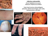
examination findings of infective endocarditis (FROM JANE C)
- Fever
- Roth spots on retina
- Osler’s nodes (painful)
- new heart Murmur
- Janeway lesions (painless)
- Anaemia (pallor)
- Nail-splinter haemorrages
- Emboli
- Clubbing
investigations for infective endocarditis
- Urinalysis
- Bloods: FBC-low Hb, high WCC, high CRP, U&Es, LFTs, +ve rheumatoid factor
- Blood cultures (x3)
- CXR- cardiomegaly
- ECHO - check for vegetation (>2mm)
Management of Infective endocarditis
What’s the diagnostic tool for infective endocarditis
Duke’s criteria
- 2 major (+ve blood cultures, new murmur)
- 1 major + 2 minor
- 5 minor (temp, IV drug user, pre-existing heart disease, R/O/J/S)
Management of infective endocarditis
- 4-6 week ANTIBIOTICs (empirical =benzylpenicillin/amoxicillin)
- if not surgery
Complications of infective endocarditis
- aneurysm
- heart failure
- valve incompetence
define hypertension
peristently raised high blood pressure >140/90
- essential (idiopathic/unknown)- 90%
- secondary (medical cause)
Risk factors for primary (essential) hypertension
- older age
- males (<65), females (>65)
- high salt diet
- poor exercise
- obesity
- high alcohol intake
- black afro-carribean ethnicity
- stress/ anxiety
Causes of secondary hypertension (medical)
renal
- diabetic nephropathy (proteinurea, microalbuminuria)
- polycystic kidney disease
- glomerulonephritis (microscopic haematuria)
- pyelonephritits
- RCC (haematuria, loin mass/pain)
endocrine
- primary hyperaldosteronism (hypokalemia, alkalosis/XS bicarbonate)
- phaechromocytoma (XS adrenaline- intermittent high bp/sweating/headaches/postural hypotension)
- Cushing’s (XS cortisol=> moon face, striae, central obesity)
- acromegaly (XS GH => enlarged hands/feet, macroglossia, sweating)
- hypothyroidism
- hyperthyroidism
vascular
- coarcation of aorta (radio-femoral delay)
- renal artery stenosis (abdominal bruit + peripheral vascular disease)
drugs
- ALCOHOL
- OCP
- corticosteroids
- cocaine
How to classify hypertension?
- stage 1 - 140/90 in clinic or 135/85 ABPM/home
- stage 2- 160/100 in clinic or 150/90 ABPM/home
- stage 3/ severe- 180/120
Why does Ambulatory blood pressure monitoring (ABPM) have a lower threshold for hypertension diagnosis?
- avoids blood pressure rise due to white coat syndrome (anxious in clinic can increase bp)
Presenting symptoms of hypertension
- severe:
- headaches
- bleeding nose
examination signs for hypertension
- signs of heart failure (raised JVP, pitting oedema)
end organ damage (fundoscopy)
- retinal haemorrhages
- papilloedema
- proteinuria (AKI)
Investigations for hypertension
- ABPM/HBPM
Check for secondary cause
- ECG (check for left ventricular hypertrophy)
- Urine dipstick- haematuria, proteinurea (AKI), microalbuminuria (nephropathy)
Bloods:
- FBC
- U&Es
- urine albumin:creatine ratio
- eGFR - CKD
- TFTs (hypo/perthyroidism)
- lipid profile (check CV risk)
- HbA1c- diabetes (check CV risk)
Calculate Q-risk
Imaging
- renal USS + CT
Management of hypertension
- lifestyle (low salt diet, exercise, stop smoking, reduce alcohol)
- medical (A=ACE-inhibitor rampiril, C= CCB amlodipine, D= thiazide-diuretics indapamide)
<55 / T2DM
- A
- A+C/ A+D
- A+C+D
>55/ afro-carribean descent
- C
- C+A/ C+D
- A+C+D
- Still uncontrolled bp
- repeat bp=> ABPM, check for postural hypertension
- add low dose loop diuretic (spiranolactone)/ alpha or beta-blocker if they have hyperkalemia (>4.5mmol/L)
- if bp still not controlled refer to specialist
complications of hypertension
- heart (coronary artery disease, heart failure)
- vascular (vascula dementia, peripheral artery disease, stroke)
- renal (CKD)
What score should be calculated for hypertensive patients?
QRISK - risk of developing CVD in 10yrs
define pericardial disease
pericarditis = inflammation of pericardium
constrictive pericarditis = chronic inflammation of pericardium (rigid/thickened so heart is restricted)
causes of pericarditis
- mainly viral (coxsacchie virus, echovirus,mumps)
- bacterial (streptococci, staph.)
- POST-MI
- Dressler’s syndrome
presenting symptoms of pericarditis
- central chest pain
- may radiate to shoulder/arm
- pleuitic chest pain relieved by leaning forward
- nausea
examination findings of pericarditis
- fever
- friction rub (like leather rubbing together)
- faint heart sounds
Signs of constrictive pericarditis
Right heart failure signs:
- pulsus paradoxus (>10mmHg drop in SBP on inspiration)
- Kussmaul’s sign (paradoxical raise in JVP on inspiration)
- hepatomegaly
- ascities
- pericardial knock (early diastolic knock)
- peripheral oedema
- AF
investigations for pericarditis
- ECG - widespread saddle-shaped ST elevation
- Echo - pericardial effusion
- Bloods: FBC, CRP, troponins, U&Es,
- CXR - cardiomegaly

Management for pericarditis
acute:
- treat underlying cause
- NSAIDs for pain/fever
- aspirin
recurrent
- low-dose steroids
- immunosuppressants
surgical (if constricitve pericarditis) => pericardiectomy (cut part of pericardium)
complications of pericarditis
- pericardial effusion (fluid in pericardium)
- cardiac tamponade (fluid build up compresses heart)
- cardiac arrythmias
symptoms + signs of cardiac tamponade
symptoms:
signs: (BECK’S TRIAD)
- raised JVP
- hypotension
- muffled heart sounds
define cardiac failure
inability of cardiac output to meet body’s demands despite normal venous pressures
how to classify heart failure
- acute/chronic
- LHF/ RHF or congestive heart failure (LHF +RHF)
- low cardiac output or high cardiac output
Causes of LHF (low cardiac output)
Valvular
- aortic stenosis
- aortic regurgitaiton
- mitral regurgitation
Muscular
- ischaemic heart disease (IHD)- most common
- arrythmias
- myocarditis
Systemic:
- sarcoidosis
- amyloidosis
Causes of RHF
Secondary to LH failure (congestive cardiac failure)
Lungs:
- Pulmonary hypertension (cor pulmonale)
- Pulmonary embolism
- Chronic lung disease (Cystic fibrosis, ILD)
Valvular disease:
- tricuspid regurgitation
- pulmonar valve disease
Causes of high output failure (NAPMEALS)
*higher CO demand than normal
- Nutritional (thiamine/ B1 deficiency)
- Anaemia
- Pregnancy
- Malignancy
- Endocrine (hyperthyroidism)
- AV malformation
- Liver cirrhosis
- Sepsis
symptoms + signs of left heart failure
Symptoms:
- dyspnoea/ SOB- paroxysmal nocturnal dyspnoea, exertional dyspnoea, orthopnoea
1 - none
2 - on ordinary activities
3 - less than ordinary activities
4 - at rest
- nocturnal cough (+ pink frothy sputum)
- fatigue
Signs:
- raised RR/HR
- S3 gallop- rapid ventricular filling, S4
- displaced apex beat
- pluses alternans- arterial pulse waveforms with alternating strong and weak beats
- bilateral basal crackles (pulmonary oedema)
- wheeze
symptoms + signs of right heart failure
symptoms:
- swollen ankles
- fatigue
- reduced exercise tolerance
- anorexia
- nausea
signs:
- face swelling
- raised JVP
- pan-systolic murmur (tricuspid regurgitation)
- ascites, hepatomegaly
- pitting oedema
explain why pulsus alternans occurs
LV dysfunction
= significant decrease in EF
= reduced SV
= increased EDV
= LV stretched more for next contraction
starling’s law: increased stretch = increased strength of myocardial contraction
= stronger systolic pulse
Investigations for cardiac failure
Bedside: ECG
Bloods:
- BNP (specific)
- FBC (exclude anaemia)
- U&E
- LFT
- TFT (exclude hypothyroidism)
Imaging:
- CXR (ABCDE)
- Alveolar shadowing
- kerley B lines
- Cardiomegaly
- upper lobe Diversion
- Effusion
- Transthoracic echocardiogram (DIAGNOSTIC)
- assess ventricular contraction
- systolic dysfunction (LV EF < 40%)
- diastolic dysfunction (due to decreased compliance which causes restrictive filling defect)
Management of acute heart failure
- upright position
- 60-100% oxygen (consider CPAP)
- IV diamorphine (venodilator + anxiolytic)
- GTN infusion (venodilator- reduces preload)
- IV furosemide (venodilator- reduces preload)
Management of chronic heart failure
- treat cause and exacerbating factors
- Lifestyle- low salt diet, exercise
- Drugs (ABD)
- ACE inhibitors or angiotensin receptor blockers (if intolerant/ cough)
- Beta blockers
- loop Diuretics -furosemide
- aldosterone antagonists (monitor K+)
- hydralazine and nitrate- for afro-carribeans
- digoxin
- cardiac resynchronisation therapy
complications of cardiac failure
- respiratory failure
- renal failure
- cardiogenic shock
- death
prognosis= 50% die within 2 yrs
define myocarditis
inflammation of the myocardium (heart muscle)
causes of myocarditis
- Coxsaccie virus
- infection
- drugs- cocaine
- radiation
- metal
presenting symptoms of myocarditis
- flu-like
- SOB
- chest pain (worse on lying down)
- palpitations
investigations for myocarditis
- ECG - ST/T wave changes
- cardiac biomarkers: CK, troponin
- endomyocardial biopsy (diagnostic but very invasive so rarely done)
management of myocarditis
- supportive care
- conventional heart failure therapy
define pulmonary hypertension
increase in pulmonary arterial pressure (PAP)
Causes of pulmonary hypertension
- Pulmonary arterial hypertension - changes to pulmonary arteries (thick/stiff)
- connective tissue disorders (scleroderma)
- liver disease
- congenital heart defects
- HIV
- Idiopathic
- PH associated with Left ventricular failure
- PH associated with Lung disease (less oxygen)
- COPD
- interstitial lung disease- pulmonary fibrosis
- Obstructive sleep apnoea
- obesity hyperventillation syndrome
- PH associated with blood clots Thromboses/emboli in lungs
- HIV
- Liver disease/ portal hypertension
- congenital heart defects
- PH associated with other rare causes
- sarcoidosis
- tumours (compression of pulmonary vessels)
Presenting symptoms of pulmonary hypertension
- progressive breathlessness
- weakness/tiredness
- exertional dizziness and syncope
Late stage:
- angina
- oedema
- ascites
- tachyarrhythmias
Examination findings of pulmonary hypertension
Inspection:
- raised JVP
- peripheral oedema
- ascites
Palpation:
- right ventricular heave
Auscultation:
- loud pulmonary second heart sound
- pulmonary regurgitation murmur (early diastolic)
- tricuspid regurg
Investigations for pulmonary hypertension
Bedside:
- ECG- right ventricular hypertophy + strain
Bloods:
- LFTs - liver disease/portal hypertension
Imaging:
- CXR- exclude other lung problems
- ECHO- check right ventricular function
Other:
- Right heart catheteristion (directly measures pulmonary pressures) - DIAGNOSTIC
- Pulmonary function tests
- Lung biopsy - interstitial lung disease
Management of pulmonary hypertension
Supportive treatment
- diuretics- removes excess fluid (treat ankle swelling)
- oxygen (reduce breathlessness)
- pulmonary rehabilitiation (exercise to help with breathlessness)
Specialist treatment varies on type of PH
- Pulmonary arterial hypertension (group 1) =>pulmonary vasodilators
- caused by left heart disease/lung conditions(group 2/3) => as PH is secondary treat the primary condition
- caused by blood clots (group 4) => anticoagulants(warfarin, DOACS)
Complications of pulmonary hypertension
- Cor pulmonale - right ventricle enlargement => failure
- Blood clots
- Arrythmias (palpitations, dizziness, fainting)
- Bleeding in lungs (haemoptysis)
- Pregnancy complications
types of aortic valve disease
- aortic stenosis- aortic valve becomes thick and fuses together, narrowing the opening causing reduced blood flow to body
- aortic regurgitation- dilation of aortic valve causing backflow of blood back into left ventricle

causes/risk factors for aortic valve disease
aortic stenosis
- calcified aortic valve (elderly/CKD) most common
- congenital bicuspid aortic valve
- inflammation after rheumatic fever (Strep. A causes inflammation + calcification to valve endothelium)
- Others: SLE/paget’s
aortic regurgitation
- rheumatic heart disease (valves damaged after rheumatic fever)
- congenital bicupsid aortic valve
- endocarditis
- connective tissue disorder -MARFAN’S, aortitis
symptoms + signs of aortic stenosis
Symptoms:
- exertional SOB
- exertional syncope
- angina (chest pain)
- RISK FACTORS
Signs:
- A: ejection-systolic murmur (loudest over aortic valve and radiates to carotid)
- narrow pulse pressure
symptoms + signs of aortic regurgitation
symptoms:
- usually asymptomatic until they develop heart failure
- palpitations
- fatigue/ dyspnoea/ chest pain
signs:
- diastolic murmur
investigations for aortic valve disease
- Transthoracic Echocardiogram (DIAGNOSTIC)
- ECG- aortic stenosis =>LVH (high S voltage V1-V3, high R voltage V4-V6)
- CXR
Management of aortic valve disease
- unstable - balloon valvuloplasty (valve repair)- aortic stenosis
- stable
- surgical aortic valve replacement (TAVI)
- warfarin + antibiotics (prevent infective endocarditis)
*transcatheter aortic valve replacement (for high risk patients)
complications of aortic valve disease
- heart failure
- infective endocarditis
- sudden death
- arrythmias
Types of mitral valve disease
- mitral stenosis - thick mitral valve fuses together causing narrowed opening => reduced blood flow from left atrium to left ventricle
- mitral regurgitation- dilated mitral valve so blood leaks back into left atrium
causes of mitral valve disease
mitral stenosis:
- rheumatic heart disease (most common)
- congenital
- endocarditis
mitral regurgitation: (anything damages valves)
- rheumatic heart disease (most common)
- infective endocarditis
- mitral valve prolapse
- Hx of MI, heart trauma, IHD, Hypertrophic cardiomyopathy
symptoms + signs of mitral stenosis
symptoms:
- dyspnoea
- fatigue
- orthopnoea
- risk factors (FEMALE, Rheumatic fever)
signs:
- diastolic murmur (loudest over mitral valve)
- Loud S1
- opening snap on auscultation
- signs of pulmonary hypertension/ right heart failure (raised JVP, oedema, ascites)
symptoms + signs of mitral regurgitation
symptoms:
- dyspnoea on exertion
- reduced exercise tolerance
- fatigue
- risk factors
signs:
- pansystolic murmur loudest over mitral valve that radiates to axilla
investigations for mitral valve disease
- Transthoracic echocardiogram (diagnostic)
- ECG- could have AF/ LVH
- CXR- kerly B lines (mitral stenosis -pulmonary hypertension)

management for mitral valve disease
mitral stenosis
- usually do nothing
severe disease:
- diuretic (reduce atrial pressure)
- balloon valvotomy (open valve)
- OR surgical valve replacement
- beta blockers (reduce symptoms)
mitral regurg
- mitral valve repair (balloon vavluloplasty/ annuloplasty)
- mitral valve replacement
- ACE-inhibitor + beta-blockers
Complications of mitral valve disease
- AF
- stroke
- infective endocarditis
types of right sided valve disease
- tricuspid regurgitation- backflow of blood from RV => RA during systole
- tricuspid stenosis- reduced blood flow from RA => RV during diastole
- pulmonary stenosis- narrowing of valve between RV and pulmonary atery => blocked blood flow
- pulomonary regurgitation- valve flaps don’t close properly so blood leaks back into right ventricle
causes of right valve diseases
tricuspid regurg:
- Infective endocarditis-IV drug user (most common)
- congenital (Ebstein’s anomaly - malpositioned TV)
- right ventricle dilation due to pulmonary hypertension
- Rheumatic heart disease
tricuspid stenosis:
- Hx of rheumatic fever (most common)
- carcinoid tumours
- IV drug use
pulmonary stenosis:
- congenital heart defect (most common)
- rubella
- rheumatic fever
- carcinoid syndrome (rare tumour)
pulmonary regurgitation:
- pulmonary hypertension => RV dilation
- endocarditis
- left-sided heart disease
- surgical repair of tetralogy of Fallot (pulmonary stenosis, VSD , RVH, misplaced aorta)
Symptoms + signs of tricuspid regurgitation
Symptoms:
- dyspnoea
- fatigue
- palpitations
- headaches
- nausea
- epigastric pain worse on exercise
Signs:
- Inspection: RHF signs (raised JVP, ascites, oedema)
- Palpation: parasternal heave
- Auscultation: pan-systolic murmur louder on inspiration, loud P2 of second heart sound
- Chest exam: signs of pleural effusion/ causes of pulmonary hypertension
- Abdo exam: palpable liver,ascites, jaundice
- Limbs: pitting oedema
Symptoms + signs of tricuspid stenosis
Symptoms:
- dyspnoea (SOB)
- reduced exercise tolerance
- fatigue
Signs:
- diastolic murmur at lower left sternal border
- RHF signs (raised JVP (with A wave), ascites, oedema )
Symptoms + signs of pulmonary valve stenosis
Symptoms
- fatigue
- SOB on activity
- chest pain
- fainting (loss of consciusness)
Signs:
- systollic murmur
Symptoms + signs of pulmonary regurgitation
Symptoms:
- dyspnoea (SOB)
- decreased exercise tolerance
- orthopnoea (painful breathing)
- fatigue
- palpitations
Signs:
- diastolic murmur
Investigations for right sided heart murmurs
Bloods:
- FBC
- LFTs
- Cardiac enzymes
- blood cultures
Imaging
- CXR- right side enlargment
- ECHO- show ventricle dilation/ valve proplapse
Other:
- ECG
Management for right sided heart valves
Stenosis: (surgical)
- TV: balloon valvuloplasty (repair valves) / valvotomy
- PV: vavuloplasty (balloon to pulmonary valve) or pulmonary valve replacement (open heart surgery/catheter)
Regurgitation: (surgical)
- TV: open-heart surgery to patch holes/tears, separating valve flaps
- PV: valve replacement, placing a tube with a valve between right ventricle and pulmonary artery
complications of right valve diseases
PV stenosis:
- infective endocarditis
- arrhythmias
- thickened heart muscle (RV needs to pump harder to force blood into pulmonary artery => RVH)
- heart failure
define gangrene
poor vascular supply => tissue necrosis
- dry gangrene- necrosis without infection
- wet gangrene - tissue death and infection
Rarer:
- gas gangrene - type of necrotising myositis (Clostridia perfringens)
- necrotising fasciitis- life-threatening infection of deep fasciaa causing necrosis of subcutaneous tissue
causes and risk factors for gangrene
gangrene:
- acute limb ischaemia
- trauma
- thermal injury
Necrotising fasciitis
- microbial infection (strep. , stap. , bacteriodes)
Risk factors:
- diabetes
- peripheral vascular disease
- leg ulcers
- malignancy
- immunosuppression
- steroid use
- surgical wounds
Symptoms + signs of gangrene
Symptoms:
- VERY painful
- gas gangrene- rapid onset, muscle swelling, gas production, severe pain
Signs:
- redness around gangrenous tissue
- gangrenous tissue = black (Hb breakdwn products)
- wet gangrene - swelling, blistering + pus discharge and strong odour
- gas gangrene - oedema, discoluration, crepitus (clicking joint)

symptoms + signs of necrotising fasciitis
Symptoms:
- redness and oedema
Signs:
- haemorrhagic blisters
- sepsis (high/low temp, tachypnoea, hypotension)

Investigations for gangrene
Bloods:
- FBC
- U&Es
- glucose (diabetes)
- CRP (inflammation)
- blood culture (check for microbes)
Wound swab, pus/fluid aspirate
X-ray (could show gas produced in gas gangrene)
management of gangrene
- remove affected/ dead tissue (debridement)
- To speed up recovery: IV fluids, nutrients/blood transfusions
- Amputation (if very severe)
Treat cause:
- Teat infection with antibiotics
- restore blood flow to affected area (bypass surgery/ angioplasty- balloon or stent opens up vessel)
define hyperlipidemia
- high levels of cholesterol +/- triglycerides
- total cholesterol (>200mg/dL = abnormal)
causes/risk factors for hyperlipidemia
- high fat diet
- low exercise
- older age - higher cholesterol
- Familial hypercholesterelmia
- Secondary hyperlipdemia - cushing’s, hypothyroidism, nephrotic syndrome
- Mixed hyperlipedima (high TG + cholesterol)- T2DM, metabolic syndrome, chronic renal failure,
symptoms + signs of hyperlipidemia
- usually asymptomatic until complications develop
- HYPERTENSION
- corneal arcus, xanthelesmas, xanthoma’s

investigations for hyperlipidemia
Lipid profile
- LDL - bad cholesterol
- HDL - good cholesterol that protect from CVD
- triglycerides
- total cholesterol (>200mg/dL = abnormal)
Fundoscopy
Test for secondary causes:
- HbA1c- diabetes
- TFTs- hypothyroidism
- U&Es/creatinie - kidney disease
- LFTs - check liver function
management of hyperlipidemia
- Lifestyle modification - less fatty diets, more exercise, weight loss, stop smoking
- Medicaiton
- Statins - stops liver making cholesterol
- cholesterol absorption inibitors- ezetimibe
- nicotinic acids- affect liver and raise HDL, lower LDL
- resins - binds to bile, so liver uses cholesterol to make more bile
*triglyceridemia => fibrates, nicotinic acid, fish oil
complications of hyperlipidema
- CVD/ ACS (athersclerosis causing artery narrowing)
- hypertension
- stroke
- peripheral vascular disease
- high TGs => pancreatitis
