Cardiopulmonary Resuscitation Flashcards
what is cardiopulmonary arrest
the sudden cessation of functional ventilation and effective circulation
what are signs of arrest (10)
- no heart sounds
- ECG shows asystole or arrhythmia
- no palpable pulse
- apnea or jerky, gasping breathing
- blood looks thick, dark and is not freely
- mucous membrane colour
- prolonged CRT
- no cranial nerve relfexes
- eye central with a dilated pupil
- dry cornea
what are causes of arrest (9)
- myocardial hypoxia
- toxins: includes anethetics
- pH extremes
- electrolyte imbalances (potassium)
- temperature extremes
- hypoxmia/hypercapnia
- pre-existing cardiac disease
- acute hypotension
- vagal reflexes (traction on extra-ocular muscles, during enucleation)
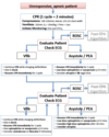
what is ABCDEF
A = airway
B = breathing
C = circulation
D = drugs
E = electrical defibrillation/ECG
F = follow-up
what are the steps in assessing airway
check for physical obstruction
ET intubation (use narrow catheter or tracheostomy)
what is done during breathing resuscitation
IPPV with preferably 100% O2 at approx 10 breaths per minute
watch chest rise and allow adequate time for deflation
what is done to assess circulation
- check pulses/heart sounds ASAP
- monitor these continuously
- maintain continuous compressions
what chest compressions are done on small dogs and narrow chested breeds
cardiac pump in right lateral recumbency
compress 3rd-6th intercostal space
100-120 bpm
what compressions are done on cats and tiny dogs
cardiac pump
finger and thumb across heart
compress at 100-120 bpm
what compressions are done on large, barrel chested breeds
thoracic pump
compress over highest point of thorax
what is the difference between cardiac pump and thoracic pump
cardiac: indirect compression of heart
thoracic: chest compression increase intrathoracic pressure –> increases pressure on outside of heart, lungs and great vessles –> blood to flow forward in arteries and backwards in veins (minimized by venous valves/collapse)
what categories can be used to decide compression technique
- cats/dog under 15 kg
- dogs over 15kg except sighthound type build
what things ensure successful external compressions (5)
- rate of 100-120 bpm
- table position
- compress approx 1/3 of thorax
- allow adequate time to refill
- change staff freq.
how are internal cardiac compressions done (6)
- rapid clip (3-6 IC spaces)
- in expiration, incise 5th interspace
- locate by using olecranon to point
- open pleura and pericardium
- milk ventricles into arteries
- continue ventilation
when is ICC preferred
- if thorax is already open
- large dogs (ECC less likely to be effective)
- disease process mean ECC unlikely to be effective (rib fractures, pleural effusion, diaphragmatic rupture)
- if ECC ineffective
- may be suitable to enter via diaphragm
what needs to be done to ensure circulation is corrected
rapid IV fluids
but rapid fluids not suitable for all cases
what route of drugs is best for successful resuscitaiton and why
- central/jugular injection
peripheral injection may not reach heart (flush saline after)
what other routes can be used for drugs
- intracardiac if done properly (risk of myocardial damage, avoid unless open thorax)
- transtracheal: need high doses (min 2x of atropine or adrenaline)
- intraosseous: exotics
when would adrenaline be used
- asystole
- to coarsen “fine” VF
- increase ionotropy
- increase systemic vascular resistance
what is the dose of adrenaline used
0.01 mg/kg IV (high dose of 0.1 has been used esp after prolonged CPR)
what is atropine used for
- atrioventricular block
- asystole which has been preceded by bradycardia (vagus mediated)
what is the dose of atropine
0.04 mg/kg IV
what is lidocaine used for
ventricular arrhythmias
what is the dose of lidocaine used
2-4 mg/kg (as a bolus) or 25-75 microgram/kg/min infusion
what is ECG used for
determine arrest rhythm
what are the types of arrest rhythms (4)
- asystole
- ventricular fibrillation
- ventricular tachycardia
- pulseless electrical activity
what arrest rhythm is this

asystole
what arrest rhythm is this

ventricular fibrillation
what arrest rhythm is this

coarse ventricular fibrillation
what arrest rhythm is this

fine ventricular fibrillation
what arrest rhythm is this

pulseless electrical activity
electromechanical dissociation
slightly wider complexes
what arrest rhythm is this

pulseless ventricular tachycardia
what arrest rhythm is this

ventricular tachycardia
no cardiac output
what rhythms is electrical defibrilation not effective for
less likely to be effective in asystole or PEA
what are the steps to electrical defibrillation
- charge
- apply conducting gel to paddles and dog
- current applied across heart
- stand clear
- 1-5 joules/kg external
- 0.1-0.5 joules/kg if using internal paddles
if monphasic upper end of range, biphasic lower end of range
what needs to be done in follow up (5)
- fluids
- ensure renal perfusion (urine production)
- warmth (caution)
- analgesics
- may need other drugs to support (dopamine)
what are signs of effective CPR (5)
- palpable pulse during cardiac compression
- retinal blood flow (as detected by using a doppler probe on the cornea)
- carbon dioxide detectable on capnography (gold standard)
- improvement in colour of mucous membranes
- ECG changes (normal rhythm)
what are signs of CPR recovery (5)
- lacrimation
- pupillary constriction
- return of cranial nerve reflexes
- return of other neurological function (response to sound, righting reflexes)
- return of spontaneous ventilation
when should you give up CPR
why did animal arrest in the first place
are we able to correct the reason for arrest
are there disease processes which we cant correct
15-20 mins is adequate to start seeing a response in most animals that are going to respond
what equipment is needed
crash cart handy
stocked and checked frequently
what are the actions during an arrest
- directing situation, making decisions, assessing patient
- compressions
- ventilation
- drawing up drugs, attaching ECG, keeping records
ideally 3-4 people
CAB?
recite the CPR algorithm