BODY Flashcards

1
Q

quadrate lobe

A

segment IV

2
Q

caudate lobe

A

segment 1

3
Q

hot quadrate sign

A

SVC obstruction

4
Q

hot spot in segment 1

A

Budd-Chiari

5
Q

dorsal pancreatic duct

A

Santorini

6
Q

ventral pancreatic duct

A

Wirsung

7
Q

entrance air kerma for abdominal radiograph

A

3 mGy

8
Q

MC duodenal malignancy

A

adenocarcinoma

9
Q
A

HIV cholangiopathy papillary stenosis

10
Q

4 phases of renal contrast

A
  • non-contrast
  • corticomedullary
  • nephrographic
  • excretory (pyelographic)
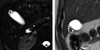
11
Q
A

pancreas divisum

  • main pancreatic duct (solid arrow, Santorini) drains into minor papilla
  • CBD and smaller ventral duct (Wirsung, curved arrow) drain inferiorly into major papilla
12
Q

caudate lobe hypertrophy

A
  • Budd Chiari
  • PSC
  • PBC
13
Q
A
  • clover leaf sign
  • healed peptic ulcer of duodenal bulb
14
Q

MC locations of GIST

A

stomach > duodenum > anorectum

15
Q

median survival after successful surgical resection of pancreatic cancer

A

1.5 years

16
Q
A

septate uterus

  • horizontal/normal uterine fundal contour
17
Q
A

bicornuate uterus

  • heart-shaped fundus, with indentation of outer contour
18
Q

MC associated abnormality with Mullerian Duct Anomalies

A
  • ipsilateral renal agenesis
19
Q
A

goblet sign

  • urothelial neoplasm
20
Q

MC MDA

A

septate uterus

21
Q

Zuckerkandl fascia

A

posterior perirenal fascia

22
Q

Gerota fascia

A

anterior perirenal fascia

23
Q

epiphrenic diverticula usually occur on which side?

A

right

24
Q
A

cone shaped cecum

  • Entamoeba histolytica
25
what is the finding and what does it indicate?
- pear-shaped bladder - retroperitoneal lipomatosis
26
interruption of IVC with hemiazygos continuation is due to..?
failure of right subcardinal vein to communicate with hepatic sinusoids
27
MR contrast agents contraindicated in GFR \< 30
- gadodiamide - gadopentate dimeglumine - gadoverstamide
28
low-osmolality iodinated contrast media t 1/2
2 hours
29
which low-osmolality iodinated contrast media are approved for intrathecal use? why?
- iohexol (Omnipaque) - iopamidol (Isovue) Because they are ***non***ionic
30
how long to wait after IV iodinated contrast material before therapeutic I-131?
3-4 weeks
31
which iodinated contrast material is preferred for angiography of small arteries, ie hand?
iodixanol (iso-osmolal)
32
macrocyclic Gd contrast agents
- gadobutrol - gadoteridol - gadoterate meglumine
33
dose of IV epi
0.1 mg (1 mL)
34
- inverted M waveform in renal artery (reversed diastolic flow) - renal vein thrombosis
35
associations with duplicated IVC
GU anomalies: - cross-fused ectopia - horseshoe kidney - cloacal exstrophy
36
area of kidney most vulnerable to ischemia
inner cortex
37
renal artery stenotic segment velocity and ratio of stenotic:pre-stenotic segments
\> 200 cm/s \> 2:1
38
Meckel Gruber
- renal cysts - encephalocele - polydactyly
39
medullary sponge kidney associations
- Caroli's - Ehlers-Danlos - Beckwith-Wiedeman
40
vessels arising from false lumen in aortic dissection
- right coronary - left renal - left iliac
41
renal vein thrombus with infarction and cortical rim sign
42
when is intradecidual sign seen?
4.5 weeks
43
embryo is visible when?
6 weeks
44
dx of pregnancy failure
- CRL \> 7 mm + NO heartbeat - MSD 25 mm + NO embryo - NO embryo with hearbeat \> 2 WEEKS after a scan that showed a GS WITHOUT yolk sac - NO embryo with heartbeat \> 11 DAYS after a scan that showed a GS WITH a yolk sac
45
oligohydramnios
AFI \< 5 cm
46
polyhydramnios
AFI \> 20 - OR- single pocket \> 8 cm
47
fetal bowel should never be more echogenic than what?
bone
48
normal midgut herniation occurs when?
9 - 11 weeks
49
increased risk with circumvallate placenta
- preterm
50
increased risk with succenturiate lobe
- vasa previa - retained placental tissue (--\> postpartum hemorrhage)
51
septate uterus
52
Zinner syndrome
- renal agensis - seminal vesicle cyst - ejaculatory duct obstruction
53
horseshoe kidney increases risk of what cancer?
- TCC - renal carcinoid - Wilms
54
thickenss of enodmetrial strip on endovaginal US in post-menopausal woman that requires biopsy
\> 5 mm
55
parametrial invasion - stage of cervical cancer?
IIB
56
when is cervical cancer treated non-operatively?
- stage IIB or higher - size \> 4cm
57
granulosa cell tumor may be associated with what findings in uterus?
endometrial hyperplasia (due to increased estrogen)
58
cancers associated with DES exposure
- vaginal clear cell adenocarcinoma - cervical clear cell adenocarcinoma
59
reproductive age woman, simple cyst 5-7 cm. follow up?
US in 1 year
60
reproductive age woman, simple cyst \<5 cm. follow up?
none
61
US findings supportive of PCOS
\>12 follicles - AND/OR - ovarian volume \> 10 cc
62
likely benign cyst in *post*menopausal female and follow up
\> 1 cm, \< 7 cm 1 year US follow up
63
thickened septation in adnexal mass on US
\> 3 mm
64
which radiographic detector results in sharpest image?
photoconductor (Selenium)
65
which radiographic detector results in lowest patient dose?
scintillator (CsI) | (highest x-ray absorption efficiency)
66
which radiographic detectors are indirect?
- scintillator (CeI) - PSP (BaFBr)
67
what is effect of increasing detector thickness have on (a)patient dose and (b)blur
(a) dose decreases (b) blur increases
68
which gastric volvulus occurs in old ladies with paraesophageal hernia?
organoaxial
69
Carman meniscus
70
SV cyst associations
- ipsilateral renal agenesis - vas deferens agenesis - ectopic ureter - ADPKD (bilateral SV cysts)
71
Gardner
- facial osteoma - extra-abdominal desmoid - colonic adenocarcinoma - thyroid, liver, ampulla, adrenal gland carcinoma
72
post-menopausal normal endometrial thickness, no hormone replacement
\< 5 mm
73
MC site of ectopic preg
ampulla
74
1st trimester maternal screen in Down's
- decreased PAPP-A - increased beta-hCG - increased nuchal translucency
75
alkali ingestion causes what kind of necrosis?
liquefactive
76
cystic thickened uterus related to tamoxifen
77
HASTE
Half Fourier Aquisition Single-shot Turbo Spine Echo
78
Dx
bicornuate uterus
79
what kind of weighting? orange arrow points to what?
- T2 - junctional zone
80
normal cervical length
\> 30 mm
81
cervical incompetence (2nd trimester)
\< 25 mm
82
involvement of which vessels precludes resection of pancreatic adenoca? what stage is this?
- celiac artery, SMA - T4
83
most accurate 2nd trimester measurements
- BPD - head circumference
84
timing of renal artery stenosis post-transplant
1st month (usually immediate post-op)
85
timing of hepatic artery stenosis post-transplant
\> 1 month
86
timing of renal vein thrombosis post-transplant
1st week
87
what is 1?
common hepatic duct
88
what is 2?
proper hepatic artery
89
- name of sign - dx
- pearl necklace - adenomyomatosis
90
- dx - mode of inheritance - other names
- Caroli disease - autosomal recessive - communicating cavernous biliary ectasia, Todani V choledochal cyst
91
- imaging appearance - dx
- pruned tree - PSC
92
morphologic classification of cholangiocarcinoma (3)
- mass forming - periductal infiltrating - intraductal growing (IPMN)
93
anatomic classification of cholangiocarcinoma (location)
- intrahepatic - hilar (Klatskin tumor) - distal extrahepatic
94
MC cholangiocarcinoma location/name
hilar (Klatskin)
95
Caroli *syndrome*
Caroli disease + congential hepatic fibrosis
96
ampullary neoplasm occurs distal to what?
confluence of CBD and pancreatic duct
97
MC biliary ductal variant anatomy
posterior right hepatic duct draining into left hepatic duct
98
ovarian cancer stage: distant spread or malignant pleural effusion
stage 4
99
ovarian cancer stage: lymph node mets or peritoneal implants outside of pelvis
stage 3
100
direction of MPV in TIPS
hepatopetal
101
direction of right and left portal veins in TIPS
hepato***fugal***
102
1st clinical signs of NSF develop when?
1 month
103
definition of oligohydramnios
vertical pocket \< 2 cm
104
Sm-153 t 1/2
1.9 days
105
gamma photon energy of Sm-153
103 keV
106
increased liver attenuation *without* increased splenic attenuation
- primary hemochormatosis - amiodarone toxicity - glycogen storage
107
what keeps PDA open
Prostaglandin
108
PDA associations
- Holt-Oram - maternal rubella - Trisomy-21
109
MC location for gastric diverticulum
juxtacardiac posterior wall
110