Basic Tissue Types Flashcards
Levels of Organization: cells interact to gorm, etc
Cells interact to form tissues, tissues interact to form organs and organ systems of the body
Body constructed of four basic tissues:
Epithelia. Connective Tissue. Muscle. Nerve.
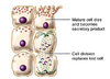
Epithelial Tissue Function (4)
Forms barriers (between inside and outside world, and between compartments within body). Covers exposed body surfaces. Lines hollow organs, body cavities and ducts of glands. Forms glands.
Connective Tissue (4)
Links tissues and organs together. Provides structural and metabolic support for other tissues/organs. Stores energy as adipose tissue. Forms the immune system (cells produced by lymphatic tissue then migrate to connective tissue)
Epithelium is avascular?
it has no blood vessels, so exchange must happen in connective tissue aka structural and metabolic support
Muscle Tissue (3)
`Specialized for contraction. Generates force to produce motion of body parts, move substances through blood vessels and hollow organs. Maintains body temperature (heat loss from contraction)
Nervous tissue (2)
Receives, processes and integrates signals from within body and external environment. Generates and transmits impulses that control and integrate various functions of the body.
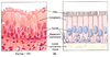
Characteristics of Epithelia (5)
Continuous, avascular, rest on basal lamina, little extracellular matrix, polarized.
Epithelia: continuous and avascular?
never begin or end; no blood vessels
Epithelia: rest on basal lamina?
basal lamina is a specialized layer of connective tissue, the epithelia binds to BL and other things in BL attach to what’s under
Epithelia: little extracellular matrix?
Extra = outside of cell. Cells secrete matrix that connect other cells to each other
Epithelia: polarized?
apical and basolateral surfaces differ - apical faces outside, there are junctions between cells. lateral + basal = basolateral
Tight junction
area where two cells are held so tightly together that things can’t really pass through - no diffusion: has to go THROUGH the cell
Types of Epithelia and Naming
different types serve different functions - named according to number of cells, 1 = simple 2+ = stratified. Shape of cells in outermost layer


Simple Squamous

Simple Cuboidal

Pseudostratified Columnar

Simple Columnar

Stratified Columnar

Stratified Squamous

Transitional
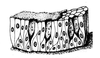
Simple Squamous Epithelium: properties, locations
Single layer of flat cells, very delicate so only found in places without a lot of force: lines body cavities, heart and blood vessels, and site of gas exchange in lungs
Simple Squamous Epithelium: Locations and Functions

Stratified Squamous Epithelium: properties, found where
Stratified cells with outermost layer squamous (aka flat) - forms outer layer of skin, lines mouth, esophagus and vagina - found near abrasive forces
Stratified Squamous Epithelium: Locations (7) and Functions (1)
Locations: surface of skin, lining of mouth, throat, esophagus, rectum, anus and vagina. Functions: physical protection against abrasion, pathogens and chemical attack


Simple Cuboidal Epithelium: properties and location
Single layer of cells, cell height and width equal, found in glands and forms ducts of glands, forms portions of kidney (tubules out of cuboidal cells because they can make walls of small tubes), lines small intestines since it is the max amount of membrane but still simple to move things from lumen
Simple Cuboidal Epithelium: Functions (3) and Locations (4)
Locations: glands, ducts, portions of kidney tubules, thyroid gland. Functions: limited protection, secretion, absorption
Simple Columnar Epithelium: properties, found where
Single layer of cells, cells taller than they are wide - lines digestive tract, fallopian tubes and gall blader


Simple Columnar Epithelium: Locations (5) and Functions (3)
L: lining of stomach, intestine, gallbladder, uterine tubes, collecting ducts of kidneys. F: Protection, secretion, absorption.
Stratified Cuboidal Epithelium: properties and found where
two to three layers of cells, outmost layer is cuboidal - are like bricks, so they line large ducts (think about it..tiny ducks, single layer is kay, but for larger ducts, more layers makes them stronger)
Stratified Columnar Epithelium: Locations (1) and Functions (3)
rare, found in lining of some ducts ex. sweat gland duct - protection, secretion and absortpion (same as simple columnar)


Stratified Columnar Epithelium: properties and found where
Multiple layers of cells, outer layer is columnar - found in very large ducts where they are so big that cuboidal isn’t enough: ex pancreatic duct
Stratified Columnar Epithelium: Locations (6) and Functions (1)
L: small areas of pharynx, epiglottis, anus, mammary gland, salivary gland ducts, urethra. F: protection


Pseudostratified Columnar epithelium: properties and found where
nuclei appear at multiple levels, all cells touch basal lamina but may not all reach luminal surface - found where cells might be damaged since an abrasive substance can kill large cell, but little one can take its place - respiratory system, male repro tract
Pseudostratified Columnar Ciliated Epithelium: Locations (4) and Functions (2)
L: lining of nasal cavity, trachea, bronchi, portions of male repro tract. F: protection and secretion
Transitional Epithelium: properties and found where and why
multiple layers of cell, outermost are large and dome shaped so is stretch - found only in urinary tract (bladder, renal pelvis, ureters) to permit expansion and recoil after stretching
Glands: formed of what and how
composed of epithelial cells: form by growth of epi cells into underlying connective tissue
Exo vs Endocrine glands
Exo: release secretions through ducts onto epithelial surfaces. Endo: ductless, secrete hormones directly into blood.
Formation of Glands: exo vs endocrine

Formation of Glands: exo vs endocrine (words)
cells both develop the same way: epithelial cells grow into underlying connective tisuue, cells at top form a channel and cells at bottom differentiate into secretory cells. If secretory cells remain: exocrine. If they degenerate: endocrine.
Three mechanisms of Secretion
Merocrine (normal exocytosis), Apocrine (apical surface is pinched off) and Holocrine (entire cell released)
Merocrine Secretion Diagram

Apocrine Secretion

Holocrine Secretion

Apocrine Secretion and Holocrine Secretion Examples
Apo: apical surface is pinched off like in mammary glands. Holo: entire cell is released like in sebaaceous glands