Bacteriology Flashcards

1
Q

Sites that should not be cultured for anaerobes

A

stool

skin

vagina

urethra

oropharynx

2
Q

Characteristics of blood culture

A

Each bottle has 10 ml blood (adults)

2 bottle set (1 aerobic and 1 anaerobic) collected from one site

multiple sets obtained from multiple sites: need 3 sets

3
Q

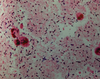
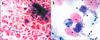
Cell wall in gram + and - organisms

A
  • Gram positive: thick peptidoglycan cell wall without outer membrane; retains purple crystal violet/iodine stain during decolorization
  • Gram negative: thin peptidoglycan cell wall with outer membrane; lose crystal violet/iodine during decolorization and are visualized with the red safranin stain
4
Q

Appearance of the following organisms on gram stain

  1. Strep pyogenes
  2. Nonpyogenic strep
  3. Strep pneumoniae
  4. Enterococcus species
  5. Neisseria
  6. vibrio
  7. campylobacter
  8. yersinia
  9. legionella
A

Gram positive cocci:

  1. pairs and short chains
  2. long chains
  3. lancet shaped diplococci
  4. diplococci

Gram negative cocci

Neisseria - coffee bean shaped diplococci

Gram negative rods

  1. curved, comma shaped, short
  2. curved, thin (seagulls or corkscrew)
  3. closed safety pin
  4. invisible in primary smear
5
Q

Pleomorphic gram positive rods

  1. Aerobic
  2. Aerotolerant
  3. Anaerobic, nonspore forming
A
  1. Aerobic
    1. Corynebacterium
    2. Other coryneforms (e.g., Rothia, arthrobacter)
    3. Non-nocardia actinomycetes
  2. Aerotolerant
    1. Actinomyces
    2. Bifidobacterium
  3. Anaerobic, nonspore forming
    1. Propionibacterium
6
Q

Small to medium gram positive rods

  1. Aerobic
  2. Aerotolerant
  3. Anaerobic, nonspore forming
  4. Anaerobic, spore forming
A
  1. Aerobic
    1. Listeria
    2. Erysipelothrix
  2. Aerotolerant
    1. Lactobacillus strains
  3. Anaerobic, non spore forming
    1. Eubacterium
    2. Lactobacillus
  4. Anaerobic, spore forming
    1. Clostridium ramosum
7
Q

Large gram positive rods

  1. Aerobic
  2. Anaerobic, nonspore forming
  3. Anaerobic, spore forming
A
  1. Aerobic
    1. Bacillus
  2. Anaerobic, nonspore forming
    1. Clostridium perfringens
  3. Anaerobic, spore forming
    1. Other clostridia
8
Q

Branching, filamentous aerobic and anaerobic nonspore forming gram positive rods

A

Aerobic:

Nocardia

Anaerobic, nonspore forming:
Actinomyces israelii

9
Q

Acid Fast Stains

A

Mycobateria:

  • cell wall structure of gram positive bacteria but contain mycolic acids, which repel crystal violet/iodine; thus are usually gram invisible
  • dye penetrates hydrophobic barrier to stain mycolic acids in cell wall
  1. Ziehl-Neelsen: red carbol fuschin dye containing phenol is heated so it can penetrate cell wall; strong acid (3% HCl) decolorizes
  2. Kinyoun (cold technique): higher concentration of phenol and no heat; otherwise same as ZN
  3. Fite (modified) acid-fast: weaker decolorizing reagent; good for:
    1. nocardia
    2. cryptosporidium
    3. isospora
    4. cyclospora
    5. sarcocystis
    6. legionella
10
Q

Alternative stain for acid fast bacilli

A

Fluorescent auramine rhodamine stain

11
Q

Buffered charcoal yeast agar

A

Legionella grows because media has cysteine and iron
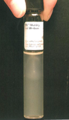
12
Q

Thioglycolate broth

A

Cultivation of bacteria, including microaerophilic and obligately anaerobic bacteria; oxygen tension decreases at bottom of tube, allowing for growth of anaerobes

13
Q

MAC agar

A
  • bile salts and crystal violet inhibit growth of Gram+ bacteria and allow for growth of Gram-
  • Lactose fermentation results in pink color
14
Q

Eosin methylene blue agar

A
  • aniline dyes inhibit growth of gram+ bacteria and allow for growth of gram negative rods
  • lactose fermentation results in purple-black colonies with a green metalic sheen
  • differentiates lactose fermenting and non-fermenting enterics
15
Q

campylobacter blood agar

A

contains cephalothin, vanc, trimethoprim, amphotericin B, polymyxin B

allows for recovery of campylobacter

16
Q

Hektoen enteric agar

A
  • Bile salts and the dyes bromthymol blue and acid fuchsin inhibit Gram+ and some Gram- strains
  • recover salmonella and shigella better than MAC
  • lactose and/or sucrose fermentation results in yellow or orange color, otherwise translucent
  • H2S production results in black coloration
17
Q

Selenite broth

A

Sodium selenite inhibits growth of gram+ bacteria and many enterics other than salmonella

  • allows for enrichment of salmonella
18
Q

Thiosulfate citrate bile salts sucrose agar (TCBS)

A
  • Bile salts inhibit gram+ bacteria
  • Alkaline pH inhibits most enterics and enhances growth of vibrio species
  • sucrose fermentation results in yellow colonies
  • differentiated between sucrose fermenting vibrio (e.g., V cholerae) and nonfermenters (other vibrio)
19
Q

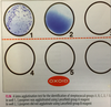
Cefsulodin-Irgasan-novobiocin (CIN) agar

A
  • antimicrobial and crystal violet inhibit most gram negative bacteria other than yersinia
  • recover yersinia
  • mannitol fermentation results in bull’s eye colonies (colorless with red center), otherwise translucent
  • differentiates between manitol fermenters (yersinia enterocolitica) and nonfermenters (other yersinia)
20
Q

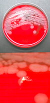
Anaerobic colistin nalidixic acid (CNA) agar

A
  • antimicrobials inhibit gram negative bacteria
  • recover anaerobic streptococci; blood in agar allows differentiation based on hemolytic reactions
21
Q

Lim broth

A

antimicrobials inhibit gram negative bacteria

  • recovers Group B streptococci (agalactiae)
22
Q

Regan-Low medium

A

contains antimicrobials that allow for selective growth of Bordetella pertussis and bordetella parapertussis

23
Q

Thayer-martin medium

A

Allows for growth of Neisseria

Vanc, colistin, nystatin, and SXT block growth of orgs

24
Q

Salmonella-shigella agar

A
  • Lactose fermentation results in pink or red colonies
  • H2S production makes black color
  • good for enterics
25
Motility test
Semisolid Motile organisms show growth spreading away from the line of inoculation, clouding the agar Nonmotile organisms remain in the stabbed inoculation site, so the rest of the agar remains clear
26
Bacteria that grow optimally at the following temperatures: 1. 25-40 degrees 2. 42 degrees 3. 37 degrees 4. 4 degrees
1. 25-40 1. Yersinia enterocolitica 2. pseudomonas fluorescens and putida 2. 42 1. campylobacter jejuni and coli 2. other pseudomonas species 3. 37 1. Listeria monocytogenes grows optimally at 37 degrees, but is motile at 25 degrees 4. 4: 1. Listeria monocytogenes can multiple at 4 degrees
27
Catalase test - purpose - how - avoid? - catalase positive organisms?
* **categorizes gram positive cocci:** differentiating staphylococci (catalase positive) from streptococci and enterococci (catalase negative) * **3% H2O2** * avoid contaminating with blood agar: **red blood cells make catalase** * Other catalase positive orgs: * **Campylobacter** * **Listeria** * **Bacillus**
28
Coagulase test - types - purpose
* Distinguish coag positive staph (Staph aureus) from other staph * Types * **Tube coagulase test** * detects free (secreted) coagulase and is the definitive test * isolate + plasma in a tube; free coagulase complexes with **coagulase reacting factor** (CRF) in plasma to convert fibrinogen to **fibrin**, forming a clot * examine at **4 and 24** hours (some S aureus make fibrinolysin that dissolves clot giving false negative after 24 hours and others produced delayed reaction that give false negative if only examined after 4 hours) * **Slide coagulase test**: detects bound coagulase (i.e., clumping factor); reaction is immediate * false negative results when there is a free, not bound, coagulase * **Staph lugdunensis** and **S schleiferi** produce **bound**, not free coagulase, so are positive with slide test and negative with tube test * **Strep intermedius is coag +**
29
Novobiocin susceptibilty test
S **saprophyticu**s is novobiocin **resistant**; S epidermidis and other coagulase negative staph are susceptible
30
Optochin test (P disk)
S **pneumoniae** is **susceptible** to optochin; viridans strep are resistant
31
CAMP test
* identifies **streptococci agalactiae** and **listeria monocytogenes** * **CAMP factor** is produced by both of the above * CAMP factor increases **staph beta hemolysin** * isolate sreaked at perpendicular angle to a S aureus streak; **rectangular** enhancement of beta hemolysis at intersection is seen with **listeria** and **arrowhea**d shaped increase in hemolysis is seen with **GBS** * **Turicella otitidis is also positive, but is non-hemolytic unlike listeria which is beta hemolytic**
32
PYR test
* identifies **enterococci** and **group A strep** * enzymatically hydrolyze PYR, yielding **beta naphthylamide** with a **red** color change
33
Bile esculin test
* identifies **enterococci (also tolerant to 6.5% NaCl)** and **group D streptococci (S bovis)** * both able to **hydrolyze esculin in presence of bile** * bile esculin agar slant inoculated and incubated for **24 hours**; **diffuse blackening** of the medium indicates esculin hydrolysis * **rapid spot test** versions of this assay available
34
Oxidase test
* Classifies gram negatives * positve: * Neisseria * Moraxella * Pseudomonas * Legionella * Brucella * Aeromonas * Pasteurella * curved gram negative rods (campylobacter, vibrio, and helicobacter) * [one can hallucinate on **PCP** a **VAN** drive by an OX named "**Bruc**e" * negative * enterobacteriaceae * the reagent (**P phenylenediamine**) is oxidized by **cytochrome oxidase**, forming **iodophenol blue**
35
Indole test
Identifies E coli (positive) * Determines organism's ability to produce indole via deamination of tryptophan, using Kovac reagent, with which indole combines to produce a pink color * H influenzae and Pasteurella are also positive, but they are lactose non-fermenters
36
Rapid urease test
* identifies proteus and H pylori * splitting (hydrolysis) of urea alkalinizes the medium, leading to color change (red or pink) * many bacteria have urease activity, but few can degrade it within 4 hours and are designated "rapid urease positive" organisms
37
Beta glucuronidase enzyme activity
* presumptive ID of **E coli** members of the anginosus group strep * **fluorometric substrate** (4-methylumbelliferyl-beta-D-glucuronide) is used, which makes a fluorescent compound and can be added to **MacConkey** * E coli 0157:H7 does not have this activity
38
Hippurate hydrolysis test
* presumptive ID of * campylobacter jejuni * listeria monocytogenes * Streptococcus agalactiae * gardenerella vaginalis * legionella pneumophila * All of the above are hippurate hydrolysis positive * Uses ninhydrin reagent; blue = positive
39
Lysozyme test
* ID nocardia * most bacteria are inhibited by lysozyme * growth in lysozyme containing broth medium indicative of nocardia
40
Anaerobic gram+ cocci - organisms - colonizes what sites? - diseases
* Peptostreptococcus * Finegoldia * Peptococcus * Parvimonas * Anaerococcus Commensal flora of the oropharynx, GI, vagina, skin * endogenous polymicrobial infections (e.g., abscess of the skin, soft tissues, lung, or brain) * aspiration pneumonia * necrotizing pneumonia * empyema * diabetic foot infections * crepitant cellulitis * synergistic gangrene or necrotizing fasciitis * septic abortion * intraabdominal infections
41
Staphylococcus Colony morphology and gram stain
Creamy colonies Grapelike clusters and tetrads
42
S aureus - biochemicals - resistance mechanisms - susceptible to what abx? - ID of MRSA
* coagulase positive * most express a **beta lactamase**, conferring resistance to penicillin, ampicillin, and early generation cephalosporins * Susceptible to **nafcillin, dicloxacillin, or oxacillin** * MRSA - carries **mecA** gene, which expresses **PBP2A**, a penicillin target with little affinity for the drug * MRSA ID * **disk diffusion (cefoxitin)** * **DNA** hybridization probes to detect mecA * **PBP2A latex agglutination**
43
Coagulase negative staphylococci
**S epidermidis** * skin commensal * bloodstream infection related to IV catheter, prosthetic vascular grafts, or prosthetic cardiac valves * prosthetic joint infections * CNS infection related to ventricular shunts **S saprophyticus** * uropathogen * novobiocin resistant **S lugdunensis** * skin commensal * infections similar in severity to S aureus * positive by slide coagulase test, but negative by tube coagulase test * PYR+ * ornithine decarboxylase+
44
Strep pyogenes - colony morphology - gram stain - biochemicals - colonizes - infections - treatment
* **beta hemolytic (wide)** * **catalase negative** * gram+ cocci single, in pairs and short chains * susceptible to **bacitracin (A disk)** * **PYR+** * **Lancefield group A antigens** * colonizes **oropharynx and skin** * Infections * **pharyngitis** * **retropharyngeal abscess** * **bacteremia** * **skin and soft tissue** infections * impetigo * erysipelas * necrotizing fasciitis * **toxin mediated** illnesses (toxic shock syndrome and scarlet fever) * **immune mediated** disease (acute rheumatic fever, poststreptococcal glomerulonephritis) * **pediatric autoimmune neuropsychiatric** disorders * **Likelihood of ARF decreases with treatment, but poststrep GN does not improve**
45
Streptococcus dysgalactiae
* **PYR-** * **bacitracin resistant** * **Lancefield group C or G**
46
Jones criteria
**Major** * carditis * polyarthritis * chorea * erythema marginatum * subcutaneous nodules * gram negative cocci **Minor** * arthralgia * fever * increased ESR or CRP * prolonged PR interval
47
Streptococcus agalactiae - gram stain - colony morphology - biochemicals
* gram+ cocci in pairs and short chains * **gray white** colonies * **Beta hemolytic with narrow zone** of hemolysis * **CAMP test positive** * **Hippurate hydrolysis positive** * Pregnant women screened at 35-37 weeks (**culture or molecular** methods following **enrichment broth step**)
48
Beta hemolytic strep
GAS (wide zone of hemolysis) and GBS (narrow zone of hemolysis)
49
alpha hemolytic streptococci
**Streptococcus pneumoniae** * Lancet shaped, gram+ diplococci * susceptible to optochin (P disk) * infectious strains have polysaccharide capsule that results in smooth colonies * Diseases * community acquired pneumonia * bacteremia * meningitis * otitis media * sinusitis * spontaneous bacterial peritonitis **Viridans streptococci** * S mitis group: native valve endocarditis * S mutans group: dental caries can cause endocarditis * S salivarius: bacteremia and endocarditis * S anginosus: abscesses (particularly S intermedius)
50
Nonhemolytic, catalase negative gram+ cocci
Enterococcus Streptococcus bovis
51
Enterococcus - gram stain - colony morphology - biochemicals - Lancefield group - colonize what site? - diseases - Resistance patterns and mechanisms
* Diplococci in short chains * small, **gray**, nonhemolytic colonies * hydrolyze **bile esculin** (esculin gets hydrolyzed in presence of bile) * Lancefield Group **D** * normal inhabitants of **lower GI** * Diseases: * **UTI** (often nosocomial and related to catheters) * **bacteremia** * **endocarditis** * **intraabdominal and pelvic infections** * **E faecalis**: susceptible to vancomycin * **E faecium** resistant to: * **vancomycin** (due to **acquisition of vanA**, **vanB**, or **vanD** gene clusters) * **penicillin, ampicilin, and carbapenem** due to altered **PBP** * E gallinarum and E casseliflavus have intrinsic, low level vanc resistance due to chromosomal **vanC** gene
52
Streptococcus bovis group - gram stain - colony morphology - biochemicals - diseases - colonizes - subgroup associated with malignancy
* **gram+ cocci in pairs or short chains** * **gray white, nonhemolytic colonies** * Lancefield **group D** * **hydrolyze bile esculin** * **PYR negative** * **cannot grow in 6.5% NaCl** * Colonizes the **GI tract** * Diseases: **bacteremia and endocarditis** * S bovis **gallolyticus** subgroup is associated with colonic malignancy
53
Anaerobic gram negative cocci
**Veillonella** and other gram negative cocci are normal inhabitants of oral cavity, GI, GU, respiratory tracts (sometimes found in polymicrobial anaerobic infections)
54
Neisseria species - gram stain - culture agar - other biochemicals
* fastidious, requiring incubation in CO2 at 35 degrees * intracellular coffee bean shaped gram negative diplococci * cultured on chocolate agar with antibiotics to inhibit indigenous flora (Thayer-Martin, Martin-Lewis, or New York City agar) * cotton swabs inhibit growth * oxidase positive
55
Neisseria gonorrheae - how to identify - rate of asymptomatic men and women - infections
* identified by gram stain and colony morphology + biochemical reactions or hybridization probes * 10% of affected males and 50% of affected females asymptomatic * Infections: * epididymitis * PID * urethritis in women * disseminated infection manifests as Fitz Hugh Curtis syndrome or purulent arthritis
56
Neisseria meningitides - how to identify - serogroups defined by \_\_\_ - diseases caused by what serogroups - vaccine includes which serogroups - colonization - infections
* identified by culture * capsular polysaccharide antigens define serogroups * serogroups **B, C, and Y** cause majority of disease in the US * serogroup **B is not in the tetravalent vaccines** * nasopharyngeal carriage is common * invasive infection is most often meningitis
57
Moraxella catarrhalis - gram stain - culture morphology - colonizes - biochemical - diseases
* gram stain similar to neisseria; can be coccobacillary * oxidase positive * DNAse positive (unlike neisseria) * grows well on blood agar and does "hockey puck" * colonizer of upper respiratory tract * diseases: * otitis media * COPD exacerbation
58
Kingella kingae
* gram negative coccobacillus * pairs and short chains * HACEK organism * Diseases * endocarditis in adults * septic arthritis in kids * osteomyelitis in kids * occult bacteremia (in kids?)
59
Anaerobic spore forming gram positive rods
Clostridium species
60
Nonspore forming anaerobic gram positive rods
Actinomyces israelii Propionibacterium
61
Aerobic gram+ rods spore formers
Bacillus species
62
Aerobic gram+ rods non-spore formers
Listeria monocytogenes Erysipelothrix rhusiopathiae Nocardia Rhodococcus equi Corynebacterium diphtheriae Tropheryma whipplei
63
C perfringens - morphology - ID
* Large boxcar shape gram+ rod, anaerobic * short chains * double zone of beta hemolysis * lecithinase activity
64
Clostridium septicum
* **large, gram+ rod in chains** * **swarm** rather than form discrete colonies * **bacteremia** related to **colonic carcinoma** or other GI pathology
65
Botulism - source - diagnosis
* Botulism dx relies on detection of **toxin** in serum, stool, vomitus, or food * raw honey = source of infant botulism
66
Clostridium tetani - tetanus diagnosis
Clinical diagnosis; no useful tests for toxin available
67
Clostridium difficile - culture medium - colony morphology and other characteristics - gram stain - disease and diagnosis
* culture in cycloserine cefoxitin egg yolk fructose agar (**CCFA**) under **anaerobic** conditions * CCFA colonies are **yellow** with ground glass appearance, and **fluoresce** under UV light * **"horse manure"** odor * Gram stain shows thin, regularly shaped gram+ or gram variable rods sometimes with **subterminal and free spores** * nontoxigenic strains of C difficile are common in the bowel, so must isolate the toxin to show disease: * **cytotoxic culture** is gold standard, but outdated * molecular tests for **toxin A** and **toxin B** genes (toxin B much more likely to cause disease) * **ELISA** for **stool toxin** relatively insensitive; ELISA for stool glutamate dehydrogenase **(GDH)** more sensitive * Severe C difficile colitis associated with **BI/NAP1/027** strain; can detect toxin genes and mutated form of **tcdC** gene that typifies the 027 strain
68
Actinomyces - gram stain - culture conditions - diseases
* Anaerobic gram+ rod, non-spore former * branching, filamentous * A israelii forms white **cerebriform** (molar tooth) colonies * same gram stain (almost) as nocardia * unlike nocardia, actinomyces are **anaerobes** and **not acid-fast** * disease * **cervicofacial actinomycosis** as a result of trauma * **thoracic actinomycosis** 2/2 aspiration * **IUDs** * **intraabdominal actinomycosis** related to appendicitis, diverticulitis, or surgery
69
Proprionibacterium
* anaerobic gram+ rod, non-spore forming * diphtheroid gram stain morphology * prionic acid production * disease * skin commensal causing prosthetic joint infection * infection of other foreign bodies
70
Bacillius anthracis - gram stain - colony morphology - biochemical - motility - diseases
* forms **chains and subterminal spores** * **nonhemolytic** colonies with irregular borders **(Medusa heads)** that **stand up** when teased with a loop * **catalase+** * **nonmotile** * **Edema factor, protective factor, lethal factor** * Disease * **cutaneous anthrax** (inoculation of spores into skin wound while handling animals or hides) * **GI** anthrax causes **ulcers** * **inhalation anthrax** (woolsorter disease): widened mediastinum on AP film caused by **hemorrhagic mediastinitus**
71
Bacillus cereus
* chains and demonstrates subterminal spores * large, beta hemolytic colonies * nontenacious colonies (do not stand up when teased) * motile * foodborne illness: Asian rice (lack of refrigeration)
72
Listereria monocytogenes - gram stain - biochemicals - temperature - motility - diseases
* Aerobic gram+ rod, non-spore former * small colonies in primary culture after 1-2 days * **beta hemolytic** with **narrow zone** of hemolysis like GBS * gram stain shows short, nonbranching gram+ rods, single or in short chains * **catalase+** * **CAMP+**: rectangular zone of enhanced hemolysis at Staph intersection with the listeria * temperature dependent **tumbling motility** in wet mounts (best at room temperature) * semisolid agar motility: **umbrella shaped motility** * can grow at 4 degrees (can contaminate refrigerated foods * Disease * chorioamnionitis * neonate may have granulomatosis infantiseptica - multiple abscesses * elderly/IC: bacteremia, meningitis, encephalitis
73
Erysipelothrix rhusiopathiae - motility - gram stain - biochemicals - treatment - disease
* nonbranching, nonspore forming gram+ rod * short (coccobacillary) single or short chains, but can be long, thin, and filamentous * **nonmotile** * **catalase negative** * produces **H2S on TSI** slant, causing black butt (unlike listeria and lactobacillus) * **intrinsically resistant to vancomycin** * Disease * erysipeloid: a cellulitis acquired when a wound comes in contact with animal carrying organisms (anglers, butchers, farmers)
74
Nocardia - gram stain - culture medium - colony morphology - other characteristics - disease
* long, thin, filamentous, branching, beaded gram+ rods * **modified acid-fast** positive * **BCYE** is best for primary culture * chalky **white** colonies; become **salmon orange** pink when mature * **musty basement** odor * Disease * indolent soft tissue infections (**mycetoma**, forming macroscopic granules (similar to sulfur granules of actinomycosis)) * **invasive pulmonary** infection and disseminated infection, often in **CNS**
75
Rhodococcus equi
* gram+ cocci, coccobacilli, or coryneform rod * often within histiocytes * modified acid-fast+ * salmon colored and slimy * affects IC: pulmonary infections
76
Corynebacterium diphtheriae - gram stain - culture medium and morphology - biochemical - disease
* coryneform or diphtheroid pleomorphic, club shaped, gram+ rods that **palisade** and cluster together * grows well on **blood agar** * isolation better on **Tinsdale** agar: **brown black** colonies surrounded by brown black **halos** * ID involves **reduction of potassium tellurite to metallic tellurite** * Disease: * fever, sore throat, white/glossy pseudomembrane (turns gray) on tonsils
77
Tropheryma whipplei
* aerobic gram+ rod * ubiquitous in **environment**; disease develops in those with selective **immune deficiency** * older males (10:1= **M**:F) * **foamy histiocytes** in tissue * **PAS+** * **AFB negative** * Whipple disease
78
Anaerobic gram negative rods
Bacteroides fragilis Porphyromonas/Prevotella Fusobacterium
79
B fragilis - gram stain - colony morphology - treatment - disease
* gram stain: pleomorphic; bacilli with bipolar vacuoles (safety pin) on liquid media gram stain * small, shiny colonies on blood agar * beta lactamase producer: resistant to penicillin * polymicrobial infections
80
Porphyromonas/Prevotella
* Brick red fluorescence under UV light * after several days of growth appear brown/black in natural light
81
Fusobacterium
* fusiform gram negative rod, anaerobic * diseases: * F necrophorum colonizes tonsils and is associated with Lemierre syndrome (tonsilitis + septic thrombophlebitis of the internal jugular vein) * placental infection
82
Enterobacteriaceae characteristics - biochemicals - growth on agar - colonization - diseases - virulence - classification - abx
* **aerobic** on MAC agar * + for **glucose fermentation** * reduce **nitrate to nitrite** * **oxidase negative** * many are natural inhibitants of the **colon** * **nasopharyngeal carriage** in * hospitalized patients * patients with uncontrolled DM * chronic alcoholics * isolated from **urine, blood, wounds, abscesses, and respiratory tract** * **Lipid A**, component of LPS is an endotoxin * **O antigen**, a second LPS component forms basis for serogrouping * abx **resistance** is common
83
strong lactose fermenters
E coli Klebsiella Enterobacter
84
Enterobacteriaceae that produce H2S
Salmonella Edwardsiella Citrobacter freundii Proteus **(SPEC)** [Erysopelothrix also makes H2S]
85
Enterobacteriaceae that are strongly urease+
Proteus Morganella Providencia rettgeri
86
Nonmotile enterobacteriaceae
Shigella Klebsiella Yersinia are not motile at 37 degrees, but motile at 22 degrees
87
VP positive enterobacteriaceae
Klebsiella Enterobacter Hafnia Serratia Pantoea
88
Penylalanine deaminase (PAD) + enterobacteriaceae
Proteus Morganella Providencia
89
List of enterobacteriaceae
1. Escherichieae 1. E coli 2. Shigella: sonnei, flexneri, dysenteriae, boydii 2. Ewardsiellae tarda 3. Salmonellae: enteritidis, typhi, paratyphi, choleraesuis 4. Citrobacterae: freundii 5. Klebsiellae 1. K pneumoniae 2. Enterobacter: aerogenes, cloacae 3. Hafnia alvei 4. Pantoea agglomerans 5. Serratia marcescens 6. Proteae 1. Proteus: mirabilis, vulgaris 2. Morganella: M morganii 3. Providencia stuartii 7. Yersinia enterocolitica
90
Describe Kligler iron agar slant
(KIA) * **10:1 lactose:glucose** * phenol red indicator is yellow when pH is **\<6.8** and red when \>6.8 * Fermentation occurs with lowering of pH (yellowing of indicator) * Interpret tubes at 24 hours * **"K" = alkaline** * **"A" = acid** * Initially a bacterium that ferments glucose but not lactose will produce acidic (yellow) slant over acidic butt * after glucose consumed, organism will oxidatively metabolize proteins where there is oxygen, turning the slant back to red by 24 hours, while the butt is yellow * organisms that ferment will continue to ferment lactose throughout the incubation, mainting the yellow slant and butt through the **24 hours** * An H**2**S indicator is also present (black) * In short: * **Glucose fermenters: K/A** * **Lactose fermenters: A/A**
91
Escherichia coli
* lactose fermenting * indole + * Beta glucuronidase + (excetp EHEC o157) * **EHEC**: produces shiga toxin (0157 does not ferment sorbitol in the sorbitol MAC agar unlike other E coli) * **ETEC**: watery diarrhea from cholera like toxin: stimulates the G proteins in the small intestine resulting in secretory diarrhea * **EIEC**: produces shiga-like toxin; nonmotile and lactose **non**fermenting (like shigella) * EPEC: dysentery; no shiga toxin; person to person spread * EAggEC (enteroaggressive E coli): infant diarrhea in underdeveloped countries and chronic refractory diarrhea in HIV
92
Salmonella diseases and strains that cause them
1. Salmonellosis * enteritis * bacteremia: * S cholerasuis * S paratyphi * S typhi * children with sickle cell disease prone to osteomyelitis 2. Typhoid fever * S typhi or S paratyphi * bacteria traverse bowel wall and entrench within reticuloendothelial cells of liver, spleen, and gallbladder * gallbladder a reservoir continually reinfecting bowel
93
Shigella
* **S sonnei** most common cause of shigellosis in US * **S flexneri** most common cause in underdeveloped countries; causes postinfectious arthritis in HLA-B27 patients * **Shigella dysenteriae** can cause HUS * requires smallest inoculum of any bacterial gastroenteritis (100-200 bacteria)
94
Klebsiella
* pneumonia: necrotizing and productive of currant jelly sputum * K oxytoca causes neonatal sepsis * rhinoscleroma (chronic granulomatous disease) of upper airways caused by K rhinoscleromatis
95
Yersinia
* isolation at room temperature in CIN agar * humans are accidental host; typically in cold climates * transfusion related sepsis * Y pseudotuberculosis leads to necrotizing granulomata resembling TB * Y pestis causes plague
96
Nonenteric fermenters list
Growth on MacConkey Oxidase+ Glucose fermenters 1. Vibrio cholerae 2. V parahemolyticus 3. V vulnificus 4. Aeromonas and Plesiomonas
97
V cholerae
* sucrose positive on TCBS agar (yellow colonies) * string test positive: added to 0.5% of bile salt (deoxycholate), V cholerae will string out from the mixture and stay that way; other species will cease to string out after a minute * categorized as O1 or non-O1
98
V parahemolyticus
* sucrose negative on TCBS (green colonies) * most common cause of foodborne illness in Japan (seafood) * wound infection with sea water exposure
99
V vulnificus
* warm, low salinity marine environments (e.g, Gulf of Mexico) * vomiting and diarrhea * skin infection following trauma with water exposure
100
Aeromonas and plesiomonas
* fresh and brackish water (hospital or household water, e.g., fishtanks) * not all strains pathogenic * presence in stool culture does not necessarily indicate infection
101
Nonenteric gram negative rods; does not ferment glucose and does grow on MAC
Pseudomonas aeruginosa * oxidase + * **motile** 2/2 polar monotrichous flagella * **beta hemolytic**; grapelike odor * green diffusable pigment * few strains produce other pigment * pyorubicin (red) * pyomelanin (black) * able to grow at 42 degrees in culture and in environment * associated with water * **endocarditis in IVDU** * CF patients * K/K on KIA
102
Fastidious gram negative bacilli
no growth on MAC Slow growth of pinpoint colonies on enriched media 1. Francisella tularensis 2. Brucella 3. Bordetella 4. Pasturella 5. Legionella 6. Campylobacter jejuni 7. Capnocytophaga canimorsus 8. Streptobacillus moniliformis 9. Haemophilus 10. HACEK 11. Helicobacter pylori 12. Coxiella
103
Francisella tularensis
* in vitro growth requires **S compounds** (e.g., cysteine and cystine) * can be isolated on routine media after extended isolation * **oxidase negative** * **urease negative** * **Beta lactamase** positive * Tularemia (rabbit fever, deer fly fever) from arthropods or infected animals [deer fly - **Chysops**; Lone star tick - **Amblyomma americanum**; **Dermacentor** tick]
104
Brucella
* can be isolated using routine media after extended incubation or chocolate agar with CO2 supplementation * infection from livestock: * cattle (brucella abortus) * goats and sheep (brucella melitensis) * swine (brucella suis) * abattoir workers * foodborne also * Diseases * fever with LAD, HSM, malodorous perspiration * spontaneous abortion * hepatitis with granulomas * endocarditis
105
Bordetella - growth needs - media - colony morphology - disease
* requires **nicotinic acid, cysteine, and methionine** * does not require X or V factor * grows in 2-4 days on **Bordet-Gengou** or **Regan-Lowe** media at **35 degrees with CO2** * colonies are small **smooth and shiny** (like a drop of mercury) * B pertussis causes whooping cough
106
Pasturella
wound infection from animal bites oxidase+ catalase+ strongly indole+
107
Legionella - gram stain - growth needs - colony morphology and plate type - diagnosis
* gram invisible in primary gram stain * Needs L cysteine for in vitro culture * Whitish colonies in 2-5 days on BCYE * ID confirmed by direct fluorescent antibody or latex agglutination * L pneumophila serogroup 1 can be detected using urinary antigen test
108
Campylobacter jejuni
Tiny seagull shaped gram negative rods growth on Campy BAP at 42 degrees Oxidase+ Catalase+ Hippurate hydrolysis+ (key to distinguishing jejuni from other campylobacter species)
109
Capnocytophaga canimorsus
* spindle shaped, thin gram negative rod * requires **CO2** * may not grow on blood agar from blood sample * may need **blind subculture onto chocolate** agar * **catalase+** * **oxidase+** * Soft tissue infections after dog or cat bites
110
Streptobacillus moniloformis
* **puffball or cottonball** colonies in serum supplemented **thioglycolate broth** * gram stain shows **tangles of gram variable, filamentous rods** with moniliform swellings * **rat bite fever**
111
Haemophilus species
* no growth on MAC or SBA, but small colonies on chocolate agar * classified according to hemolysis and growth
112
HACEK group
* fastidious gram negative organisms causing endocarditis: * aggregatibacter aphrophilus * aggregatibacter actinomycetemcomitans * cardiobacterium hominis * eikenella corrodens * kingella * fastidious orgs needing 48-72 hours in CO2 on chocolate
113
Helicobacter pylori - gram stain - diagnosis - treatment/monitoring
* gram negative, spiral bacillus * Diagnosis * anti **IgG** antibodies detectable after 4 weeks of infection; remains positive after successful tx * Diagnosis and to confirm eradication * **urea breath test** used diagnostically and to confirm eradication * **histologic exam** used for diagnosis and to confirm eradication * treatment has high failure rate; need at least **4-12 weeks between cessation of treatment** and testing to confirm eradication
114
Coxiella - classification - gram stain - disease (hosts, transmission, acute versus chronic, histologic findings, other risk) - diagnosis
* **obligate intracellular pathogen** classified as **gamma proteobacteria** with francisella and legionella * **pleomorphic gram negative coccobacillus** * C burnetii is causative agent of **Q fever** * hosts: livestock, pets, birds, fish, ticks (shed orgs into milk, feces, birthing fluids) * human infection is a result of **inhalation** during slaughter or while attending birth * **Acute** Q fever nonspecific with 1-2% fatality, often related to **myocarditis** * **Chronic** Q fever takes form of **endocarditis, endovasculitis, or infection of bone and joint; high mortality** * **granulomata** found in liver and bone marrow and have fibrin ring * recrudescence of Q fever may occur during **pregnancy** * Diagnosis based on **serology or PCR** of tissue * Small cell form can live extracellularly or within monocytes, but the large cell form only lives in monocytes
115
Spirochetes - list - common features
T pallidum Borrelia Leptospira interrogans Brachyspira aalborgi * Triphasic clinical course * Primary phase: site of inoculation changes * Secondary phase: systemic * Tertiary (latent) phase
116
T pallidum - phases - motility - diagnosis
* gram negative microaerophilic organism with flexing and bending **motility** * venereal syphilis * **Primary**: regional LAD, Jarisch-Herxheimer reaction; obliterative endarteritis and plasma cells * **Secondary**: dissemination (rash, condyloma lata, aseptic meningitis, hepatitis, Jarisch-Herxheimer reaction, **immune complex GN**; obliterative endarteritis and plasma cells * **Tertiary**: * Neurosyphilis: tabes dorsalis, paresis, otitis, optic neuritis * Cardiovascular: aortic insufficiency or coronary artery stenosis * Gummatous: bones or skin * Diagnosis * **direct visualization** with darkfield microscopy, direct fluorescent antibodies, IHC, or silver stains * **Serologic tests: most sensitive during secondary syphilis and lowest sensitivity occurs during treated and primary disease** * **Treponemal** * FTA-ABS * TP-PA * syphilis IgG by EIA * **Nontreponemal** * RPR * VDRL * **Nontreponemal antibodies tend to wane with treatment** or in late infection, but treponemal antibodies remain positive forever * **Screening with treponemal** specific test (syphilis IgG by EIA) * **Confirm with nontreponemal test** (this is used as screening at Barnes)
117
Organism causing yaws
T pallidum pertenue
118
Organism causing endemic, nonvenerial syphilis (Bejel) in Africa, Asia, Middle East
T pallidum endemicum
119
Organism causing pinta
T carateum (South America) From Wikipedia: limited to the skin, is thought to be transmitted by skin-to-skin contact (similar to bejel and yaws), and after an incubation period of two to three weeks, produces a raised hyperkeratotic papule
120
Borrelia - gram stain - motility - Disease: - organisms and vectors - coinfection - stages - diagnosis
* Loosely **coiled** * **Corkscrew motility** * **Lyme disease** * **B burgdorferi** in North America and B **afzeli** and B **garini**i in Europe * Northeast, Upper midwest, West Coast * **White footed mouse and deer** * Vector = **Ixodes** scapularis and Ixodes pacificus * Coinfection with **Babesia and/or anaplasma** common * **Stages** * **Early**: erythema migrans * **Second stage**: **neurologic** or **cardiac** involvement; most commonly facial nerve palsy and/or atrioventricular (AV) block * **Third stage**: joints * **Diagnosis: serology**
121
Leptospira interrogans - gram stain - disease - transmission - geographic distribution - diagnosis
* **tightly coiled, motile** bacteria with hooked ends * **leptospiroses** * **Triad** * meningitis * hepatitis * nephritis (**Weil** disease) * human are infected through contact with **urine of infected animal** (e.g., rats contacting conjunctiva or abrasions) * Hawaii * Diagnosis: serology
122
Brachyspira aalborgi
Intestinal spirochetosis
123
C trachomatis - epidemiology - diseases - diagnosis
* **most common STD in US** * **Elementary body is extracellular and represents infectious form** * **Reticulate body is larger and found only within infected cells** * **Diseases** * trachoma * **lymphogranuloma venereum** * urethritis * epididymitis * prostatitis * proctitis * cervicitis * neonatal infections (inclusion body **conjunctivitis, pneumonitis, otitis**) * Diagnosis: * Cell culture **(McCoy** cells) * **NAAT** on urine
124
C psittaci
Psittacosis: - contact with birds - pneumonitis
125
C pneumoniae (TWAR bacillus)
Atypical pneumonia
126
R rickettsii - disease - geographic distribution - vector - disease phases - fulminant disease in \_\_\_
* Rocky Mountain spotted fever * **southeast** US * American dog tick: **Dermacentor** variabilis * RMSF * Early: **fever, rash** (wrists and ankles and spreads proximally), **headache** * Later: **renal failure, DIC, CNS** involvement * Fulminant disease in **G6PD** deficiency
127
Ehrlichiosis and anaplasmosis - geographic distribution - microscopic appearance - diseases associated with which organisms
* **North America** * Wright stained blood: organisms have **intracellular vacuoles (mulberries)** * **Human monocytic ehrlichiosis** caused by E chaffeensis * **Human granulocytic anaplasmosis** (HGA) caused by anaplasma phagocytophilum
128
Bartonella species - diseases and associated vectors and organisms - diagnosis
* **Trench fever (B quintana)** transmitted by human body **louse** (pediculus humanus corporis) * **Oroya fever** and **verruga peruana** (B **bacilliformis**) transmitted by **sandfly** * The above disease = **Carrion** disease * **Cat scratch disease and HIV** **patients with** **bacillary angiomatosis** in skin, viscera, LN (**B henselae**) * Diagnosis: serology
129
Mycoplasma
**M pneumoniae** * **atypical pneumonia** * associated with **autoimmune hemolytic anemia** due to **cold** agglutinins **(IgM anti I** antibodies) **Ureaplasma urealyticum and Mycoplasma hominis** * nongonococcal **urethritis** * M hominis has **fried egg colonies**
130
131
132
133
134
135
136
137
138
139
140
141
142
143
144
145
146
147
148
149
150
151
152
153
154
155
156
Bartonella henselae, francisella tularensis, and chlamydia trachomatis have what histologic feature in common
Stellate microabscesses with parasitized palisading histiocytes
157
158
159
160
161
162
163
164
165
166
167
168
169
170
171
172
173
174
175
176
177
178
179
180
List organism and vector
181
182