Anatomy of the biliary tract and the spleen Flashcards
Name the gut associated organs
Liver
Gallbladder
Pancreas
Spleen
How is bile produced?
Produced by hepatocytes
Secreted into canaliculi
Canaliculi join to enter bile ductules and ducts in the portal triad.
Label the lobule


What type of epithelium are ductules and ducts made of?
Ductules = cuboidal epithelium
Ducts = columnar epithelium
Complete the diagram of the biliary tree


What is the structure of the biliary tree?
Smaller ducts continuously join together.
Liver, gallbladder and pancreas secretions enter the duodenum.
Where are the Hepatic, Cystic and Bile Ducts?
Left and right hepatic ducts leave via porta hepatis.
-Join to form the common hepatic duct.
Cystic ducts joins to form the common bile duct.
Complete the diagram on the biliary tree


What is the structure of the Extrahepatic Bile Duct?
Duct wall now contains fibrous connective tissue and smooth muscle.
Meets the pancreatic duct to form the (hepatopancreatic) ampulla of Vater.
Finally, the sphincter of Oddi moderates emptying into the duodenum.
What are the anatomical relations of the bile duct and portal triad?
Anatomical relations in this area are a crucial surgical consideration.
Bile duct is anterior to the portal vein.
Bile duct is to the right of the hepatic artery.
Label the portal triad


What are the functions on the gallbladder?
●Store and concentrate bile
●Selectively absorb bile salts
●Excrete cholesterol and mucous
What is bile?
Bile is an important digestive fluid which emulsifies fats.
What is the gross anatomy of the gallbladder?
Characteristically conical/pear-shaped.
Divided into the fundus, body and neck
Where is the gallbladder located?
Located on the inferior surface of the right lobe of the liver.


Blockages of the biliary tree can occur and inhibit the movement of bile to the duodenum. Which of the following is most likely to worsen symptoms related to a blockage such as this?
A.Emotional stress
B.Heavy exercise
C.Ingesting fatty foods
D.Pregnancy
E.Smoking
C.Ingesting fatty foods
Where is the pancreas?
Located posterior to the stomach
What is the function of the pancreas?
Exocrine and endocrine gland
- Exocrine: secrete digestive enzymes into the duodenum
- Endocrine: secretes hormones such as insulin
What is the pancreas divided into?
Divided into the head, body and tail.
What supplies the pancreas (artery, vein and nerve)?
Arteries - Splenic, Coeliac trunk, pacreaticduodenal
Veins - Pancreatic (drain into the portal vein)
Nerves: Coeliac ganglia and Vagus
Label the pancreas


What is the exocrine pathway of the pancreas?
- Pancreatic secretions collect in small ducts
- These ducts join to form the Wirsung
- The Wirsung meets the common bile duct to form the hepatopancreatic ampulla / ampulla of Vater
- This then empties into the duodenum at the major duodenal papilla

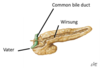
What does this show?


What are the causes of pancreatitis?
Inflammation (Pancreatitis)
- Causes:
- Gallstones
- High alcohol intake
- Cystic fibrosis
- High levels of calcium or blood fats.
What are the causes of pancreatic cancer?
- Obstructive jaundice (gallstones)
- High alcohol intake
- Smoking
Genetics
Whats the diagnosis?


Type 1 Diabetes Mellitus is caused by what issue within the pancreas?
A.Blockage of the Wirsung
B.Ischaemia of the pancreas
C.Lack of insulin production
D.Lack of glucagon production
E.Trauma to the organ
C. Lack of insulin production
What are the functions of the spleen?
Lymphoid organ
-Associated with the lymphatic system, and has an immune role
Blood gland
- Removes old blood cells
- Stores platelets
- Produces blood cells during foetal life
What does friable mean?
Spleen
Delicate and friable
-Friable means “easily crumbled”
What is the anatomy of the spleen?
Surrounded by a connective tissue (CT) capsule
The inner portion is known as parenchyma
-This contains red and white pulp
Red pulp - blood filled sinuses
White pulp - lymphatic tissue (think white blood cells)


What type of capillaries would you expect to see within the spleen?
A.Continuous
B.Sinusoidal
C.Fenestrated
Answer: B
Sinusoidal capillaries will allow easier movement of larger molecules from the blood.
What is visceral pain?
Pain felt from the organs (viscera)
Described as dull, aching, pressure etc.
What is the difference between visceral pain and somatic pain?
Visceral pain is poorly localised in comparison to somatic pain
-Due to innervation of structures
Somatic vs autonomic innervation
What is referred pain and what is it caused by?
-Where pain is felt in a different location to where organ or structure injured
Caused by visceral pain
A patient presents to A&E with pain in their shoulder. There is no surface lesion or MSK injury visible. Which of the abdominal organs could cause referred pain in the shoulder?
A.Ileum
B.Liver
C.Pancreas
D.Stomach
E.Spleen
Liver
What lobes do the caudate and quatrate lobes belong to?
Despite the obvious demarcation produced by the lesser omentum and falciform ligaments that suggest the quadrate and caudate lobes are part of the right lobe, functionally the quadrate lobe is regarded as part of the left lobe, since its arterial supply is from the left hepatic artery and bile from this lobe flows to the left hepatic duct. As for the caudate lobe, some sources consider it as a third lobe, since it receives blood (arterial and portal) from both the left and right side, and drains straight into the IVC rather than the left or right hepatic veins
The gall bladder and cystic duct are supplied by the _____ artery, usually a branch from the _______ artery. The lower part of the biliary tree – the common bile duct – is in turn, supplied by the _________ artery.
The gall bladder and cystic duct are supplied by the cystic artery, usually a branch from the right hepatic artery. The lower part of the biliary tree – the common bile duct – is in turn, supplied by the gastroduodenal artery.
The pancreas is a very useful landmark to not just locate but also distinguish between ______ and _________ arteries, the former branching from above and the latter from below this organ
The pancreas is a very useful landmark to not just locate but also distinguish between coeliac and superior mesenteric arteries, the former branching from above and the latter from below this organ
