Anatomy of hand Flashcards

1
Q

Palmar branch of both the median and ulnar Travels Superficial to the ______ _________ this portion of the median nerve usually remains functioning during carpal tunnel syndrome

A

flexor retinaculum

2
Q

The radial nerve: superficial branch to dorsal aspect of the

A

proximal 3 ½ lateral fingers (to about middle phalanges)

3
Q

The median nerve supplies the

A

proper palmar digital nerves to distal 3 ½ lateral fingers

4
Q

The ulnar nerve supplies the

A

Dorsal branch of the ulnar nerve: emerges from under the flexor carpi ulnaris tendon, on the medial aspect of the distal forearm, and winds around the wrist to provide cutaneous innervation to the dorsal aspect of the 1½ medial fingers

5
Q

Recall that the cephalic vein originates from the lateral aspect and the ______ from the medial aspect of the dorsal venous arch

A

basilic

6
Q

Median cubital vein joins the _______ vein to the basilic vein in the superficial fascia of the cubital fossa.

A

cephalic

7
Q

lymphatics: Lateral side: empty into the __________ which then drain to the apical nodes of the axilla

A

infraclavicular nodes

8
Q

lymphatics: Medial side of the hand and forearm can drain directly into the

A

lateral group of axillary nodes or to the cubital (supratrochlear) nodes before draining in the lateral group of nodes.

9
Q

Dupuytren’s Contracture

A

Is a localized thickening and contracture of the palmar aponeurosis. •A condition in which the medial part of the palmar aponeurosis undergoes progressive shortening •It commonly starts near the root of the ring finger and draws that finger into the palm, flexing it at the metacarpophalangeal joint. •Later, the condition involves the little finger in the same manner. •In long standing cases, the pull on the fibrous sheaths of these fingers (little and ring) results in flexion deformity of the proximal interphalangeal joints . •The distal interphalangeal joints are not involved and are actually extended by the pressure of the fingers against the palm. •Surgical division of the shortened part of the aponeurosis is done to straighten the bent fingers

10
Q

Attachments of Flexor retinaculum

A

Medially: hook of the hamate & pisiform bones, but volar carpal ligament encloses ulnar nerve and vessels. Laterally: to scaphoid & trapezium, but splits to enclose tendon of flexor carpi radialis tendon

11
Q

Structures passing superficial to Flexor retinaculum:

A

Ulnar nerve & vessels, palmaris longus tendon, palmar cutaneous branch of median and ulnar nerves, superficial palmar branch of radial artery

12
Q

Structures passing deep to Flexor retinaculum:

A

Median nerve, tendons of flexor digitorum superficialis and profundus with ulnar bursa, tendon of flexor pollicis longus with radial bursa.

13
Q

Extrinsic muscles are responsible for _____ movements of hand

A

crude

14
Q

intrinsic muscles control ____ movements of hand

A

fine

15
Q

Intrinsic Muscles of the Hand

A

-Thenar (thumb) -Hypothenar (little finger) muscles -Interossei muscles (four dorsally and three anteriorly) originating between the metacarpal bones -Lumbrical muscles arising from the deep flexor (and are special because they have no bony origin) to insert on the dorsal extensor hood mechanism.

16
Q

Thenar Eminence (Intrinsic Muscles of the Hand)

A
  • Abductor pollicis brevis - Flexor pollicis brevis - Opponens pollicis brevis All these muscles act on the thumb. They are all supplied by the median nerve
17
Q

Hypothenar Eminence (Intrinsic Muscles of the Hand)

A
  • Abductor digiti minimi - Flexor digiti minimi - Opponens digiti minimi All act on little finger They are supplied by ulnar nerve
18
Q

Lumbricals (Intrinsic Muscles of the Hand)

A

• Each lumbrical tendon inserts on the lateral side of the corresponding extensor (dorsal digital) expansion (on the dorsal aspect of the MCP joint). • First 2 lumbricals have one head whereas the 3rd and the 4th have 2 heads •Flex the MP joints and extend the IP joints. •1st and the 2nd are supplied by the median nerve, 3rd and 4th are supplied by the ulnar nerve.

19
Q

Dorsal Interossei (Intrinsic Muscles of the Hand)

A
  • bipennate - 4 dorsal and are numbered from lateral to medial side - Abduct the index, middle and ring fingers ( DAB), also flex the MP joints and extend the IP joints of these 3 fingers. - Innervated by ulnar nerve.
20
Q

Palmar Interossei (Intrinsic Muscles of the Hand)

A
  • unipennate - Are 4 (sometimes 3), numbered from lateral to medial side. -Adduct the index, ring and little fingers (PAD), also flex the MCP joints and extend the IP joints of these 3 fingers. - Innervated by ulnar nerve.
21
Q

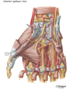
Superficial palmar arch: Formed by (Palmar Arches Deep & Superficial)

A

superficial branches of ulnar (2/3 rd ) and radial arteries (1/3 rd)

22
Q

Deep arch: Formed by (Palmar Arches Deep & Superficial)

A

deep branches of radial (2/3rd and ulnar arteries (1/3 rd)

23
Q

The superficial arch is very superficial, just deep to the __________ (Palmar Arches Deep & Superficial)

A

palmar aponeurosis.

24
Q

Laceration of palm can cause severe bleeding. -Bleeding can be controlled by clamping (Palmar Arches Deep & Superficial)

A

the brachial artery against humerus, not by compressing the radial or ulnar or both arteries (because of connection between palmar & dorsal arches)

25
Ulnar artery passes (superficial/deep) to the flexor retinaculum, (medial/lateral) to the ulnar nerve and pisiform (Superficial Palmar Arch)
superficial lateral
26
Radial artery swings from the anterior aspect of the wrist to the lateral side and then posterior aspect of the hand by passing beneath the tendons of ______________ and \_\_\_\_\_\_\_\_\_\_\_\_\_\_\_\_
abductor pollicis longus and extensor pollicis muscles (both longus and brevis)
27
Divides between the two heads of the first dorsal interosseous muscle to pass between the ________ and ________ heads of the adductor pollicis muscle.
transverse and oblique
28
Fascial spaces of the hand
Palmar spaces: 1) Superficial palmar spaces 2) Deep palmar spaces (divides into: Mid palmar space and Thenar space) 3) Pulp spaces
29
Deep Palmar Arch: deep to the _______ tendons but anterior to the metacarpal bones and \_\_\_\_\_\_
long flexor tendons interosseous muscles
30
Mid-palmar space Superficial Boundaries:
Palmar aponeurosis Superficial palmar arch Flexor tendons of medial 3 fingers
31
Mid-palmar space deep Boundaries:
Fascia covering 3rd & 4th interosseus space
32
Mid-palmar space medial Boundaries:
Medial palmar septum
33
Mid-palmar space lateral Boundaries:
Mid palmar septum
34
Mid-palmar space proximal Boundaries:
distal margin of the flexor retinaculum
35
Mid-palmar space distal Boundaries:
Distal palmar crease & continues with 3 rd & 4 th web space
36
Mid-palmar space distal Boundaries:
Distal palmar crease & continues with 3rd & 4th web space
37
Thenar space Superficial boundaries:
Palmar aponeurosis Superficial palmar arch Flexor tendons index finger
38
Thenar space deep boundaries:
Fascia covering adductor pollicis muscle
39
Thenar space medial boundaries:
Mid palmar septum
40
Thenar space lateral boundaries:
Lateral palmar septum
41
Thenar space proximal boundaries:
distal margin of the flexor retinaculum
42
Thenar space distal boundaries:
Distal palmar crease & continues with 1st web space
43
Incision for draining the abscess if Pus in the mid palmar space
drain 3rd or 4th web space
44
Incision for draining the abscess if Pus in the thenar space
drain 1st web space along the radial side of index finger.
45
Digital Pulp Spaces is the space between
skin & distal phalanx of each finger
46
Digital Pulp Spaces is the space between
skin & distal phalanx of each finger
47
Digital Pulp Spaces is Traversed by
fibrous septa & contain fat & blood vessels
48
Infection of the pulp space is known as
WHITLOW/FELON which is associated with severe throbbing pain
49
Infection of the pulp space is drained by (lateral/medial) incision.
lateral incision.
50
If Infection of the pulp space is neglected, it leads to _______________ of the distal 4/5th of distal phalanx (proximal 1/5 th remains unaffected).
“avascular necrosis”
51
Median Nerve Passes between the __________ and \_\_\_\_\_\_\_\_\_\_, deep to the palmaris tendon
flexor digitorum superficialis and the flexor carpi radialis
52
Median Nerve Passes under the \_\_\_\_\_\_\_\_
flexor retinaculum
53
Median Nerve divides into 2:
Common palmar digital nerves
54
Two common palmar digital branches dividing into
proper palmar digital nerves
55
Innervation to 1st and 2nd lumbrical
Median Nerve
56
Injury of median nerve at different levels cause different syndromes: At the wrist:
Injury by compression at the carpal tunnel causes carpal tunnel syndrome
57
Injury of median nerve at different levels cause different syndromes: In the hand,
thenar muscles are paralyzed and will atrophy over time.
58
Ulnar Nerve is Superficial to the \_\_\_\_\_\_\_\_but runs distally in a groove between the pisiform and the \_\_\_\_\_\_\_\_\_\_\_\_\_\_\_\_\_\_
flexor retinaculum hook of the hamate (tunnel of Guyon)
59
Ulnar Nerve In the palm, it divides into a _________ branch and a ______ branch
superficial and deep
60
Superficial branch of ulnar nerve: (innervations)
motor to a small muscle, the palmaris brevis, and cutaneous to the medial aspect of the medial 1 ½ fingers
61
Deep branch of ulnar nerve: to the (innervations)
3 hypothenar muscles, all dorsal and palmar interossei muscles, the 2 most medial lumbrical muscles and the adductor pollicis
62
Injury to Ulnar Nerve In the tunnel of Guyon:
lose the cutaneous innervation to the palmar aspect of their medial 1 ½ fingers and ability to contract all the intrinsic muscles of the hand, except for the 5 muscles innervated by the median nerve Flexor and extensor extrinsic muscles will be unopposed and the fingers will take the very characteristic position of the “clawed • All interossei are paralyzed: the MCP joint present in an extended position with both PIP and DIP joints in flexion • Only 2 medial lumbrical muscles are paralyzed: contribute to this position but note that the extension at the MCP and flexion at both the DIP and PIP of the index or ring and middle fingers will be less dramatic. Why? • Adductor pollicis brevis is non
63
label the following:
64
label the following:
65
label the following:
66
label the following:
67
label the following:
68
label the following:
69
label the following:
70
label the following:
71
label the following:
72
label the following:
73
label the following:
74
label the following:
75
label the following:
76
label the following: