Advanced 12-Lead EKG Flashcards
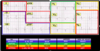
Bipolar Limb Leads

Augmented Limb Leads

Precordial Leads

Atrial hypertrophy

Diagnosis of right atrial hypertrophy
could be caused by tricuspid stenosis

Diagnosis of left atrial hypertrophy
caused by left heart failure d/t systemic HTN
mitral stenosis
aortic stenosis
massive MI
2nd half of humpity larger

Ventricular hypertrophy

Diagnosis of right ventricular hypertrophy

Diagnosis of left ventricular hypertrophy

Ischemia, Injury, and Infarct

Myocardial ischemia

Myocardial injury
- Signifies that the MI is “acute” as in right now! This is a STEMI; pain is heavy from arm to jaw, hard to breathe
- Pericarditis- looks like ST elevation but its all across the 12-lead and maybe can hear rub and the pain is different
- Prinzmetal’s- can look like ST elevation; but it’s more of a spasm; responds well to CCB’s

Reciprocal changes
- Supports diagnosis of STEMI
- Increased likelihood of complications
- High specificity
- Looking at opposite direction

Myocardial infarction (non-transmural) NSTEMI a little less severe than STEMI
- Temporarily/resolving
- Excessive supply/demand: stress test
- Prolonged
- St- depression
- If they have the MI story

Myocardial infarct (transmural
- “old” will have Q waves: (-) deflection (away from a (+) electrode
- Dead muscle doesn’t contract or depolarize
- Must be significant…1mm wide or 2mm deep or 1/3 QRS tall and 2 contiguous leads

Poor R wave progression-
old mi

Location of infarct
Coronary arteries of infarct

Axis deviation

right axis deviation
- QRS is negative in Lead I
- Vertical heart
- RVH
- Anterolateral MI

left axis deviation
- QRS positive in Lead I But negative in AVF
- Inferior MI
- Left ventricular hypertrophy
- Obesity
pregnancy

look at P wave for all things related to the
atrai
look at QRS for all things related to the
ventricle

left atrial hypertrophy


