Abnormal labour Flashcards
(45 cards)
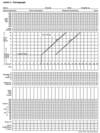
What do the top and bottom lines on this tracing indicate?

Top line - fetal heart rate
Bottom line - uterine contractions
How many contractions in ten minutes is this lady having?

3
How is the strength of contractions measured during labour?
Placement of an intrauterine pressure catheter
How is the strength of uterine contractions measured on the tracing?

Strength of contraction = amplitude of each wave

What pain relief options are avaliable in labour?
Support
Massage / relaxation techniques
Inhalational agents - Entonox
TENS (T10-L1, S2-S4)
Water immersion
IM opiate analgesia e.g. Morphine
IV Remifentanil PCA
Regional anaesthesia
What is TENS?
Transcutaneous electrical nerve stimulation that can provide pain relief in labour

What is the ‘attitude’ of the passenger/baby?
Flexion/extension of the baby

What observations are recorded in the partogram?
- Fetal Heart
- Amniotic Fluid
- Cervical Dilatation
- Descent
- Contractions
- Obstruction - Moulding
- Maternal Observations
Why is this labour failing to progress?

Contractions are weak, incoordinated and infrequent (only just reached 3 every ten mins)
Why is this labour failing to progress?

Cervix won’t dilate past 6cm
How often during stage one and stage two of labour should doppler auscultation of the fetal heart be performed?
•Stage 1:
During and after a contraction
Every 15 minutes
•Stage 2:
Every 5-10 minutes
What are the risk factors for fetal hypoxia?
Small fetus
Preterm / Post Dates
Antepartum haemorrhage
Hypertension / Pre-eclampsia
Diabetes
Meconium
Epidural analgesia
VBAC
PROM >24h
Sepsis (Temp > 38C)
Induction / Augmentation of labour
What are some of the acute causes for fetal distress?
- Abruption
- Vasa Praevia
- Cord Prolapse
- Uterine Rupture
- Feto-maternal Haemorrhage
- Uterine Hyperstimulation
- Regional Anaesthesia
What should be assessed on cardiotogograph (CTG)??

What is the normal fetal heartrate? What are normal and pathological variations seen on CTG?

What feature is being pointed out on this CTG?

Accelerations
What is the difference between early and late decelerations?
Early decelerations are normal and occur with contractions: this is due to increased foetal intracranial pressure causing increased vagal tone, and so resolves quickly after contraction
Late decelerations begin at the peak of uterine contraction & recover after the contraction ends: this type of deceleration indicates there is insufficient blood flow through the uterus & placenta, as a result blood flow to the foetus is significantly reduced causing foetal hypoxia & acidosis
.

What are variable decelerations?
Variable decelerations are seen as a rapid fall in baseline rate with a variable recovery phase
They are variable in their duration & may not have any relationship to uterine contractions

When are variable decelerations most often seen?
In labour and in patients with reduced amniotic fluid volume
What is the usual cause of variable decelerations?
Variable decelerations are usually caused by umbilical cord compression:
- the umbilical vein is often occluded first causing an acceleration in response
- then the umbilical artery is occluded causing a subsequent rapid deceleration
- when pressure on the cord is reduced another acceleration occurs & then the baseline rate returns
- accelerations before & after a variable deceleration are known as the “shoulders of deceleration”
- their presence indicates the foetus is not yet hypoxic & is adapting to the reduced blood flow
What does DR C BRAVADO stand for?
D ETERMINE
R ISK
C ONTRACTIONS
B ASELINE
R
A TE
V ARIABILITY
A CCELERATIONS
D ECELERATIONS
O VERALL IMPRESSION
What factors must be taken into consideration when ‘definining risk’ during a CTG reading?
Maternal medical illness e.g. asthma, diabetes, hypertension
Obstetric complications e.g. multiple gestation, post-date gestation, previous cesarean section, IUGR, PROM, congenital malformations, induction of labour, pre-eclampsia
Other risk factors e.g. no prenatal care, smoking, drugs
What should be recorded in the ‘contractions’ part of the CTG assessment?
Record the number of contractions present in a 10 minute period – e.g. 3 in 10
Each big square is equal to 1 minute, so you look how many contractions occurred in 10 squares
Individual contractions are seen as peaks on the part of the CTG monitoring uterine activity
You should assess contractions for the following:
- Duration: how long do the contractions last?
- Intensity: how strong are the contractions? (assessed using palpation)

How is the baseline fetal heartrate assessed?
Ignore accelerations or decelerations, just assess the average heart rate over the last 10 minutes


