A first look at the ECG Flashcards
What are your standard limb leads?

What does an approaching wave of depolarisation cause?
Upward going blip
Which events are better transmitted, fast or slow?
Fast
What is the PR interval and how long does it usually last?
Time from atrial depolarisation to ventricular depolarisation
Reflects transmission through AV node (0.12-0.20 sec)

What is the QRS interval?
Time for the whole ventricle to depolarise
(normally about 0.08 sec)
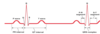
What is the QT interval?
Time for ventricles to depolarise and repolarise
(varies with heart rate, normally 0.42 sec at 60 bpm)

Why can’t you see atrial repolarisation?
Coincides with ventricular depolaristion
Ventricular depolarisation involves much more tissue depolarising much faster so it swamps any signal from atrial repolarisation

How do you explain the three stages of the QRS complex?

Different parts of the ventricle depolarise at different times and directions
- Interventricular septum depolarises from left to right
- Bulk of ventricle depolarises from endocardial to epicardial surface – travels towards electrode on left leg – (hence upwards spike)
- Upper interventricular septum depolarises
Why is the T - wave positive?
AP longer in endocardial cells than epicardial cells, wave of repolarisation runs in opposite direction to wave of depolarisation
i.e. wave of repolarisation moving away from recording electrode produces another positive-going blip

Why is the R-wave bigger in SLL II than in SLL I or SLL III?
Main vector of depolarisation is in line with the axis of recording from left leg with respect to the right arm.
What are the augmented limb leads?

What extra information do the augmented limb leads give you?
Gives you 3 other perspective on events in the heart
ie recordings from SLLs I, II, III and aVR, aVL, aVF give you 6 different views of events occurring in the frontal (or vertical) plane
Can you correctly label the vectors to the correct limb leads?


Should AVR be positive or negative?
Negative, travels away from the heart
Which wave is:
- aVL
- aVF
- aVR


What extra information do the precordial (chest) leads give you?
Main vector of depolarisation is shown by the arrow, will produce negative going blip when recorded from V1, positive going blip from V6, and flip over around V3/V4. “Progression”

Look

On which plane is the spread of depolarisation measured when using:
- Limb leads
- Precordial leads
- Frontal
- Transverse

What does the rhythm strip tell you?
Paper should run at 25mm/sec
Calibrating pulse is 0.2 sec = 1 large square (5mm) (5 arge squares per second)
can determine heart rate:
Measure R-R interval and work out how many occur in 60 sec
Count R waves in 30 large squares (= 6 sec) and multiply by 10
60-100 beats per min = normal
Below 60 beats per minute = bradycardia
Above 100 beats per minute = tachycardia

What does STEMI or NSTEMI stand for?
ST elevated myocardial infarction or non-ST elevated myocardial infarction.
ST should be flat because cells are in refractory state – st elevation is indication of severity of heart attack
More dead tissue means more elevation

