9/13 Posterior thigh and popliteal fossa Flashcards
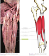
posterior thigh muscles

Posterior Thigh Muscles
Hamstrings: – Tendons posterior to knee are used to hang hams of pigs – “Hamstringing” enemy & their horse during ancient times
Common proximal attachment: – Ischial tuberosity, except short head of biceps femoris
Common innervation: – Tibial division of sciatic nerve, except short head of biceps femoris (common fibular portion)
Functions: – Thigh extension, except short head of biceps femoris – Leg flexion (all four)
posterior thigh muscles

Semitendinosus
- Long, cordlike tendon that begins ~2/3 of the way down thigh
- Function: Extend thigh; flex leg (med. rotate)
- Innervation: tibial division of sciatic nerve
Semitendinosus Attachments
- Ischial tuberosity
- Medial surface of superior aspect of tibia

Pes Anserinus

Semimembranosus
- Function: Extend thigh; flex leg
- Innervation: tibial division of sciatic nerve

- Ischial tuberosity: – Flattened membranous proximal attachment
- Posterior part of medial condyle of tibia
Semimembranosus Attachments
Distal tendon divides into 2 parts:
which are?
– Medial tibial condyle
– Part blends with popliteal fascia and becomes oblique popliteal ligament (reinforces the intercondylar part of joint capsule of knee)

Biceps Femoris
what are the two parts?
what is the innovation?
Long head: Flex leg; extend thigh, tibial division of sciatic nerve
Short head: Only flexed leg, common fibular branch of the sciatic

biceps femoris short and long heads

Biceps Femoris Attachments
Two heads:
- Long head – Ischial tuberosity
- Short head – Distal lateral lip of linea aspera
Common insertion on the head of the fibula
Adductor Magnus: “Hamstring” Portion
- Attaches proximally at ischial tuberosity and distally to the adductor tubercle on the medial epicondyle of femur
- Function: Extend thigh
- Innervation: Tibial division of sciatic nerve

Posterior Thigh Muscle Schematic

popliteal fossa
• Boundaries:
- Superomedially (semitendinosus & semimembranosus)
- Superolaterally (biceps femoris)
- Inferolaterally (lateral head gastrocnemius)
- Inferomedially (medial head gastrocnemius)
Popliteal fossa contents (superficial to deep):
– Nerves
– Popliteal vein, lymph nodes and branches
– Popliteal artery and branches

what is the deepest structure in the popliteal region?
from which artery? and gives rise to which artery?

Popliteal Artery
• Continuation of femoral artery:
– Becomes popliteal artery after passing through adductor hiatus
• Runs close to knee joint capsule:
– Gives rise to genicular branches…

Popliteal Artery: Genicular Branches
- Participate in formation of genicular anastomosis: – Important collateral circulation bypassing popliteal artery:
- Knee fully flexed too long
- Narrowed or occluded popliteal vessels
- Supplies articular capsule and ligaments of knee joint

genicular anastomosis

Femoral Artery Branches
“Put My Leg Down Please”
Profundus femoris (deep femoral artery)
Medial circumflex femoral artery
Lateral circumflex femoral artery
Descending genicular artery
Perforating Artery
Popliteal Artery: Termination Ends by dividing into:
- ______
- ______

Anterior tibial artery
Posterior tibial artery

Popliteal artery to termination

Popliteal Vein
Formed by union of anterior & posterior tibial veins, usually near inferior border of popliteus muscle.

Small saphenous vein terminates into the popliteal vein
- Lies superficial to and in same fibrous sheath as popliteal artery
- Becomes femoral vein after traversing adductor hiatus
Nerves in the Popliteal Fossa
Sciatic nerve usually ends at superior angle of popliteal fossa where it divides into the…
_____ and the _____ (peroneal) Nerve

Tibial Nerve and Common fibular

Tibial Nerve
- Most superficial (relative to popliteal artery and vein)
- Distribution:
– Superficial and deep posterior leg muscles
– Knee joint

Common Fibular (Peroneal) Nerve
- Leaves popliteal fossa by passing superficial to lateral head of gastrocnemius
- Winds around head and neck of fibula (susceptible to injury)
- Deep to fibularis longus it terminates into:
- Deep fibular nerve
- Superficial fibular nerve

what is the cutaneous nerve in the lower thigh region?
sural nerve
• Composed of…
– medial sural cutaneous nerve from the tibial nerve
– sural (or fibular) communicating branch from the common fibular nerve or the lateral sural cutaneous nerve
- Runs inferiorly with small saphenous vein
- Supplies distal posterior aspect of leg and lateral aspect of ankle & foot
sural nerve


sural nerve

