3a & 3b.) Somatosensory System Flashcards
State the three main ascending ascending somatosensory tracts in the spinal cord that you need to be aware of
- Dorsal column system
- Spinothalamic (anterolateral) system
- Spinocerebellar tract
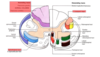
Which side of this diagram represents motor systems and which side represents sensory systems?

- Left= sensory
- Right= motor
State the two general types of senses
Describe any subdivisions of each
-
General sensory system : relying sensory information from body wall and viscera e.g. pain, pressure
- Somatic (input from body that is then consciously dealt with)
- Visceral (input from bdy that is not conciously/unconcioulsy dealt with)
- Special sensory system e.g. taste, smell
What do we mean by modalities?
Smallest unit/subtype of sensation
e..g. temperature, pain, crude touch etc… we can combine modalities to get different sensations e.g. stickiness
Each modality has a single type of receptor; true or false?
True
State the 7 modalities (of sensation)
Mediated by spinothalamic system:
- Temperature
- Pain
- Crude touch
Mediated by dorsal column system:
- Vibration
- Fine touch
- Proprioception
- 2-point discrimination
Describe the difference between crude touch and fine touch
- Crude touch refers to sensations from stimulation of tactile receptors of low sensitivity with large receptive fields e.g. elbow someone in arm
- In contrast, fine touch refers to tactile receptors of high sensitivity with small receptive fields e.g. draw number on somones hand
Describe how you can test two point discrimination
- Get paperclip and spread apart
- Touch person with it
- Keep moving the ends closer together and re-touching person
- Determine at which point they can’t tell that it is two separate points

State which modalities are mediated by:
- Spinothalamic (anterolateral) system
- Dorsal column system
Mediated by spinothalamic system:
- Temperature
- Pain
- Crude touch
Mediated by dorsal column system:
- Vibration
- Fine touch
- Proprioception
- 2-point discrimination

Given that the intensity of a sensory signal (e.g. someone touching you) is an analogue signal- what must happen, and where, to enable nervous system to convey this signal?
- The intensity of a sensory signal e.g. someone touching you is analogue (as it is related to ion influx during generator potential)
- Receptor smust covert this into a digital signal since the nervous system uses digital signals
What is meant by the receptor potential?
Descirbe how the signal will differ for a low pessure touch and a high presure touch
- Receptor/generator potential is the depolarisation that occurs in a sensory receptor in response to a stimulus and causes an action potential to be formed.
- Receptor potential is graded according to it’s intensity e.g. hard/high pressure touch will have high intensity, light/low pressure touch will have low intensity
- Strong receptor activation results in high frequency of action potentials
- Weak receptor activation results in low freqency of action potentials

State which type of receptor is involved in each of the modalities of somatic sensation
Spinothalamic (anterolateral) system:
- Temperature: thermoreceptors
- Pain: nociceptors
- Pressure/crude touch: mechanoreceptors
Dorsal column-medial lemniscus system:
- Vibration: mechanoreceptors
- Proprioception/joint poisition sense, kinaesthetic sense: variety of receptors e.g. muscle spindles, golgi tendon organs
- Fine touch: mechanoreceptors
- Two point discrimination: mechanoreceptors
Receptors can be rapidly adapating or slowly adapting; describe the difference between the two and provide an example for each
Rapidly Adapting
- Frequency of firing diminishes rapidly after initial stimulus e.g. mechanoreceptorss- why you are not aware of the clothes you are wearing
Slowly Adapting
- Change their frequency of firing very little after the initial stimulus e.g. nociceptors- this explains why pain is persistent and you don’t really get used to having pain (protective mechanism!)

What is meant by a receptive field?
Given area of skin that a single primary sensory neurone supplies
*NOTE: receptive fields can overlap

Describe the relationship between acuity and size of receptive field
Acuity is inversely proportional to the size of the receptive field
E.g. if a sensory neurone has a relatively large receptive field it will have low sensory acuity (this could be confirmed by poor two point discrimination)
Describe the relationship between acuity and the number of sensory neurones
Acuity is proportional to the number of sensory neurones
*Think, this kind of makes sense, if there are a large number of sensory neurones to an area you would assume they all have relatively small receptive fields and we know receptive field size in inversely proportional to acuity
Receptive fields of primary sensory neurones can overlap; what are the consequences of this?
Adjacent dermatomes can have ‘fuzzy’ boundaries
*This is why when testing a dermatome you must always test an autonomous region i.e. region you know belongs to that dermatome and is not near a boundary
There is a chain of three neurones in the somatosensory system; state these three neurones
- First order sensory neurones
- Second order sensory neurones
- Third order sensory neurones
For first order sensory neurones, describe:
- Whether they communicate directly with receptor
- Where their cell body is
- Where their central axon projects to in spinal cord
- What neurones they project on to
- Communicate directly with receptor
- Cell body is in a dorsal root ganglia (DRG)
- Central axon projects into cord ipsilaterally
- Projects onto secondary neurone

For second order sensory neurones, describe:
- Where their cell body is
- Whether they decussate
- What they project onto
- Cell bodies in either the dorsal horn of the spinal cord (spinothalamic system) or the medulla (dorsal column system)
- Decussate
- Project onto third order neurones

For third order sensory neurones, describe:
- Where their cell bodies are
- Where they project to
- Cell boies in thalamus
- Project to primary somatosensory cortex through internal capsule (post central gyrus) *NOTE: whether they project onto medial side of primamry somatosensory cortex or lateral side depends on whether they are upper or lower limb: due to homunculus

Remind yourself of the cortical homunculus (think about subtle difference between motor and sensory homunculus)

As we move upwards through the neuraxis what happens to information *HINT: some reorganisation occurs
Information becomes reorganised such that at level of spinal nerves and spinal cord we have dermatomal organisation but at the levels of the thalamus and above we have homuncular pattern
At the level of the sensory homunculus what happens to all modalities?
All modalities converge at level of the sensory homunculus
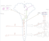
Describe the path of each of the neurones involved in the dorsal column-medial lemniscus system (DCML)
Primary order neurone
- Cell body in DRG
- Axons enter spinal cord on ipsilateral side and ascend to medulla
- 1st order neurones from T7 and below ascend through gracile fasciculus to gracile nucleus in medulla)
- 1st order neurones from T6 and above ascend through cuneate fasciculus to cuneate nucleus in the medulla
Second order neurone
- Cell body in medulla (in either gracile or cuneate nucleus)
- Decussate to contralateral side
- Ascend in contralateral side through the medial lemniscus
Third order neurone
- Cell body in thalamus
- Project to post-central gyrus/primary somatosensory cortex through the internal capsule (P.S.C):
- Neurones from lower limb project to medial part of P.S.C
- Neurones from upper limb project to lateral part of P.S.C

Describe the topographical organisation of dorsal columns (e.g. in dorsal column where do axons from lower limb and axons from uppper limb run?)
- Axons from lower parts of body: run most medially
- Axons form upper parts of body: run most laterally

Describe the topographical organisation in the medial lemniscus (e.g. in medial lemniscus where do axons from lower limb and axons from uppper limb run?)
- Lower limb: laterally (were medial in dorsal column but now they have decussated they are lateral)
- Upper limb: medially (were lateral in dorsal column but now they have decussated they are medial)
In diagrams of both the dorsal column medial lemniscus pathway and spinothalamic pathway you see the third order neurones cross each other; is this a decussation?
NOT a decussation, represents how fibres twist as they ascend the internal capsule
Describe the difference between sensory ataxia and cerebellar ataxia
- Sensory ataxia: loss of voluntary co-ordination due to sensory dysfunction
- Cerebellar ataxia: loss of voluntary co-ordination due to cerebellar dysfunction
Describe how a patient with severe sensory ataxia may stand and walk
- Can only stand unsupported with feet well apart and gaze directed downward to include feet
- Gait is broad based with stomping action that maximises any conscioius proprioceptive function that remains
How can you differentiate between sensory ataxia and cerebellar ataxia?
- Romberg sign: get patient to stand with feet together and eyes closed; if they start to sway severely this is a positive Romberg’s sign and indicates loss of proprioception
Suggest why spinothalamic tract is well developed in most animals
Spinothalamic tract is responsible for life presevering modalities e.g. pain, temperature and crude touch. These are senses that all animals need to survive
Describe the path of each of the neurons in the spinothalamic pathway
First order neuron
- Cell body in DRG
- Axons project to ipsilateral spinal cord
- Project onto second order neurones in the dorsal horn in the segment at which they enter the cord
Second order neuron
- Cell bodies in dorsal horn (in segment/at level the 1st order neurone entered the cord)
- Axons decussate in ventral white commissure of cord and then go on to form the spinothalamic tract
- Spinothalamic tract projects to thalamus
Third order neuron
- Thalamic neurones receiving information from more inferior/lower body parts projects to medial part of primary somatosensory cortex/post-central gyrus
- Thalamic neurones receiving information from more superior/upper body parts projects to lateral part of primary somatosensory cortex/post-central gyrus

Describe the topographical organisation in the spinothalamic tract (e.g. in spinothalamic tract where do axons from lower limb and axons from uppper limb run?)
- Lower parts of body= most laterally
- Upper parts of body= most medially
*Axons form more superior body parts are progressively added medially

In the dorsal column medial lemniscus pathway, axons from lower limb are found more medially and axons from upper limb are found more laterally. However, in spinothalamic tract, axons from lower limb are found more laterally and axons from upper limb are found more medially. Explain this difference
- In dorsal column medial lemniscus pathway, secondary neurones decussate higher up/in or just above medulla
- In spinothalamic pathway, secondary neurones decussate at level of entry of first order neurones
What is the relevance of Lissauer’s tract in the spinothalamic system?
Some of primary neurones in spinothalamic system can ascend a few of segment and project onto a secondary neuron a few segments higher e.g. C5 primary neuron could travel in Lissauer’s tract to project onto C3 secondary neuron

Lissauer’s tract contains both ascending and descending pathways; true or false?
True
What is the dorsal spinocerebellar tract responsible for?
Proprioception/joint position sense
What is meant by dissociated sensory loss?
Defined as a pattern of neurological damage which involves selective loss of fine touch and proprioception without loss of pain and temperature, or vice versa.
What is Brown-Sequard syndrome?
What structures will be destroyed?
What will the symptoms be?
- Injury to one side of spinal cord in which spinal cord on that side is damaged but the spinal cord itself is not severed completely (in othe words a complete cord hemi-section which could be of a single segment or a more)
- Following structures would be destroyed unilaterally:
- Dorsal horn
- Ventral horn
- All other cord greymatter
- All white matter pathways
- Dorsal and ventral roots
- Symptoms/signs:
- Ipsilateral complete segmental anaesthesia affecting singel dermatome (due to destruction of dorsal root & horn)
- Ipsilateral loss of dorsal column modalities below destroyed segment
- Ipsilateral loss of motor function
- Contralateral loss of spinothalamic modalities at and below the destroyed segment (although level can be up to a couple of segments lower due to ascent of some of primary afferents in Lissauer’s tract)

In the control of pain, what do A fibres carry and what do C fibres carry?
- A fibres carry impulses from mechanoreceptors
- C fibres carry impulses from nociceptive receptors (hence carry pain)

Explain why rubbing a sore area can relieve pain
- A fibres from mechanoreceptors project onto inhibitory encephalinergic interneurones
- Impulses from mechanoreceptors can activate these inhitory encephalinergic interneurones causing them to release the endorphin encephalin
- They can then inhibit signal transduction of pain in secondary neuron (remember afferent/first order C fibres carry pain signal to secondary neurone)

Can higher centres in CNS prevent body from feeling pain?
Encephalinergic inhibitory interneurons can be activated by descending inputs from higher centres such as the periaqueductal grey matter or nucleus of raphe magnus

Where do secondary neurones in spinothalmic pathway decussate?
Ventral white commisure
