2 - Stroke Related Neuroanatomy-Neurophysiology Flashcards

1
Q

What anatomical direction is this?

A

Axial

2
Q

What anatomical direction is this?

A

Coronal

3
Q

What anatomical direction is this?

A

Sagittal

4
Q

What anatomical direction are these?

A

A = Superior/Dorsal

B = Posterior/Caudal

C = Inferior/Ventral

D = Anterior/Rostral

5
Q

Name the Layers of the Brain.

A

A = Scalp

B = Cranium

C = Dura Mater

D = Arachnoid

E = Subarchnoid Space

F = Pia Mater

G = Cerebral Cortex

6
Q

Name the 4 Major Lobes of the Brain.

A

Frontal

Parietal

Temporal

Occipital

7
Q

What Is “Controlled” By These Specific Areas Of The Brain?

A

A = Motor Control

B = Cognition, planning, + problem solving

C = Speech

D = Smell

E = Hearing

F = Facial recognition

8
Q

What Is “Controlled” By These Specific Areas Of The Brain?

A

A = Touch + pressure

B = Taste

C = Body awareness

D = Language

E = Reading

F = Vision

G = Cerebellum

9
Q

What are these neruo sulci?

A

A = Precentral

B = Superior frontal

C = Inferior Frontal

D = Lateral frontal (Sylvian Fissure)

E = Superior temporal

10
Q

What are these neruo sulci?

A

A = Central (Rolandic)

B = Postcentral

C = Intraparietal

D = Lateral occipital

E = Lunate

F = Interior temporal

11
Q

What is the homunculus?

A

An metaphorical representation of the way motor + sensory information is organized neurologically

12
Q

What is the motor area of the brain called?

A

Precentral Gyrus

13
Q

What is the sensory area of the brain?

A

Postcentral Gyrus

14
Q

Which of Brodmann’s Areas are important to SLPs?

(4)

A

44 + 45 (Broca’s)

22 + 40 (Wernicke’s)

41 + 42 (Auditory Association)

39 (Angular Gyrus)

15
Q

What happens when Brodmann’s Areas 44 + 45 are injured?

(2)

A

Broca’s Aphasia

Apraxia of Speech

16
Q

What happens when Brodmann’s Areas 22 + 40 are injured?

A

Wernicke’s Aphasia

17
Q

What happens when Brodmann’s Areas 41 + 42 are injured?

A

Processing issues in Wernicke’s Aphasia

18
Q

What happens when Brodmann’s Area 39 is injured?

(2)

A

Acalculia

Agraphia

19
Q

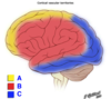
Label the following neruo landmarks.

A

A = Anterior cingulate cortex

B = Ventromedial prefrontal cortex

C = Orbitofrontal prefrontal cortex

D = Dorsolateral prefrontal cortex

20
Q

What is the ACC?

A

Anterior cingulate cortex

21
Q

What does the ACC do?

(4)

A

Reward anticipation

Decision making

Empathy

Emotions

22
Q

What happens when there is damage to the ACC?

(2)

A

Apathy

Poor motivation

23
Q

What is the VMPC?

A

Ventromedial prefrontal cortex

24
Q

What does the VMPC do?

A

Processing of risk + fear

25
What happens when there is damage to the VMPC? (2)
Poor inhibition Poor decision making
26
What is the OFPC?
Orbitofrontal prefrontal cortex
27
What does the OFPC do? | (2)
Empathy Civil + appropriate social behavior
28
What happens when there is damage to the OFPC? (5)
Impaired emotional reactivity + processing Personality change Poor impulse control Increased aggression Mood issues
29
What is the DLPC?
Dorsolateral prefrontal cortex
30
What does the DLPC do?
Organizing behavior to solve complex problems (new learning, searching memory)
31
What happens when there is damage to the DLPC? (5)
Poor organization strategies Poor word list generation Poor word fluency Poor sorting behavior Poor concreteness
32
What is the Insula Cortex? What does it play a large role in?
Patch of cortex behind the frontal, parietal, and temporal lobes Expressive speech + motor output
33
What is the Arculate Fasciculus? What is it important for?
Association fibers connecting the temporal lobe (Wernicke's) to the frontal lobe (Broca's) Language processing
34
What is the Basal Ganglia? What does it do?
Area within the diencephalon (top of brain stem) that receives input from multiple sites in cortex Refines muscle movements
35
What can happen if the Basal Ganglia is damaged? (3)
Problems with movement + sensation Loss of voluntary movement Change in appearance of involuntary movements
36
What is the Thalamus? What is it believed to do? (2)
Two egg shaped nuclei located in the diencephalon (top of brain stem) next to basal ganglia Major relay center for descending motor information and ascending sensory information Regulates overall electrical energy in cortex
37
What does the Thalamus play an important role in? (3)
Maintaining consciousness, alertness, + attention
38
What does the Internal Capsule contain? (2) What supplies blood to this area?
Both ascending + descending axons Fibers coming to and from the cerebral cortex Lenticulostriate arteries
39
Label the following arteries.
A = Anterior cerebral artery B = Posterior cerebral artery C = Internal carotid artery D = Basilar artery E = Vertebral artery
40
Label the following arteries.
A = Circle of Willis B = (Right) Middle Cerebral Artery C = Basilar Artery D = (External) Carotid Arteries E = Vertebral Arteries
41
Label the following arteries.
A = Anterior Cerebral Artery B = (Left) Middle Cerbral Artery C = Posterior Cerebral Artery D = Internal Carotid Arteries
42
What major arteries suppy the following areas?
A = Anterior cerebral artery B = Middle cerebral artery C = Posterior cerebral artery
43
What major arteries supply the following areas.
A = Anterior cerebral artery B = Middle cerebral artery C = Posterior cerebral artery
44
Label the following arteries.
A = Anterior cerbral artery (ACA) B = Lenticulostriate arteries C = MCA inferior division D = MCA superior division
45
What are the following areas called? What are they?
Watershed areas Border zones between the territories between two major arteries in the brain
46
Fill in the following chart.
A = Silent CNS Infarction B = Ischemic Stroke C = TIA D = Ischemic Stroke
47
What is a CNS Infarction? | (2)
Brain, spinal cord, or retinal cell death due to ischemia. Can also be cause by hemorrhage
48
What is an Intracerebral Hemorrhage?
Focal collection of blood within the brain
49
What is a Subarchnoid Hemorrhage?
Bleeding within the subarchnoid space
50
What is the difference between Ischemia + Infarction?
Ischemia = Physiological tissue changes Infarction = Tissue death
51
Do similiar neuro injuries always result in the same loss of function?
No
52
What area of the brain will result in Expressive Language deficits?
Anterior damage
53
What area of the brain will result in Receptive Language deficits?
Posterior damage
54
What is an Ischemic Stroke?
One cause by blockage
55
What is an Hemorrhagic Stroke?
One cause by bleeding
56
What is a Thrombosis Ischemia? How fast do these occur? What is an added risk?
Stroke caused by a build up of plaque or a blood clot Slower since the build up of the clot is gradual An area of the blockage can break off and cause an embolic stroke
57
What is a Embolism (Ischemia)? | (2)
Stroke cause when a piece of plaque or a blood clot breaks off This piece travels to a vessel that is too small for it to pass and it causes a blockage
58
What is Hypoperfusion (Ischemia)? What will it cause? What specific areas may also be affected?
Damage that occurs when blood flow is decreased to all parts of body, but is not fully stopped Brain damage Watershed areas
59
What is an Intracerebral Hemorrhage?
Bleeding within the brain
60
What is an Extracerebral Hemorrhage?
Bleeding occuring within the meninges
61
What kind of stroke is this?
Ischemic
62
How common are Ischemic Strokes? Intracerbral Hemorrhage? Subarchnoid Hemorrhage?
87% 10% 3%
63
What kind of strokes are the following?
A = Ischemic B - Hemorrhage
64
Where do Infarctions tend to occur?
Closest to the clot
65
Where do Ischemias tend to occur?
Further from the clot where it might receive a secondary blood supply from a nearby artery
66
What is the Physiological response to an Ischemic Stroke? (5)
Edema Transneural degeneration Denervation hypersensitivity Diaschisis Collateral sprouting
67
What is Edema?
Tissue swelling
68
What is Transneural Degeneration?
Neuron death due to disruption of input/output of nearby neurons
69
What is Denervation Hypersensitivity?
Extreme sensitivity due to interruptions of neural supply/activity
70
What is Diaschisis?
Distal loss of function due to connection in damaged area
71
What is Collateral Sprouting?
When neurons adjacent to damaged area take over some of the lost function
72
Are stroke a singular event? | (2)
No - they often reoccur They can last over several days
73
What is an Ischaemic Core?
The brain tissue near a blockage that is destined to die
74
What is a Penumbra?
The brain area near a blockage that is salageable
75
What do clots normally form around?
Atherosclerotic plaques
76
What is a clot called?
Thrombus
77
What is a traveling particle in the blood stream called?
Embolus
78
What are treatment options of Ischemic Strokes? (3)
Carotid Endarterectomy Carotid Stenting Tissue Plasminogen Activator (tPA - medication)
79
What is a Carotid Endarterectomy?
A surgical removal of the plaque causing a blockage
80
What is a Carotid Stent? What are the risks?
A device inserted at the plaque build up that expands the vessel Part of the clot breaks off or the vessel wall is weakened
81
What is Tissue Plasminogen Activator (tPA )? When is it a treatment option?
A clot dissolving medication Within 2-4 hours of an ischemic stroke
82
What is the most common Extracerebral Hemorrhagic Stroke?
Subarachoid (3%)
83
How often do Intracerbral Strokes occur?
7-9%
84
What are the major risk factors for Hemorrhagic Stroke? (4)
AA ethnicity Hypertension (HTN) High blood pressure LDL (bad) cholesterol + triglyerides inversely related to ICH (good)
85
What are some risk factors NOT associate with Hemorrhagic Stroke? (7)
Sex Smoking Alcohol intake BMI Waist-to-hip ratio Waist circumference Diabetes
86
What is the BIGGEST risk factor for Hemorrhagic Stroke?
Blood pressure
87
Can infraction occur with a hemorrhagic stroke?
Yes due to pressure on the tissue
88
Where are Intracerebral Hemorrhages most likely to occur? (5)
Central lobes Basal ganglia Thalamus Pons Cerebellum
89
What is an Aneursym?
A swelling of a blood vessel
90
What is an Anterior Venus Malformation (AVM)? What does this increase the risk of?
A congenital condition where arteries and veins grow together into a web Risk of vessel rupture
91
What are some medical treatments for Hemorrhagic Stroke? (2)
Microsurgical clipping Cerebral Stenting
92
What is Microsurgical Clipping? How is it inserted?
Small metal clip is place at the neck of the aneurysm to stop blood flow Inserted via craniotomy (opening the skull)
93
What is Cerebral Stenting? Why does it work?
Coils of platinum wire are passed through a catheter to fill the aneurysm The coils fill the aneursym and cause blood to clot obliterating the aneursym
94
What are TIA's also referred to as?
Mild Strokes (May need to clarify with patient)
95
What is CSF Leakage? How is it treated?
Too much cerebral spinal fluid is produce putting excessive pressure on the brain Extra fluid is removed via a shunt