2. Myology (Olinger) Flashcards
Identify the blacked out structures.


Which nerves innervate the anterior, middle, and posterior scalene muscles?
Anterior scalene: C4-C6.
Middle scalene: C3-C8.
Posterior scalene: C6-C8.
What innervates the anterior belly of the digastric muscle?
The nerve to the mylohyoid.
What are the infrahyoid muscles?
Sternothyroid muscle.
Thyrohyoid muscle.
Sternohyoid muscle.
Omohyoid muscle.
What is the innervation to the geniohyoid?
Fibers from C1, which travel with the hypoglossal nerve.
What is the innervation to the stylohyoid?
The facial nerve.
What is the innervation to the thyrohyoid muscle?
Fibers from C1 by the hypoglossal nerve.
What are the anterior borders for the superior, middle, and inferior pharyngeal constrictor muscles?
Where do they all attach posteriorly?
Anterior borders.
Superior – pterygomandibular raphae
Middle – hyoid
Inferior – thyroid cartilage
All three attach to the pharyngeal raphae posteriorly.
Identify the following blackedout structures.


Identify the blacked out infrahyoid muscles.


In which triangle is the thyroid gland contained?
Within the muscular triangle – a sub triangle of the anterior triangle of the neck
Label the following muscles.
In which triangle are they found?


What is congenital torticollis?
CN: Congenital Torticollis is a disorder produced by fibrous tissue tumor which forms in the Sternocleidomastoid M. which causes the head to turn and the face to look away from the affected side. A hematoma can arise and impinge on the Spinal Accessory N. which denervates the Stenocleidomastoid M.
What is the innervation to the posterior belly of the digastric muscle?
The facial nerve.
What are the two divisions for zones I, II, and III of the neck?
The angle of the mandible and the cricoid cartilage.
What innervates the sternocleidomastoid?
The spinal accessory nerve.
Identify the following posterior aspect laryngeal muscles.


What is the innervation for all of the laryngeal muscles except for the cricothyroid?
Recurrent laryngeal nerve.
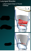
Identify the following muscles on the lateral aspect of the larynx, deep to the thyroid cartilage.


What innervates the anterior vertebral column muscles?
Anterior rami of the cervical spinal nerves.
Label the following three structures.


Where does longus capitis insert?
On the skull anterior to the foramen magnum.
Identify the following triangles and listed borders.


Identify the blacked out suprahyoid muscles.


What four muscles make up the suprahyoid muscles?
Mylohyoid M.
Geniohyoid M.
Stylohyoid M.
Anterior belly of the digastric M.
Posterior belly of the digastric M.
What are the contents of the submandibular triangle?
Facial artery.
Submental artery.
Submandibular gland.
Hypoglossal nerve.
Mylohyoid nerve.
What are the muscles of the anterior vertebral column?
Longus colli (superior oblique, vertical, inferior oblique).
Longus capitis.
Rectus capitis anterior.
Rectus capitis lateralis.
How can you tell apart the stylohyoid muscle and the posterior belly of the digastric muscle?
The stylohyoid muscle remains muscular throughout its entire presentation.
The posterior belly of the digastric muscle has a tenderness portion near its insertion.
What is the origin and insertion for rectus capitis anterior and rectus capitis lateralis?
Both go from the atlas to the occipital bone.
What are the contents of the carotid triangle?
Hypoglossal nerve.
Internal jugular vein.
Vagus nerve.
External carotid artery.
Common carotid artery.
Ansa cervicalis.
Identify the blacked out contents of the carotid triangle.


What is spasmodic torticollis?
Adult onset torticollis which involves abnormal tonicity of the cervical muscles - usually sternocleidomastoid and trapezius.
Identify the blacked out origins and insertions for the pharyngeal muscles.


Identify the blocked out muscles on the anterior vertebral column.


What is significant about zone II in terms of neck wounds?
Zona II is the most dangerous, but also the most easy to access surgically.
What are the laryngeal muscles?
Cricothyroid muscle.
Posterior cricoarytenoid muscle.
Lateral cricoarytenoid muscle.
Transverse arytenoid muscle.
Thyroarytenoid muscle.
Oblique arytenoid muscle.
(Aryepiglottic part of the oblique arytenoid muscle.)
Vocalis muscle.
What are the attachment points for the salpingopharyngeus muscle?
The pharyngotympanic tube and the pharynx.
What are the contents of the muscular triangle?
Sternohyoid muscle.
Sternothyroid muscle.
Thyroid gland.
Parathyroid gland.
What are the pharyngeal constrictor muscles?
What innervates these muscles?
The pharyngeal constrictor muscles include the superior, middle, and inferior pharyngeal constrictor muscles.
They are innervated by the pharyngeal branch of the vagus nerve and pharyngeal plexus.
What is this muscle?
What are the contents of the posterior triangle?
Levator scapulae muscle.
Anterior, middle, and posterior scalene muscles.
The inferior belly of the omohyoid muscle.
Splenius Capitis M.
Identify the blacked out contents of the anterior muscular triangle.


What is the innervation to all of the infrahyoid muscle except for the thyrohyoid?
The ansa cervicalis.
Identify the blacked out pharyngeal muscles.


What is the innervation to the platysma?
A branch of the facial nerve.
What is the innervation of the mylohyoid?
Nerve to the mylohyoid.
What are the three parts of longus colli?
A superior oblique part, a vertical part, and an inferior oblique part.
What is the innervation for the cricothyroid muscle?
The external laryngeal nerve.
What are the boundaries of the posterior triangle of the neck?
The sternocleidomastoid, the trapezius, and the clavicle.
Label the following structures of the posterior triangle.

