18 - Peripheral and Arterial Vascular Disease Flashcards
What are the three conditions in peripheral arterial disease? (PAD)
- Intermittent claudication
- Critical limb ischaemia
- Acute limb ischaemia
If a patient presents with an acutely painful limb presents with the following features, what are the main differentials you think of?
- Cold and Pale
- Hot and Swollen
- Traumatic history
- Neurological signs
- Cold and Pale: acute limb ischaemia
- Hot and Swollen: DVT, cellulitis, MSK related infections
- Traumatic Hx: fractures
- Neurological signs: radiculopathy, MS (central), disc herniation (spinal), infection (peripheral)
What are the symptoms of acute limb ischaemia?
- Pulseless
- Pain
- Pallor
- Paraesthesia
- Perishingly cold
- Paralysis
Top three are usually first to present
How do you investigate and manage a suspected acute limb ischaemia in general terms?
EARLY INVOLVMENT OF VASCULAR TEAM
Ix
- Examine contralateral limb for comparison
- Look at underlying risk factors e.g AF, DM, smoking, HTN
- Arrange CT angiogram and urgent vascular review
Mx
- Emergency as irreversible tissue damage can occur in six hours
- Start on IV heparin
- Analgesia

How do you investigate and manage a DVT in general terms?
Ix
- Swollen hot limb with pain localised to calf
- Calculate Well’s score, if 2 or more do US Doppler
- If <2 do D-dimer
Mx
- Start apixaban or rivaroxaban for 3-6 months. If Cx start LMWH for 5 days first then switch to dabigatran for 3 months
- If iliofemoral DVT then urgent vascular review

What is the clinical difference between a politeal vein DVT and an iliofemoral DVT?
- Popliteal: pain, swelling and tenderness localised to calf, conservative management with LMWH and DOACs
- Iliofemoral: pain and swelling in whole leg, may be blue or white leg, needs urgent vascular review

If a patient presents with an acutely painful limb you should consider neurological pathology like radiculopathy. What would the clinical picture be if this was the underlying cause?
- Back pain that radiates to affected area
- Pain worse on movement
- Muscle weakness
- Paraesthesia
- Altered reflexes

How do you assess, investigate and manage a patient that presents with an acutely swollen limb?
- Accurate history
- Vascular and neurological exams of both limbs
- Ensure patient haemodynamically stabilised
- Look for red flags
- CT angiography if suspect acute limb ischaemia
- Routine bloods with G+S
- Analgesia

What are the different types of lower limb ulcers?
- Venous, Arterial, Neuropathic
- Most lower limb ulcers have venous origin
- Can also be caused by trauma, vasculitis, SCC malignancy
- Can also be a pressure sore (prolonged excessive pressure over a bony prominence)

How are pressure ulcers managed in hospital generally?
- Adequate mattress
- Repositioning
- Good wound management
What is the pathophysiology of a venous ulcer?

- Due to venous insufficiency
- Shallow with irregular borders and a granulating base and often found over medial malleolus. Prone to infection and cellulitis
- Due to valvular incompetence so impaired venous return with resultant venous hypertension. Trapping of WBC in capillaries and formation of fibrin cuff around vesel hindering oxygen transport to tissue
- WBC also release inflammatory mediators so tissue injury, poor healing and necrosis

What are some risk factors for developing a venous ulcer?
- Increasing age
- Pre exiting venous incompetence (e.g varicose veins) or previous DVT
- Pregnancy
- Obesity
- Severe leg injury

What are the clinical features of a venous ulcer and how do you investigate them?
Features:
- Painful with aching, itching or burning before ulcer appears
- May have varicose veins and ankle oedema
- May have varicose eczema, thrombophlebitis, haemosiderin skin staining, lipodermatosclerosis or atrophie blanche
Ix:
- Clinical
- Do Doppler US to confirm venous insufficiency, usually at saphenofemoral or saphenopopliteal junction
- Ankle Brachial Pressure index to assess arterial component to see if compression therapy would help
- Take swab cultures if infection
- Consider thrombophilia or vasculitic screening in younger patients

How are venous ulcers managed?
Conservative
- Leg elevation and increased exercise to promote calf pump
- Lifestyle changes e.g weight loss, improved nutrition
- Abx if swabs so infection
Definitive
- Multicomponent compression bandaging changed one or twice a week for about 6/12. Need ABPI to be >0.8 before any bandaging applied
- Use emollients to keep skin intact
- If concurrent varicose veins treat with endovenous techniques or open surgery as improving venous return helps heal ulcers

What are the risk factors for developing an arterial ulcer?
Reduction in arterial blood flow so decreased perfusion of tissues.
Same risk factors for peripheral arterial disease:
- Smoking
- DM
- HTN
- Hyperlipidaemia
- Increasing age
- Obesity
- Inactivity

What are the clinical features of an arterial ulcer?
- Small deep lesions with well defined borders and a necrotic base with no granulation tissue
- Found at pressure points and sites of trauma
- Preceding history of intermittent claudication (pain on walking) or critical limb ischaemia (pain at night)
- Limbs often cold and pulseless but sensation maintained
- Often have limb hair loss

How are arterial ulcers investigated and managed?
Ix

- Do ankle brachial pressure index to quantify extent of any peripheral arterial disease. (>0.9 normal, <0.5 severe)
- Can do duplex US, CT angiography or MRA to find location of arterial disease
Mx
- Urgent vascular review
- Conservative: lifestyle changes like weight loss, stop smoking, increase exercise
- Medical: statin, antiplatelet (aspirin or clopidogrel) and optimise BP and glucose
- Surgical: angioplasty or bypass grafting if extensive
What are the risk factors for neuropathic ulcers?
Anything that causes peripheral neuropathy:
- B12 Deficiency
- Diabetes
These can precipitate:
- Any foot deformity
- Any peripheral vascular disease

What are the clinical features of a neuropathic ulcer?
- Painless as loss of peripheral neuropathy so repetitive stress and unnoticed injuries have no protective mechanism so form ulcers at pressure points
- History of peripheral neuropathy e.g glove and stocking distribution with warm feet and good pulses
- May have burning/tingling in legs (painful neuropathy) or amotrophic neuropathy (painful wasting of proximal quads)

What investigations should you do with a neuropathic ulcer?
- Blood glucose levels (either BM or HbA1c)
- Serum B12
- ABPI +/- duplex to look for arterial disease
- Swab if evidence of infection
- If signs of deep infection (e.g visible bone) do X-Ray to look for osteomyelitis
- Assess extent of neuropathy with 10g monifilament and 128Hz tuning fork

How are neuropathic ulcers managed?
Refer to Diabetic Foot Clinic
- Optimise diabetic control (HbA1c <7%)
- Improved diet and exercise
- Regular chiropody for foot hygeine
- Appropriate footwear
- Any signs of infection take swabs and give flucloxacillin (gram +ve cover)
- If ischaemic or necrotic may need surgical debridement or amputation

What is Charcot’s foot?

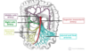
What is the pathophysiology of carotid artery disease and how is it classified?
- Bifurcation of carotid artery predisposes to atheromas and atherosclerosis
- Fatty streak
- Lipid core and formation of fibrous cap
- Classified by the degree of stenosis

What are some risk factors for carotid artery disease?
- Age >65
- Smoking
- HTN
- Hypercholesterolaemia
- Obesity
- DM
- CVD
- FHx

How does carotid artery disease present?
- Asymptomatic until it causes a stroke or TIA (focal neurological deficit)
- May hear carotid bruit on auscultation
- Even if complete occlusion, if unilateral asymptomatic due to collateral supply from contralateral ICA due to the Circle of Willis

Atherosclerosis is the most common cause of carotid artery disease leading to focal neurological deficit. What are some other causes of carotid artery disease?
- Carotid dissection
- Fibromuscular dysplasia
- Vasculitis
- Todd’s paresis (unilateral motor paralysis following seizure)
- Subdural haematoma
- Post ictal state
- Hypoglycaemia

If a patient has a stroke what are the initial and follow up investigations carried out?
Initial
- Urgent non-contrast CT to look for infarction
- If thrombectomy being considered then CT head contrast angiography
- Bloods (FBC, U+Es, clotting, lipid profile, glucose)
- ECG (AF)
Follow-Up
- Do Duplex US (Carotid US Doppler) to look for carotid artery stenosis
- Any stenosis within carotid artery can then be looked at with CT angiography

How do you manage a patient with a suspected stroke acutely?
- High flow oxygen
- Optimise blood glucose between 4 and 11
- Swallowing screen assessment

What long term management should be carried out for patients who have had a TIA or stroke?
- Antiplatelets: 300mg Aspirin for 2/52 then 75mg Clopidogrel long term
- Statin: high dose atorvastatin
- ?Carotid Endarterectomy: for acute non-disabling stroke/TIA if stenosis 50-99%
- Management of HTN and DM
- Smoking cessation
- Regular exercise and weight loss

What happens in a carotid endarterectomy and what are the complications of this?
(p.s better than stenting as less risk of long-term major adverse events)
- Done in symptomatic (TIA or stroke) 50-99% carotid artery oclusion
- Remove atheroma and damaged intima
- Reduces risk of future strokes/TIAs
- Complications: stroke, damage to hypoglossal/vagal/glossopharyngeal nerve, MI, local bleeding, infection

What are some general complications of a stroke?
- Dysphagia
- Seizures
- Bowel incontinence
- Anxiety and depression
- Cognitive decline
What is the definition of an
- aneurysm
- abdominal aortic aneurrysm
- Aneurysm: abnormal dilatation of a blood vessel more than 50% its normal diameter
- AAA: dilatation of the AA greater than 3cm, every 8mm increase there is 34% more chance of death

What are some risk factors for an AAA?
- Smoking
- HTN
- Hyperlipidaemia
- FHx
- Male
- Increasing age
- DM is negative risk factor

What are the clinical features of an AAA?
Asymptomatic: detected on screening or incidental finding
Symptomatic: see image

What is the AAA screening programme in the UK?
Abdominal US offered to men aged 65 once
If AAA detected either direct referral for surgery or 3-5 years surveillance before reaching threshold for elective repaire

What are the differentials with the pain produced in AAA?
- Renal colic (due to back pain and no other symptoms)
- IBD/IBS
- GI haemorraghe
- Appendicitis
- Ovarian rupture
- Splenic infarctions

How do you investigate a suspected AAA (not ruptured)?
- US
- Once US has confirmed then CT scan with contrast with a threshold diameter of 5.5cm

How are unruptured AAA’s managed?
Medical (<5.5cm asymptomatic)

- Monitor with Duplex USS (3-4.4cm yearly, 4.5-5.4 every 3 months)
- Smoking cessation to stop expansion and rupture
- Improve blood pressure control
- Aspirin and Statin therapy
Surgical (>5.5cm, symptomatic or expanding >1cm a year)
- If >6.5cm tell DVLA
- If unfit patient can wait until 6cm
- See image for options
What would be preferred for an AAA repair, endovascular stenting or open repair?
- Both have similar outcomes
- Endovascular repair has better short term outcomes (30 day mortality and decreased hospital stay) but higher rate of reintervention and aneurysm leaking
- Young patient open repair preferred

What are the complications of an AAA?
- Rupture
- Retroperitoneal leak
- Embolisation
- Aortoduodenal fistula

How do AAA ruptures present?
- Abdominal and back pain
- Syncope
- Vomiting
- Haemodynamicall compromised
- Pulsatile tender mass in abdomen
TRIAD OF RUPTURED AAA: flank or back pain, hypotension, pulsatile abdominal mass

How is any suspected AAA rupture managed?
Immediate: high flow O2, IV access with 2 large bore cannulas, urgent boods (FBC, U+Es, Clotting), crossmatch for minimum 6 units
Shock treatment: try to keep BP<100 as raising BP could dislodge any clot and cause further bleeding
Transfer to local vascular unit: if unstable immediate theatre for open surgical repair, if stable CT angiogram to determine if suitable for endovascular repair

Where are the common locations for aneurysms in the body?
AAA most commonly infrarenal

What is an aortic dissection?
A tear in the intimal layer of the aortic wall causing blood to flow between the tunica intima and media, splitting the two apart
Acute < or equal to 14 days to diagnosis
Chronic > 14 days to diagnosis

How can aortic dissections be classified?
Stanford Classification
A - Debakey Type I and II involving ascending aorta
B - Debakey Type III and do not involve ascending aorta
DeBakey Classification
I - originates in ascending aorta and propagates to at least aortic arch
II - confined to asending aorta
III - originates distal to subclavian artery in descending aorta

What are some risk factors for an aortic dissection?
- Hypertension
- Atherosclerotic diease
- Male
- Connective tissue disorders (Marfan’s and EDS in younger pt)
- Bicuspid aortic valve

What are the clinical features of an aortic dissection and what are some differentials?
- Tearing chest pain that usually radiates to back
- Tachycardia, hypotension, aortic regurg murmur
- Signs of end-organ hypoperfusion e.g reduced urine output, lower limb ischaemia
DD: MI, PE, Pericarditis, MSK back pain

How do you investigate a suspected aortic dissection?
- Bloods (FBC, U+Es, LFTs, troponin, coagulation) with crossmatch of at least 4 units
- ABG
- ECG to rule out cardiac pathology
- CT angiogram diagnosis gold standard 1st line
- Can do transoesophageal ECHO

How should you manage an aortic dissection generally?
- Initial: high flow oxygen, IV access with 2 large bore cannulas, fluid resus with target BP<110
- Stanford A: managed surgically as worse prognosis
- Standford B: can be managed medically if uncomplication
- Lifelong antihypertensive therapy and surveillance imaging at 1,3,12 months
How are Type A dissections managed?
- Transfer to cardiothoracic centre
- Remove ascending aorta and replace with synthetic graft
- Reimplant branches of aortic arch to graft

How are Type B dissections managed?
MEDICALLY DUE TO RISK OF RETROGRADE DISSECTION IF MANAGENED SURGICALLY
1st line: IV beta blockers (labetolol) or CCB to lower systolic pressure and minimise dissection
Complicated: if rupture, ischaemia, pain or uncontrollable HTN then surgical repair
What are some complications of aortic dissections?
- Aortic rupture
- Aortic regurgitation
- MI if coronary artery dissection
- Cardiac tamponade
- Stroke or paraplegia if cerebral or spinal artery involved
- Type B can become chronic and form aneurysm

What is a thoracic aortic aneurysm?
Aneurysm involving the aortic arch, ascending aorta or descending aorta

What is the aetiology of a thoracic aortic aneurysm?
Degradation of the tunica media that normally provides tensile strength and elasticity
Artery loses structural integrity and dilates
Usually due to connective tissue diseases (EDS and Marfan’s) or Bicuspid aortic valve

What are the risk factors for a thoracic aortic aneurysm?
- FHx
- Hypertension
- Atherosclerosis (especially descending aneurysms)
- Smoking
- High BMI
- Male gender
- Advancing age
What are the clinical features of a thoracic aortic aneurysm?
- Often asymptomatic and found incidentally
- See image for pain
- Back pain from spinal compresion by descending aorta
- Hoarse voice due to left reccurent laryngeal nerve damage in arch aneurysm
- Distended neck veins if SVC compression
- Symptoms of heart failure if aortic valve involved
- Dyspnoea if tracheal/bronchial compression

What is acute aortic syndrome and how does it present?
Acute painful and potentially life-threatening aortic pathologies that require immediate medical attention
Include: aortic dissection, aortic ulcer, intramural hematoma and unstable thoracic aortic aneurysm
Symptoms: sudden onset pain in back, chest, neck and/or abdomen

How are thoracic aneurysms investigated?
Initial: routine bloods (FBC, U+Es, clotting), ECG, CXR
Imaging: often found incidentally on CXR with widened mediastinal silhouette, enlarged aortic knob and possible tracheal deviation
- CT chest scan with contrast
- Transoesophageal echocardiography

How are thoracic aortic aneurysms managed?
Medical (initially)
- Start on aspirin and statin to reduce risk of MI
- Control BP
- Smoking cessation
Surgical (depends on location of aneurysm)
- See image for criteria
- Lower threshold for Marfan’s and previous thoracic dissection

What is the prognosis with a thoracic aortic aneurysm?
- Risk of rupture or dissection
- Development of second aneurysm common so imaging studies following surgery e.g MRI or CT
What is acute limb ischaemia and what is the aetiology of this?
Sudden decrease in limb perfusion that threatens the viability of the limb
Three groups:
- Embolisation
- Thromus in situ
- Trauma

What are the clinical features of acute limb ischaemia?
SUDDEN ONSET 6 P’s
Pain
Pallor
Pulselessness
Paraesthesia
Perishingly cold
Paralysis

What are some causes of embolisation causing acute limb ischaemia?
Normal pulsatile contralateral limb is sign of embolic occlusion
- AF
- Recent MI with mural thrombus
- Symptomatic AAA
- Peripheral aneurysms
What are the different categories of acute limb ischaemia?
Later onset more likely there is irreversible damage to neuromuscular structures (>6h post symptom onset)
Rutherford Classification

What are some differentials for acute limb ischaemia?
- Critical chronic limb ischaemia
- DVT
- Spinal cord or peripheral nerve compression
How do you investigate suspected acute limb ischaemia?
- Routine bloods (lactate to assess level of ischaemia, thrombophillia screen if <50, G+S )
- ECG to look for AF
- Doppler US on both limbs then
- CT angiography

How is acute limb ischaemia managed short term?
Initial: Surgical emergency as irreversible damage in 6 hours. High flow O2, IV access, bolus heparin dose
Conservative (Rutherford I+IIa): prolonged course of heparin with regular clinical review and APTT monitoring
Surgical (Rutherford IIb onwards): if irreversible limb ischaemia (non-blanching and woody muscles) need urgent amputation.

Why do most acute limb ischaemia post-op patients need a high level of care?
Need HDU due to ischaemia reperfusion syndrome.
Sudden increase in capillary permeability can cause:
- Compartment syndrome
- Release of substances from damaged muscle cells (K+ hyperkalaemia, H+ acidosis, Myoglobin AKI)

What is the long term management of acute limb ischaemia?
- Reduce CVS mortality risk: weight loss, smoking cessation, regular exercise
- Antiplatelet agent: low dose aspirin or clopidogrel
- Treat any predisposing conditions: e.g AF
What are some risk factors for chronic limb ischaemia?

What is chronic limb ischaemia and what is it caused by?
Peripheral arterial disease that results in symptomatic reduced blood supply to limbs
Usually due to atherosclerosis (sometimes vasculitis) and commonly affects lower limbs
What are the clinical features of chronic limb ischaemia, what test can be done?
- Early sign is intermittent claudication (cramping pain in calf, thigh or buttock after walking a set distance and relieved by rest)
- Buerger’s Test: lie patient supine and raise legs until go pale then lower until colour returns, angle less than 20 degrees is severe ischaemia

What is Leriche Syndrome?

How is critical limb ischaemia defined?
- Ischaemic rest pain for greater than 2 weeks needing opiates
- Presence of ischaemic lesions or gangrene
- ABPI <0.5

How may critical limb ischaemia present on examination?
- Pale and cold limb
- Weak or absent pulses
- Limb hair loss
- Skin changes (atrophic skin, ulceration, gangrene)
- Thickened nails

What are the differential diagnoses for patients presenting with limb ischaemia?
- Critical limb ischaemia
- Spinal stenosis (Neurogenic claudication)
- Acute limb ischaemia

How is chronic limb ischaemia investigated and diagnosed?

Dx
- Clinical diagnosis
- ABPI confirms and quantifies severity
Further Ix
- Doppler US to find site of occlusio
- CT angiography or MRA
- CVS risk assessment (BP, Lipid profile, Blood glucose, ECG)
- <50 thrombophillia screen and homocysteine levels

How is chronic limb ischaemia managed?
Medical

- Life style advice
- Statin therapy (atorvastatin 80mg)
- Antiplatelet therapy (clopidogrel 75mg)
- Optimise diabetes control
- Supervised exercise programme to improve claudication distance
Surgical (risk factor modification discussed or supervised exercise failed to improve symptoms)
- Angioplasty with or without stenting
- Bypass grafting
- Amputation if gangrene
What are the complications of chronic limb ischaemia?
- Sepsis secondary to infected gangrene
- Acute-on-chronic ischaemia
- Amputation
- Reduced quality of life
5 year mortality after diagnosis of chronic limb ischaemia is 50%!!!
What is the aetioloy of acute mesenteric ischaemia?
Sudden decrease in blood supply to the bowel resulting in bowel ischaemia and death if left
- Thrombus in situ
- Embolism
- Non-occlusvie cause
- Venous occlusion and congestion

What are some risk factors for acute mesenteric ischaemia?
Depends on underlying cause e.g AMAE
- Smoking
- Hyperlipidaemia
- Hypertension
What are the clinical features of acute mesenteric ischaemia?
- Generalised abdominal pain out of proportion to clinical findings
- Pain is diffuse and constant
- N+V
- Non-specific tenderness
- Look for AF or heart murmurs for embolic casue

What investigations should be done to diagnose acute mesenteric ischaemia?
- ABG to assess acidosis and serum lactate
- Routine bloods (FBC, U+Es, clotting, amylase, LFTs if coeliac trunk blocked)
- Definitive diagnosis: CT scan with IV contrast

Apart from pancreatitis, what can raise serum amylase levels?
- Acute mesenteric ischaemia
- Ectopic pregnancy
- Bowel perforation
- DKA
How is acute mesenteric ischaemia managed?
Initial
- Emergency so senior input
- IV fluids, catheter inserted and fluid balance chart
- Broad spectrum abx due to risk of faecal contamination if bowel perforates
- Early ITU input as will have acidosis so at high risk of multi-organ failure
Definitive
- Exicision of necrotic or non-viable bowel if not suitable for revascularisation.
- Revascularisation of the bowel and remove any thrombus or embolism

What are some complications with acute mesenteric ischaemia?
- Bowel necrosis
- Bowel perforation
- Mortality 50% even with diagnosis made
- Those that survive may have short gut syndrome

What is the pathology of chronic mesenteric ischaemia?
Reduced blood supply to the bowel which decreases over time due to atherosclerosis of coeliac, SMA, IMA
Collateral blood supply of the mesentry means two of the three vessels have to be affected to be symptomatic
Although often asymptomatic, when there is an increased demand for blood supply (after eating or after blood loss) this will exacerbate symptoms
Tends to affect females >60
What are some risk factors for chronic mesenteric ischaemia?
- Smoking
- Hypertension
- Diabetes
- Hypercholesterolaemia
What are the clinical features of chronic mesenteric ischaemia?
- Post prandial pain: 10mins-4hrs after
- Weight loss: decreased calorie intake and malabsorption
- Concurrent vascular co-morbities: previous stroke, MI
- May have: change in bowel habit loose stools, N+V, malnutrition
- On examination: usually remarkable or generalised abdominal tenderness and abdominal bruits

What investigations are done to diagnose chronic mesenteric ischaemia?
- CT angiography for diagnosis
- Blood tests (check FBC, LFTs, U+Es, Mg and Ca due to malnutrition)
- Cardiovascular risk profile (lipid profile, blood glucose)

How is chronic mesenteric ischaemia managed?
Conservative
- Modify risk factors to stop propagation e.g stop smoking
- Start anti-platelets and statins
Surgical (severe, progressive disease or debilitating symptoms)
- Endovascular procedures: mesenteric angioplasty with stenting, this is preferred due to general nutritional status of patient
- Open procedures: endartectomy or bypass proedure

What are some complications of chronic mesenteric ischaemia?
- Bowel infarction
- Malabsorption
- Concurrent CVS diease
What is a pseudoaneurysm and how do they form?

Breach to arterial wall so accumulation of blood between tunica media and tunica adventitia
Usually after damage to vessel wall (e.g following cardiac catheterisation, IVDU, inflammation or vasculitis)
Most common in femoral artery, but can occur in radial, carotid, abdominal/thoracic aorta

What is a vascular complication of acute pancreatitis?
Splenic pseudoaneurysm
What are the clinical features of a pseudoaneurysm?
- Pulsatile lump that is tender and painful
- Could be limb ischaemia due to distal arterial occlusion by compression so check pulses
- If infected will be erythematous, tender and purulent material may be discharging

If a patient reports they have had a bleed from a pseudoaneurysm but it has stopped now, what should you do?
Close monitoring as could represent a Herald Bleed so could rebleed at any time

How are pseudoaneuryms diagnosed an assessed?
- Gold standard: Duplex US which will show turbulent back and forward flow (yin-yang sign).
- Can do CT if cannot access with US
- Routine bloods (FBC, CRP, U+Es, clotting, crossmatch due to risk of rupture)
- Blood cultures and Pus MC+S if discharging

How are small and large non-infected pseudoaneurysms managed?
Small: often left alone as can thrombose and close off, however some continue to grow until perforate
Large:
Ultrasound guided compression (can be painful and needs 30 mins direct compression)
OR ultrasound guided thrombin injection (follow up imaging to check resolution)
OR endovascular stenting
OR surgical repair/ligation

Why is endovascular stenting and surgical repair of pseudoaneurysms more complication?
Endovascular stenting: sometimes insufficient space to put stent without covering a major branch, can leak so pseudoaneurysm still perfused, can migrate
Surgical repair/Ligation: need healthy proximal and distal artery, ligation can sometimes cause distal ischaemia so will need bypass graft

How are infected psuedoaneurysms managed? (usually from IVDU)
- More likely to become septic or rupture so need urgent treatment
- If any discharge pressure dressing and urgent imaging
- Surgical ligation, sometimes requiring bypass graft due to acute limb ischaemia but be careful as graft can become infection
- Small risk of amputation even with collateral blood supply

What are some risk factors for peripheral and visceral aneurysms?
- Smoking
- Hyperlipiadaemia
- HTN
- FHx
- Marfan’s
- Takayasu’s aortitis
Aneurysms are persisitent abnormal dilatations of an artery above 1.5X the normal diameter

What vessels are classed as visceral aneurysms?
- Splenic (most common)
- Hepatic (second most common)
- Renal
- Intestinal arteries
- Mesenteric arteries

How are peripheral and visceral aneurysms investigated and managed in general terms?
Ix:
- CT angiography or MRA if renal issues
- US duplex scans for detection or follow up
Mx
- Watchful waiting whilst optimising CVS factors so antiplatelet and statin therapy
- Surgical intervention (endovascular or open)
What are the most common peripheral artery aneurysms?
- Popliteal (high risk of embolisation and/or occlusion)
- Femoral
Popliteal aneurysms are the most common peripheral aneurysm. How do they present?
- Acute limb ischaemia (aneurysm thrombosis or emboli) OR
- Intermitten claudication
Can also be found incidentally during AAA work up or during TKR or from compression symptoms on popliteal vein or peritoneal nerve

How are suspected popliteal aneurysms investigation?
- Initially US duplex to rule out other popliteal swellings e.g Baker’s cyst, lymphadenopathy
- CT angiogram or MRA
How are popliteal aneurysms managed?

If symptomatic or asymptomatic and over 2.5cm should be given treatment
- If thrombosis in tibial vessel then embolectomy or thrombolysis
- Endovascular: stent insertion
- Open: ligation of aneurysm or resection with bypass graft

What are the causes of femoral pseudoaneurysms?
- Patient self-injecting e.g IVDU
- Percutaenous vascular interventions
How do femoral pseudoaneurysms present, and how are they investigated and managed?
Presentation
- Symptoms due to thrombosis, rupture, embolisation or infecton
- Varying degrees of claudication or acute limb ischaemia
Ix
- US duplex then CT angiogram or mra
Mx
- Open surgical repair

What are some risk factors for splenic pseudoaneurysms and how do they present?
Risk factors: female, multiple pregnancies, portal hypertension, acute pancreatitis, pancreatic pseudocysts
Presentation: vague epigastric or LUQ pain, if ruptured then severe abdominal pain

How are splenic and hepatic psuedoaneurysms investigated and managed?
Ix: CT angiography or MRA
Mx: 1st Line is endovascular repair wirh embolisation or stent graft once patient haemodynamically stable
What is the cause of hepatic pseudoaneurysms and how do they present?
Causes: percutaneous instrumentation, trauma, degenerative disease, post liver transplant around vessel anastomoses
Presentation: usually asymptomatic but can have vague RUQ/Epigastric pain and jaundice if biliary obstruction

How do renal artery pseudoaneurysms present, and how are they investigated and managed?
Presentation: often incidental finding/asymptomatic but can have haematuria, resistant hypertension or loin pain if renal infarction
Ix: CT angiogram or MRA
Mx: Endovascular repair with stent if affecting renal artery, or coils if affecting hilum. Can also do renal transplant rarely

What is the difference between intra and retroperitoneal AAA ruptures?

Where is rest pain usually felt and what would be the ABPI in this instance?
- Toes or foot
- <0.5
In peripheral vascular diease, why does a patients foot go redder in the diseased limb than the normal limb after Buerger’s test?
Severe ischaemia (during elevation) leads to the release of local vasodilators (e.g. ADP, potassium, hydrogen ions, lactate, carbon dioxide) that subsequently increase the perfusion of the ischaemic foot when back to normal
