12. Colorectal Disorders Flashcards
What is colorectal cancer divided into, and what are the % occurances of each?
List the treatment modalities for colorectal cancer, and indicate which one is the curative option.
How do elective colon and rectal cancer surgeries differ in tx?
What is the presentation for left sided and right sided colorectal cancers?
Colonic and rectal: 66% occur in rectum, rectosigmoid and sigmoid; 20% in right colon (ascending); remainder in L (descending) and transverse colon
Surgery (only curative option), radiotherapy (may cure some), chemotherapy
Colon: surgery then chemo were indicated.
Rectum: radiotherapy before surgery, then chemo where indicated
Left: bleeding/mucus PR, altered bowel habit/obstruction, tenesmus, mass PR
Right: weight loss, low Hb, abdo pain
Both: abdo mass, perforation, harmorrhage, fistula
What might you notice on examination if a pt has colorectal cancer?
Describe Dukes’ staging of colorectal cancer.
What is most commonly used to stage the disease?
Palpable tumour mass, hepatomegaly, large rectal mass, palpable LN
- A:** limited to muscularis mucosae (93% 5yr survival rate*)
- B:** extension through muscularis mucosae (77% 5yr survival rate*)
- C:** involvement of regional LN (48% 5yr survival rate*)
- D:** distant mets (6.6% 5yr survival rate*)
TNM criteria. Tx depends on clinical, radiological (CT/MRI) and pathological staging
Who would be present in a colorectal cancer MDT?
Distinguish between adjuvant and neo-adjuvant treatment for colorectal cancer.
What are the 3 different levels of surgical resection?
Colorectal surgeon, gastroenterologist, colorectal cancer nurse, radiologist, clinical oncologist, medical oncologist, pathologist, pallitative care physician. All pts with colorectal cancer should be discussed at MDT prior to definitive mx
Adjuvant: chemotherapy. Given to pt following putatively curative surgery for tx of occult micrometastases
Neo-adjuvant: chemoradiation. Given to pt pre-op to down-stage disease on presumption that they progress to curative surgery subsequently
R0 = no residual tumour following resection; R1 = microscopic residual tumour following surgery; R2 = macroscopic residual tumour at surgery completion
Label the colonic vascular anatomy below.
Case 1
78yo female, presenting with lethargy and wt loss, has iron deficiency anaemia, previously fit and active.
How would you investigate the colon? What might a gastroenterologist do?
Which members of the MDT would be invovled?
- *A:** middle colic artery
- *B:** R colic artery
- *C:** ileo-colic artery
- *D:** L colic artery
- *E:** sigmoid artery
- *F:** superior rectal artery
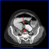
Colonoscopy/OGD, barium enema, CT pneumocolon [Pic]
Gastroenterologist = biopsy, tattooing, endomucosal resection
Tissue diagnosis - pathologist. CT chest/abdo/pelvis = radiologist

Case 2
78yo previously fit man with rectal bleeding, loose stools and tenesmus. PR firm irregular mass. What next?
Case 3
67yo lady with colicky abdo pain, intermittant distension and vomiting. Admitted directly from clinic. AXR below. What next?
How would she be treated?

- *Colonoscopy:** biopsy and exclude synchronous lesions
- *Histopathology:** tissue dx
- *CT chest/abdo/pelvis:** exclude distant mets
- *MRI pelvis:** local staging
Exclude pseudo-obstruction.
Confirm site of blockage via CT/gastrograffin enema [Pic]
Resection, stoma, stent


