1
Q

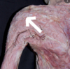
What is the significance of a Lump in the Upper Epigastric Region?

A

The Xiphoid Process has been Calcifed.

2
Q

What are the True Ribs?

A

1st - 7th Ribs
Connect to Sternum via individual Costal Cartilages.

3
Q

What are the False Ribs?

A

8th - 10th Ribs
Fuse together into a Common Costal Cartilage.

4
Q

What are the Floating Ribs?

A

11th and 12th Ribs

5
Q

How do ribs 11 - 12 differ from a typical rib?

A

Do not articulate with any bone anteriorly.

6
Q

Which ribs are most commonly fractured?

A

7th - 10th Ribs

7
Q

What is the significance of the lower ribโ€™s proximity,
to the Abdominal Cavity?

A

They protect the organs in the Thoracic Cavity.

8
Q

How many Vertebrae are in the Thorax?

A
12
Thoracic 12 (T12)
9
Q

What is Thoracic Outlet Syndrome?

A

Group of disorders causing pain and paresthesis in the neck, shoulder, arms and hands.
Caused by compression of the Brachial Plexus and/or Subclavian Vessels,
As they pass through the Thoracic Outlet.
Anatomically the location is the Thoracic Inlet.

10
Q

How are the Vessels at the Manubrium organised?

A

The Vein comes before the Artery.
The Vein is more Anterior.
The Artery is more Posterior.

11
Q

What is the weakest part of each rib?

A

Just anterior to Angle of the Rib.

12
Q

What structures are found at the Costal Groove?

A

V: Vein (Most Superior)
A: Artery
N: Nerve (Most Inferior)

13
Q

What muscle attaches to the Scalene Tubercule?

A

Anterior Scalene Muscle:

  • Attaches onto the Scalene Tubercle,
  • On the inner border of the first Rib.
14
Q

What does the Anterior Scalene Muscle divide?

A

Helps to divide the Brachial Plexus.
Also divides the Arterial and Venous supply of the First Rib.

15
Q

List the arrangement of Structures over the First Rib:

A

From Anterior to Posterior:

  1. Subclavian Vein
  2. Anterior Scalene Muscle
  3. Subclavian Artery
  4. Brachial Plexus
16
Q

What are all the Scalene Muscles?

A
  1. Anterior Scalene
  2. Middle Scalene
  3. Posterior Scalene
17
Q

Where do the Scalene Muscles come from?

A

Come from the Transverse Processes of the Cervical Vertebrae.

18
Q

Which are the atypical Ribs?

A

1st, 11th, 12th

19
Q

What is a Cervical Rib and what are its consequences?

A

An elongation of the Transverse Process of one of the Cervical Vertebrae.
Impinges on the Brachial Plexus or the structure around it,
Leading to Paresthesia in the fingertips.

20
Q

What is Costo - Chondritis?

A

Inflammation of the Cartilage that connects a Rib to the Sternum.

21
Q

Why might damage to cartilage take a long time to heal?

A

It doesnโ€™t have a very extensive vascular supply.

22
Q

Pectoralis Major Origin:

A

Clavicular Part:
Anterior Surface of Medial Half of Clavicle.

Sternocostal Part:

  • Anterior Surface of Sternum,
  • Costal Cartilages of Ribs 1 - 6

Abdominal Part:
- Anterior Layer of Rectus Sheath

23
Q

Pectoralis Major Insertion:

A

Greater Tubercle of Humerus

24
Q

Pectoralis Major Action:

A

Shoulder Joint:

  • Arm Adduction,
  • Arm Internal Rotation,
  • Arm Flexion (Clavicular Head),
  • Arm Extension (Sternocostal Head);

Scapulothoracic Joint:
- Draws Scapula Anteroinferiorly

25
Pectoralis Major Innervation:
Lateral and Medial Pectoral Nerves | (C5 - T1)
26
Pectoralis Major Blood Supply:
Pectoral and deltoid Branches of: - Thoracoacromial Artery (Second part of the Axillary Artery) Perforating Branches of: - Superior thoracic and lateral thoracic arteries
27
Pectoralis Minor Origin:
Anterior Surface, Costal Cartilages of Ribs 3 - 5
28
Pectoralis Minor Insertion:
Medial Border and Coracoid Process of Scapula
29
Pectoralis Minor Action:
Scapulothoracic Joint: - Draws Scapula Anteroinferiorly, - Stabilizes Scapula on Thoracic Wall.
30
Pectoralis Minor Innervation:
Medial Pectoral Nerves (C5-T1)
31
Pectoralis Minor Blood Supply:
Thoracoacromial Artery (Pectoral branch), Internal thoracic artery
32
External Intercostals Origin:
Inferior Border of Ribs
33
External Intercostals Insertion:
Superior Border of immediate Rib Below
34
External Intercostals Action:
Elevate Ribs during forced inspiration Supports Intercostal Spaces and Thoracic Cage
35
External Intercostals Innervation:
Intercostal Nerves
36
External Intercostals Blood Supply:
Anterior Intercostal Arteries Posterior Intercostal Arteries
37
Internal Intercostals Origin:
Costal Groove of Ribs
38
Internal Intercostals Insertion:
Superior Border of Immediate Rib below
39
Internal Intercostals Action:
Depress Ribs during forced expiration Support Intercostal spaces and Thoracic Cage
40
Internal Intercostals Innervation:
Intercostal Nerves
41
Internal Intercostals Blood Supply:
Anterior Intercostal Posterior Intercostal
42
Innermost Intercostals Origin:
Costal groove of Ribs
43
Innermost Intercostals Insertion:
Superior Border of immediate Rib below
44
Innermost Intercostals Action:
Depress Ribs during forced expiration Support Intercostal Spaces and Thoracic Cage
45
Innermost Intercostals Innervation:
Intercostal Nerves
46
Innermost Intercostals Blood Supply:
Anterior Intercostal Posterior Intercostal Internal Thoracic Musculophrenic Arteries Costocervical Trunk
47
Transversus Thoracis Origin:
Inferoposterior Surface of Body of Sternum and Xiphoid Process Sternal Ends of Costal Cartilages of Ribs 4 - 7
48
Transversus Thoracis Insertion:
Internal Surface of Costal Cartilages of Ribs 2 - 6
49
Transversus Thoracis Action:
Depresses Ribs during forces expiration Supports Intercostal Spaces and Thoracic Cage
50
Transversus Thoracis Innervation:
Intercostal Nerves
51
Transversus Thoracis Blood Supply:
Sternal Branches of Internal Thoracic Artery
52
Serratus Anterior Origin:
Superior Part: Ribs 1 - 2, Intercostal Fascia Middle Part: Ribs 3 - 6 Inferior Part: Ribs 7 - 8/9/10 (+ External Oblique Muscle)
53
Serratus Anterior Insertion:
Superior Part: Anterior and Posterior surface of Superior Angle of Scapula Middle Part: Anterior Surface of entire Medial Border of Scapula Inferior Part: Anterior and Posterior surface of Inferior Angle of Scapula
54
Serratus Anterior Action:
Scapulothoracic Joint: - Draws Scapula Anterolaterally - Suspends Scapula on Thoracic Wall - Rotates Scapula (draws inferior angle laterally)
55
Serratus Anterior Innervation:
Long Thoracic Nerve (C5 - C7) SALT: Serratus Anterior - Long Thoracic 'C5, 6, 7 raise your arms to heaven!'
56
Serratus Anterior Blood Supply:
Superior Thoracic Arteries Lateral Thoracic Arteries Thoracodorsal Artery Branches
57
Subclavius Origin:
Costal Cartilage, Sternal end of Rib 1
58
Subclavius Insertion:
Anteroinferior Surface of Middle Third of Clavicle
59
Subclavius Action:
Sternoclavicular Joint: - Anchors and Depresses Clavicle
60
Subclavius Innervation:
Subclavian Nerve (C5 - C6)
61
Subclavius Blood Supply:
Clavicular Branch of Thoracoacromial Artery, Suprascapular Artery
62
Where do the Lower 9 Posterior Intercostal Arteries arise from?
Directly from the Posterior Surface of the Thoracic Aorta.
63
Where do the Upper 2 Posterior Intercostal Arteries arise from?
Superior (Supreme) Intercostal Artery, A branch of the Costocervical Trunk
64
Where do the Lower 3 Anterior Intercostal Arteries arise from?
Musculophrenic Arteries
65
Where do the Upper 6 Anterior Intercostal Arteries arise from?
Internal Thoracic Artery
66
How are the Nerves related to Blood Vessels?
V: Vein (Most Superior) A: Artery N: Nerve (Most Inferior)
67
How is the Neurovascular bundle protected?
Protected by the Costal Groove of the Superior Rib of each Intercostal Space
68
Between which muscles does the Neurovascular bundle pass?
Internal Intercostal Muscle Innermost Intercostal Muscle
69
Into which intercostal space are chest drains inserted?
4th, 5th or 6th Intercostal Spaces in the Mid - Axillary Line.
70
What is a Dermatome?
An area of skin supplies by a single Spinal Root.
71
What is the pattern of Dermatomes in the Thorax?
T1 - T12 T1 - T9 being near Horizontal Lines T10 - T12 having the lower borders Dip Inferiorly
72
Which Dermatome is the Nipple and Umbilicus?
Nipple: T4 Umbilicus: T10
73
How many Dermatomes are there in the Thorax?
12
74
What are Shingles (Herpes Zoster)?
Reactivation of the Herpes Zoster Virus. Unilateral and affects one Dermatome.
75
What muscle does the Long Thoracic Nerve supply?
Serratus Anterior
76
What muscles does the Medial Pectoral Nerve supply?
Pectoralis Major Pectoralis Minor
77
What muscle does the Lateral Pectoral Nerve supply?
Pectoralis Minor
78
Where do Lateral and Medial Pectoral Nerves arise from?
Brachial Plexus
79
What is the clinical significance of the Mid - Axillary line?
Safe point for the insertion of Chest Drains
80
List the vertebral level of the main landmarks in the Thorax:
T3: Jugular Notch (Manubriosternal Joint) T4/5: Sternal Angle (Angle of Louis) T9: Xiphoid Process (Sterno - Xiphoid Junction)
81
What is the function of the Platysma?
Facial Expressions Lowering the corners of the mouth and lower lip.
82
What would be the effect of paralysis of the Serratus Anterior Muscle?
Winged Scapula Paralysis occurs as a result of damage to the Long Thoracic Nerve.
83
How does the Diaphragm contribute to respiration?
Upon inhalation, the Diaphragm Contracts and Flattens, Enlarging the Chest Cavity, This contraction creates a vacuum, which pulls air into the lungs.
84
What is a Hiatus Hernia?
Part of the Stomach pushes through the Oesophageal Hiatus into the Thoracic Cavity.
85
How does this differ from Congenital Diaphragmatic Hernia?
A Hiatus Hernia is through the Oesophageal Hiatus, A Congentital Hernia is through a congenital defect opening in the Diaphragm that is not normally present.
86
What are Venae Comitantes?
A vein that is usually paired, with both veins lying on the sides of an artery. They are found in close proximity to Arteries so that the pulsations of the Artery aid Venous Return.
87
What parts of the Thoracic Wall does the, Thoraco - Acromial Artery supply?
Pectoralis Major Pectoralis Minor Part of the Deltoid Muscle The skin overlying the Clavipectoral Fascia
88
What parts of the Thoracic Wall does the, Lateral Thoracic Artery supply?
Serratus Anterior Muscle Pectoralis Major Muscle Axillary Lymph Nodes Subscapularis Muscle
89
What is the site of attachment/insertion of this muscle?
Crest of tubercle of the humerus
90
What is the origin of this muscle?
* anterior surface of medial half of clavicle * anterior surface of sternum * costal cartilages of ribs 1-6 * anterior layer of rectus sheath (abdominal part)
91
What are the functions of this muscle?
* Adducts the arm * medially rotates the humerus * extends a flexed arm
92
What is the name of this muscle?
Pectoralis major
93
How is this muscle separated from pec major?
Deltopectoral groove
94
What is the origin of this muscle? (deltoid muscle)
* lateral part of clavicle * acromion and spine of scapula
95
What is the insertion of this muscle?
deltoid tuberosity of the humerus
96
what are functions of the clavicular fibres of this muscle?
Arm flexion
97
What are the functions of the posterior fibres of this muscle?
extension of arm
98
What is another function of the deltoid muscle?
Abducts the arm
99
What is the function of this muscle?
Stabililsation, depression, abduction or protraction and internal rotation of scapula
100
What is the origin of this muscle (pec minor)?
Costal cartilages of ribs 3, 4, 5
101
What is the insertion of this muscle?
Choracoid process of scapula
102
What is the origin of this muscle?
* superior part: ribs 1-2, intercostal fascia * middle part: ribs 3-6 * inferior part: ribs 7-8/9/10, (& external oblique muscle)
103
What is the insertion of this muscle?
* Superior part: anterior and posterior surface of superior angle of scapula * middle part: anterior surface of entire medial border of scapula * inferior part: anterior and posterior surface of inferior angle of scapula
104
What is the function of this muscle?
Protraction and rotation of scapula
105
What is the function of this muscle in breathing?
The interchondral portion of the internal intercostal muscles causes elevation of the ribs and sternum due to location and fibre direction. Hence it aids inspiration.
106
What is the function of the interosseous portion of the internal intercostal muscles?
Depresses the rib cage and sternum hence aids expiration
107
What is the location of the neurovascular bundle?
Located between the innermost intercostal and internal intercostal muscles. 'Protected by' costal groove of rib. Ordered as V-A-N (vein, artery, nerve)
108
What are the nerves that are part of the neruovascular bundle?
* anterior rami of the thoracic spinal nerves (T1-T11) make up the intercostal nerves * T1-T6 just run with the intercostal spce * T7-T9 run deep to costal cartilages to innervate the anterior abdominal wall (because the costal cartilages are fused together so nerves can't run alongside them and enter deep) * T10-T11 pass directly to anterior abdominal wall * T12 is not an intercostal nerve as it runs below T12 (not an intercostal space)
109
True or false, the veins highlighted in the image drain the pericardium?
True
110
Why are azygous veins important?
They drain the posterior thoracic wall and posterior abdominal wall and eventually drain into the SVC. Hence are an important anastomotic pathway for venous return in the IVC becomes blocked.
111
What does the union of these two veins form?
Azygous vein (union of right subcostal and right ascending lumbar vein)
112
What is this structure ?
Areola
113
What is the function of the areola?
It contains **many sebaceous glands** which enlarge during pregnancy and **secrete an oily substanc**e which forms a **protective lubricant** of **the areola and nipple.**
114