09.09 Kidney Flashcards

Located retroperitoneally, between T12 and L3 vertebrae
cortex: outer light granular layer from capsule to the base of pyramids.
Medulla: inner dark striated layer with piramids
Sinus/pelvis: connective and adipose tissue

calyces: minor (8-18/kidney) and major (2-3/kidney)
Pelvis: joining of the major calyces
hilum: includes vessels nerves and ureter


- Aorta → renal artery (through hilum)→ segmental artery → lobar artery → arcuate artery → interlobular artery (between columns) → afferent arteriole (surrounds glomerulus)→ glomerulus (capillaries) → efferent arteriole (surrounds glomerulus) → peritubular capillaries (surrounds convoluted tubules) and vasa recta (bundles of thin vessels that carry blood to and from medulla) → interlobular vein → arcuate vein → lobular vein → renal vein → interior vena cava.


i. Renal Corpuscles (in cortex)
a. Bowman’s capsule: parietal layer (outer wall) and visceral layer (inner wall)

Podocytes:
Modified squamous epithelial cells. They sit on top of the glomerular capillaries not quiet touching each other and the spaces between the podocytes are called slits through which blood can be filtered.

where blood and urine leave the corpuscle

where blood and urine leave corpuscle

- podocytes surround capillary loops and their foot processes form slits
- negative charge on the podychtes keep negatively charged protein from filtering through

- capillray network for filteration
- includes afferent and efferent arterioles
- capillary loops are supported by mesangium: for filteration and phagocytes
- capillaries have a fenestrated endothelium.
- urine consisting of water and waste products collects initially in Bowman’s space in the glomerulus

Juxtaglomerular apparatus (JGA) in cortex
- regulates renal blood flow and filteration rate.
- located between vascular pole and distal convulted tubule of the same nephron

granular renin-producing cells sit on afferent and efferent arteriole and can sense arterial BP, sodium concentration, or renal sympathetic nerve activity

Macula densa cells are sensitive to sodium concentration in the distal tubule

Proximal tubule in the cortex
- microvilli makes brush border
- eosinophilic cytoplasm (lots of mitochondria)
- cuboidal cells
- gets urine from Bowman’s space
- 80% of filtered urine is reabsorbed here


Distal Tubules
• Distinct apical ends ⇒ distinct lumen
- Less mitochondria ⇒ less eosinophilic (means it can be dyed pink)
- Cuboidal cells (increased mitochondria makes cytoplasm more eosinophilic.
- Secretion of ions due to hormonal control
- Aldosterone mediates reabsorption of Na+ and H2O as needed to regulate BP.

D=distal
P=proximal


Medulla has no glomeruli.
Contains portions of proximal and distal convulated tubules.

CD= collecting ducts
TS = thin segments of the Loop of Henle
DT = distal tubules

a. Descneding: thin (simple squamous)
b. Ascending : thick (simple cuboidal)
c. Absorbs Na+ ⇒ increase in [Na+] in surrounding parenchyma

a. Simple cuboidal and clear cytoplasm
b. Further adjust urine volume and solute concentration
- Antidiuretic hormone (ADH) from posterior pituitary gland makes cell more permeable to water → absorption of free H2O → more concentrated urine.
- Increase in [Na+] in medulla draws water out once cells are permeable.

series of straight capillaires that lies parellel to the Loop of Henle


- Papillary duct (of Bellini)
- convergence of collecting ducts
- tall columnar cells with clear cytoplasm
- large lumen taht open into calyces
- Calyx: transitional epithelium


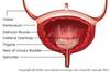
- Carry urine from renal pelvis to trigone of bladder
- Located inferior to the blood vessels
- Has transitional epithelium
- Thin lamina propria

- inner longitudinal and outer circular layer of smooth muscle
- Lower third has another outer longitudinal layer of smooth muscle
- For continuous peristalsis
- Ureter is convoluted at resting which allows for easy expansion by fluid.

- Ureters enter the bladder at an angle at the trigone, producing one-way valve . This prevents reflux of urine.
- transitional epithelium produces a mucoid secretion with natural anti-bacterial properties to prevent infection

- think lamina propria
- submucosa
- detrusor muscle contracts bladder
- inner longitudinaNext >>l, middle circular and outer longitudinal

- Exits the bladder below the trigone
- Males longer (prostatic urethra extends through the prostate, penile urethra and urethral meatus)
- Females: shorter

- Upper 1/3 is transitional epithelium. Lower 2/3 is stratified squamous epithelium.
- Lamina propria with vessels (more in males)
