03-06 Tumors of the Bowel + PATH Flashcards
At the end of this lecture, the learner should be able to: • Define and explain the terms: Neoplasia, benign, malignant, tumor, mass, hamartoma, hyperplasia • Outline the nomenclature of Neoplasms found in the intestinal tract • Describe the pathologic features, clinical presentations, prognosis and treatment options of the more common neoplasia, benign and malignant, of the small intestine • Describe the pathologic features, clinical presentations, prognosis and treatment op (42 cards)
<p>
OBJECTIVE: define hamartoma</p>
<ul>
<li>
mass of mature tissues normally found at that site, but present in an abnormal arrangement. </li>
</ul>
<p>Name the benign neoplasm and malignant neoplasm version of normal: <strong>glandular epithelium</strong></p>
<ul>
<li>Adenoma</li>
<li>Adenocarcinoma</li>
</ul>
<p>
Name the benign neoplasm and malignant neoplasm version of normal: Adipose tissue</p>
<p>
lipoma = benign</p>
<p>
liposarcoma = malignant</p>
<p>
Name the benign neoplasm and malignant neoplasm version of normal: vessels</p>
<p>
angioma, angiosarcoma</p>
<p>
Name the benign neoplasm and malignant neoplasm version of normal: neuroendocrine cells</p>
<ul>
<li>
benign = carcinoid*</li>
<li>
malignant = Neuroendocrine carcinoma</li>
</ul>
<p>
1 There is no reliable histopathologic feature to distinguish which of these tumors will behave in a benign vs. malignant fashion. Best "predictors" are size and mural invasion. </p>
<p>
Name the benign and malignant: Smooth muscle neoplasms</p>
<p>
leiomyoma, leiomyosarcoma</p>
<p>
Name the benign and malig Peripheral nerve sheath neoplasms</p>
<ul>
<li>
benign = Schwannoma</li>
<li>
Malignant Peripheral Nerve Sheath Tumor (MPNST) </li>
</ul>
<p>
Interstital cells of Cajal neoplasms</p>
<ul>
<li>
benign = GIST*</li>
<li>
malig = GIST*</li>
</ul>
<p>
Gastrointestinal Stromal Tumor, a unique mesenchymal tumor (formerly lumped in with leiomyoma/leiomyosarcomas or schwannoma/MPNSTs) that demonstrate expression and often mutation of the c-­kit receptor tyrosine kinase proto-­oncogene (Stem cell factor-­receptor; CD117). </p>
<p>
Only \_\_ % of GI tumors are in the small intestine. In fact, most are actually \_\_\_\_\_\_.</p>
<p>
Only <u>1</u> % of GI tumors are in the small intestine. In fact, most are actually <u>mets from other cancers</u>.</p>
<p>
Top 3 most common types of malignant small bowel cancer</p>
<ol>
<li>
adenocarcinoma</li>
</ol>
<p>
followed by carcinoid and lymphoma</p>
<p>
Estimated that the time interval required for progression from adenoma to carcinoma in individuals with sporadic colon cancers ranges from \_\_\_\_ yrs.</p>
<p>
Estimated that the time interval required for progression from adenoma to carcinoma in individuals with sporadic colon cancers ranges from <u><strong>5-12</strong></u> yrs.</p>
<p>
CRC is the \_\_\_\_ most common cancer</p>
<p>
CRC is the 3rd most common cancer</p>
<p>
adenoma of the small intestine</p>
<ul>
<li>
Benign or malignant?</li>
<li>a
Found in \_\_\_\_\_\_\_\_</li>
<li>
Clinical Presentation</li>
<li>
Path Features</li>
<li>
Prognosis</li>
<li>
Treatment options</li>
</ul>
<ul>
<li>
benign (makes up 25% of all benign small bowel tumors), but neoplastic</li>
<li>
found in small intesting (most often by the ampulla of vader)</li>
<li>
Presentation:
<ul>
<li>
♀ = ♂; Age = 50-85</li>
<li>
Sx: often vague & nonspecific, bleeding: active or occult, pain (colicky), n/v, weight loss, (rarely perf)</li>
<li>
Incidence 6200 cases/yr</li>
<li>
25-30% of symptomatic patients have palpable mass.</li>
</ul>
</li>
<li>
Path features
<ul>
<li>
low-grade dysplasia</li>
</ul>
</li>
<li>
Prognosis: may progress to adenocarcinoma</li>
</ul>
<p>
What are other some benign small bowel tumors besides adenoma?</p>
<p>
leiomyoma, lipoma, hamartomatous polyps, hemangioma, neurofibroma and ~carcinoid</p>
<p>SB adenocarcinoma</p>
<ul>
<li>Found in \_\_\_\_\_\_\_\_</li>
<li>Clinical Presentation</li>
<li>Risk Factors</li>
<li>Path Features</li>
<li>Prognosis</li>
<li>Treatment options</li>
</ul>
<p>malignant SB neoplasms</p>
<ul>
<li>Found in: Most occur in the duodenum, near ampulla of Vater. </li>
<li>Clinical Presentation:
<ul>
<li>pain</li>
<li>bleeding/anemia</li>
<li>biliary obstruction (b/c so often near ampulla of Vater)</li>
</ul>
</li>
<li>Risk Factors:
<ul>
<li>h/o IBD --> adenocarcinoma</li>
<li>Celiac dz --> lymphoma</li>
<li>Herid cancer syndromes like Fam. polyposis, HNPCC, Peutz-Jeghers</li>
<li>enviro exposures</li>
</ul>
</li>
<li>Path Features</li>
<li>Prognosis: generally poor
<ul>
<li>50-60% have advanced ds at time of dx</li>
<li>20-50% 5 yr survival for non-lymphoma </li>
</ul>
</li>
<li>Treatment options
<ul>
<li>Segmental resection :
<ul>
<li>small bowel carcinoids</li>
<li>confirmed adenomas/adenocarcinomas</li>
<li>symptomatic lesions & those of uncertain</li>
<li>histology</li>
</ul>
</li>
<li>Whipple resection for duodenal adenocarcinomas</li>
<li>Chemo for SB lymphomas</li>
<li>Endoscopic resection for benign lesions of duodenum or TI</li>
<li>Endoscopic surveillance for benign villous adenomas of duodenum in pts w/ FAP</li>
</ul>
</li>
</ul>
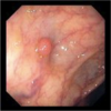
<p>SB carcinoid tumor</p>
<ul>
<li>Found in \_\_\_\_\_\_\_\_</li>
<li>Clinical Presentation</li>
<li>Path Features</li>
<li>Prognosis</li>
</ul>

<p>SB carcinoid tumor</p>
<ul>
<li>Found in: appendix #1 place; followed by followed by the small intestine (primarily ileum), rectum, colon, and stomach.
<ul>
<li>also:bronchial tree, GU & GYN tracts</li>
</ul>
</li>
<li>Clinical Presentation: MOST COMMON SB MALIG
<ul>
<li>pain/obstruction most common</li>
<li>rare: carcinoid syndrome
<ul>
<li>Occurs in <10% w/ malig. carcinoids, usu with extensive liver metastases</li>
<li>Release of serotonin into systemic circ. leads to</li>
<li>skin flushing/cyanosis</li>
<li>diarrhea & cramps</li>
<li>bronchospasm</li>
<li>R ventricular subendocardial fibrosis -></li>
<li>Pulmonic & tricuspid valve stenosis/regurge</li>
</ul>
</li>
</ul>
</li>
<li>Path Features
<ul>
<li>yellow submucosa (pic on reverse)</li>
<li>tumor invades muscularis propria, a characteristic muscle fiber stranding</li>
<li>fibrosis often severe enough to kink →obstruction. </li>
<li>organoid (seen here) or gyrating histo</li>
</ul>
</li>
<li>Prognosis:Site, depth of invasion, and size are prognostic indicators:
<ul>
<li><strong>Having symptoms is bad</strong>: 90% of symptomatic pts have mets
<ul>
<li>often found incidentally</li>
</ul>
</li>
<li>appendicial and rectal: likely benign</li>
<li>< 2cm: likely benign</li>
<li>hasn't invaded muscularis mucosa: likely benign</li>
</ul>
</li>
</ul>

<p>
Dx of carcinoid cancers</p>
<ul>
<li>
can use octreotide test: nearly all carcinoid tumors have somatostatin receptors</li>
<li>
CT, biopsy, blah blah</li>
</ul>
<p>SB lymphoma</p>
<ul>
<li>Found in \_\_\_\_\_\_\_\_</li>
<li>Clinical Presentation</li>
<li>Risk Factors</li>
<li>Path Features</li>
<li>Prognosis</li>
<li>Treatment options</li>
</ul>
<p>SB lymphoma</p>
<ul>
<li>Found in: GIT = commonest site for 1° extranodal lymphomas.
<ul>
<li>most often stomach followed by SB, ileocecal region and colon </li>
</ul>
</li>
<li>Clinical Presentation</li>
<li>Risk Factors: 1° GI lymphomas are commonly associated with:</li>
<li>
<ul>
<li>chronic infection (H. pylori gastritis)</li>
<li>celiac disease</li>
<li>immunodeficiency (AIDS, immunosuppression), and</li>
<li>Crohn's disease</li>
<li>Can be sporadic also. </li>
</ul>
</li>
<li>Path Features
<ul>
<li>2 types in both stomach and intestines: DLBCL and Marginal zone types</li>
</ul>
</li>
<li>Prognosis:
<ul>
<li>Lymphomas have a much better prognosis than carcinomas.</li>
<li>5 yr survival rate depends on the stage and histologic type, but, overall, it is >50%.</li>
</ul>
</li>
</ul>
<p>
SB GIST tumor basics</p>
<ul>
<li>
Mesenchymal tumor resembling modified smooth muscle and neural tissue
<ul>
<li>
Hypothesized origin = <u>Interstitial cells of Cajal</u></li>
</ul>
</li>
<li>
Resides in muscularis mucosa (so deep-seated tumor)</li>
<li>
CD117 positive (c-<u>kit tyrosine kinase</u>)</li>
<li>
Spectrum from benign to malignant</li>
</ul>
<p>
Cancers that met to SB</p>
<p>
Melanoma, breast carcinoma, lung carcinoma, renal cell carcinoma. </p>
<p>
#1 gene mutation in colon ca</p>
<p>
APC gene mutations are usually the earliest and, possibly, the initiating event in about 80% of sporadic colon cancers. With clonal progression, additional mutations accumulate, such as in K-­ras and p53 genes. </p>
<p>
When do most CRCs occur?</p>
<p>
90% occur in pts > 50 y/o</p>
<p>
If you see CRC in younger pts you should think</p>
<p>
think FAP, HNPCC, MUTYH, PJS, or IBD</p>
<p>
FAP</p>
<ul>
<li>
caused by?</li>
<li>
present at what age? how?</li>
<li>
natural hx?</li>
<li>
tx?</li>
</ul>
<p>
<strong>Familial Adenomatous Polyposis</strong></p>
<ul>
<li>
-Autosomal dominant disorder
<ul>
<li>
however, <u>25% of pts have no family hx</u></li>
</ul>
</li>
<li>
-mutation in the APC (Adenomatous polyposis coli) gene → > 300 mutation ID's</li>
<li>
-present in the 2nd to 3rd decades w/ 100's or 1000's of adenomatous polyps</li>
<li>
-rate of progression ~variable, but all patients will eventually develop colonic carcinoma(s) in early adult life (age 39)
<ul>
<li>
assoc'd w/ lots of other tumors</li>
</ul>
</li>
<li>
-the treatment is total colectomy.
<ul>
<li>
different options: some with more sparing of fxn than others</li>
</ul>
</li>
</ul>
MYH Polyps
- caused by?
- present at what age? how?
- natural hx?
- tx?
-
caused by: you have to have bi-allelic mutation in MutY Homolog (base excision repair gene)
- can lead to mutation of APC which is esp susceptible to these lesions
-
presentation can be the same as FAP but usually < 100 polyps
- Mean age at cancer dx = 45 yrs
- natural hx?
- tx?
HNPCC
- caused by?
- present at what age? how?
- natural hx?
- tx?
Hereditary Non-Polyposis Colon Cancer
-
Caused by A.D. inherited germline mutation in mismatch repair genes (MSH2, MLH1, MSH6, PMS2)
- correct microsatellite repeats that increas r/o mutation
- can do IHC to see qualitatively if these are missing
-
Present with two types
- Lynch I = CRC ca's only
- Lynch II = CRC ca's + Endometrial, ovarian, pancreatic, gastric, SB, transitional cell of kidney/ureter
-
Have adenomatous polyps that are slightly more dysplastic
- Larger with a villous or dysplastic component
- Flat adenomas/serrated adenomas
- More present in proximal colon
- present early avg = 45 y/o
- higher incidence of metachronous cancer
- however, no big phenotypic differences → difficult dx
-
natural hx:
- earlier dx (~45 y/o)
- tumors progress to cancer faster (~3.5 years) so need more screening
- BUT! Better cancer-specific survival!!
PJS
Peutz-Jeghers Syndrome:
-Autosomal dominant
-Multiple hamartomatous polyps of the entire GIT (esp SB)
-Mucocutaneous melanosis (lips & buccal mucosa).
-PJ polyps themselves have no malignant potential, but these patients have a higher incidence of carcinoma elsewhere in the colon
-Increased frequency of extra-GI cancer
Potential lifestyle, diet, meds that protect against CRC
- COX2 inhibition
- fiber
- diet/exercise
- Vit b6
-
Calcium and vitamin D
- Calcium believed to bind fatty and bile acids
- Folic Acid - deficiency may -> DNA hypermethylation
- Omega 3s
Are most CRCs caused by inheritied syndromes or sporadic occurence?

Polyps
- Two types
- Causes of and percent that are neoplastic
- Cause of and percent that are non-neoplastic
- sessile and pedunculated
-
90% are non-neoplastic caused by:
- Hyperplastic
- Hamartomatous polyps (Juvenile, PJS)
- Inflam. pseudopolyps
- neoplastic (adenomas) are only 10%
Tubular adenomatous polyp
- appearance
- malig risk

Tubular
- LOWEST MALIG RISK
-
small and pedunculated
- polyp only in head
- most pedunculated polyps are TAs

Tubulovilous adenoma polyp
- apperance
- malig risk
mix of other two (Combined tubular and villous architecture)
Risk of harboring in situ or invasive carcinoma generally correlates with the proportion of the lesion that is villous (i.e. more villous = worse)

vilous adenoma polyp


Hyperplastic polyp
- malig risk
- appearance
- other
- benign
-
Small (usually <5 mm) sessile polyps
- Elongated and serrated, stellate crypts lined by “hypermature” goblet cells
- Most common in the rectum/sigmoid

Juvenile or Retention Polyp
- Rare A.D. hamartomatous malformations
- Most frequent in the rectum
- Most often in children (<5yoa)
- Usually large (1 to 3 cm), round, smooth lesions with stalks
- Inflamed lamina propria with cystically dilated glands
- No malignant potential but slight incr risk of adenomas and adenocarcinoma
PJS

Peutz-Jeghers Polyp
- Hamartomatous
- Branching smooth muscle and glands lined by goblet cells
- Slight malignant potential
- (LOH at LKB1 locus)
- PJS pts at increased risk of developing CA of pancreas, breast, lung, ovary, and uterus.

OBJECTIVE: Outline the different screening options and recommendations for CRC.
Starting at age 50 USPSTF gives A-level evidence for:
- FOBT - every year, if normal; (if abnormal order colo)
- Sigmoidoscopy - Every five years if normal (more often and/or order full colo if polyp removed)
- Colonoscopy - Every ten years if normal (more often if polyps removed)
Classic presentation and pathological apperance of left- vs. right-sided colon tumors?
Left - obstructs apple core
- grow as annular, circumferential ("napkin ring"), ulcerated masses
Right - bleeds cauliflower
- grow as polypoid, exophytic masse
- May still be deeply infiltrating
- Obstruction is uncommon
- bleeding either occult or melena
Staging of tumors
Uses TNM (tumors, nodes, mets) rubric
pT is based on how far it invades, you just add one number for each layer of the colon it busts through (p stands for primary tumor)
N is number of nodes involved
M is number of distant mets
- pTis =
- Intraepithelial carcinoma (Severe dysplasia)
- No met risk
- Intramucosal carcinoma: Ltd to lamina propria.
- Very low met risk
- Intraepithelial carcinoma (Severe dysplasia)
- pT1 = Invasion into submucosa
- 95% survival
- pT2 = Into muscularis propria
- pT3 = Through muscularis propria
- pT4 = Penetration of serosa or invasion of adjacent viscera
- 30% survival

Barium enema in pt w/ ∆ in bowel habits shows the following. Thoughts? n/ame for sign?

This is the apple core sign in the sigmoid colon which is suggestive of the annular, napkin ring-type lesions typical of the L colon.
What are the main goals of surgery to resect CRC?
- to stage dz
- Resection of 1° lesion, w/ en-bloc excision of adjacent organ extension PRN
- Remove regional LNs
- They follow the regional blood supply
- Remove isolated mets
Lynch Syndrome
Hereditary nonpolyposis colorectal cancer (HNPCC)
—CRCs due to Lynch Syndrome are at higher risk for malignancy/seriousness
