XII - The Hematopoietic and Lymphoid Systems Flashcards

1
Q

Average volume per red blood cell

A

Mean cell volume (MCV)(TOPNOTCH)Robbins Basic Pathology, 8th Ed. p. 423

2
Q

A reduction in the oxygen-transporting capacity of blood.

A

Anemia(TOPNOTCH)Robbins Basic Pathology, 8th Ed. p. 422

3
Q

The average content of hemoglobin per red cell

A

Mean cell hemoglobin (MCH)(TOPNOTCH)Robbins Basic Pathology, 8th Ed. p. 423

4
Q

The average concentration of hemoglobin in a given volume of packed red cells, expressed in g/dL.

A

Mean cell hemoglobin concentration (MCHC)(TOPNOTCH)Robbins Basic Pathology, 8th Ed. p. 423

5
Q

The coefficient of variation of red cell volume.

A

Red cell distribution width (RDW)(TOPNOTCH)Robbins Basic Pathology, 8th Ed. p. 423

6
Q

Anemia of acute blood loss is described as ______.

A

Normocytic, normochromic anemia(TOPNOTCH)Robbins Basic Pathology, 8th Ed. p. 423

7
Q

Life span of a normal red cell.

A

120 days(TOPNOTCH)Robbins Basic Pathology, 8th Ed. p. 424

8
Q

Anemia characterized by an increased rate of cell destruction. There is a compensatory increase in erythropoeisis (seen as inceased reticulocyte count), and retention of cell destruction products, like iron.

A

Hemolytic anemia(TOPNOTCH)Robbins Basic Pathology, 8th Ed. p. 424

9
Q

A circulating protein that binds and clears free hemoglobin.

A

Haptoglobin(TOPNOTCH)Robbins Basic Pathology, 8th Ed. p. 424

10
Q

Hemolysis that can result from mechanical trauma, or biochemical or physical agents that damage the red cell membrane.

A

Intravascular hemolysis(TOPNOTCH)Robbins Basic Pathology, 8th Ed. p. 424

11
Q

Hemolysis which takes place largely within phagocytic cells of the spleen and liver.

A

Extravascular hemolysis(TOPNOTCH)Robbins Basic Pathology, 8th Ed. p. 424

12
Q

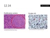
This disorder is characterized by an intrinsic defect in the red cell membrane, that renders the cells spheroidal, less defomable and vulnerable to splenic sequestration and destruction. SEE SLIDE 12.1

A

Hereditary spherocytosis(TOPNOTCH)Robbins Basic Pathology, 8th Ed. p. 424

13
Q

Small, dark nuclear remnants seen within red cells in PBS of hereditary spherocytosis. SEE SLIDE 12.2

A

Howell-Jolly bodies(TOPNOTCH)Robbins Basic Pathology, 8th Ed. p. 425

14
Q

On PBS, red cells are spherical which lack central pallor, and they show increased osmotic fragility when placed in hypotonic salt solutions. SEE SLIDE 12.1

A

Hereditary spherocytosis(TOPNOTCH)Robbins Basic Pathology, 8th Ed. p. 425

15
Q

Structural proteins that are defective in hereditary spherocytosis.

A

Spectrin and ankyrin(TOPNOTCH)Robbins Basic Pathology, 8th Ed. p. 425

16
Q

This results from substitution of valine for glutamic acid at the 6th position of the B-chain, producing HbS.

A

Sickle cell anemia(TOPNOTCH)Robbins Basic Pathology, 8th Ed. p. 426

17
Q

Bizarre, elongated, spindled or boat-shaped cells on PBS. SEE SLIDE 12.3

A

Sickle cell anemia(TOPNOTCH)Robbins Basic Pathology, 8th Ed. p. 427

18
Q

Prominent cheekbones and changes in skull resembling a “crew-cut” skull x-ray.

A

Sickle cell anemia(TOPNOTCH)Robbins Basic Pathology, 8th Ed. p. 427

19
Q

Patients with sickle cell disease are predisposed to infections caused by these type of bacteria.

A

Encapsulated bacteria(TOPNOTCH)Robbins Basic Pathology, 8th Ed. p. 428

20
Q

Treatment for sickle cell disease by increasing levels of HbF.

A

Hydroxyurea(TOPNOTCH)

21
Q

Treatment for sickle cell disease by increasing levels of HbF.

A

Hydroxyurea(TOPNOTCH)Robbins Basic Pathology, 8th Ed. p. 428

22
Q

Feared complication of sickle cell disease which can be trigerred by pulmonary infections or fat emboli from necrotic marrow that secondarily involve the lung.

A

Acute chest syndrome(TOPNOTCH)Robbins Basic Pathology, 8th Ed. p. 428

23
Q

Major complication of sickle cell disease which occurs in the setting of acute chest syndrome, causing ischemic injury to the CNS.

A

CNS stroke(TOPNOTCH)Robbins Basic Pathology, 8th Ed. p. 428

24
Q

Represents a sudden but usually temporary cessation of erythropoeisis, usually trigerred by parvovirus B19 infections in patients with sickle cell disease.

A

Aplastic crises(TOPNOTCH)Robbins Basic Pathology, 8th Ed. p. 428

25
In beta thalassemia, an individual who inherits one abnormal allele (out of 2) has this asymptomatic to mildly symptomatic condition.
B- Thalassemia minor/trait(TOPNOTCH)Robbins Basic Pathology, 8th Ed. p. 429
26
Individuals with B-thalassemia who inherit two abnormal alleles, with severe anemia requiring regular blood tranfusions.
B- Thalassemia major(TOPNOTCH)Robbins Basic Pathology, 8th Ed. p. 429
27
Red cells with a central, dark-red puddle due to collection of hemoglobin.
Target cells(TOPNOTCH)Robbins Basic Pathology, 8th Ed. p. 430
28
Target cells are often seen in this condition.
B-thalassemia minor(TOPNOTCH)Robbins Basic Pathology, 8th Ed. p. 430
29
In the PBS of this condition, nucleated red cells (normoblasts) are seen, which reflect underlying erythropoeisis.
B-thalassemia major(TOPNOTCH)Robbins Basic Pathology, 8th Ed. p. 430
30
Anemia of beta thalassemia.
Microcytic, hypochromic(TOPNOTCH)Robbins Basic Pathology, 8th Ed. p. 430
31
Disease caused by deletion of 3 alpha globin genes.
Hemoglobin H disease(TOPNOTCH)Robbins Basic Pathology, 8th Ed. p. 431
32
Condition caused by deletion of 1 alpha globin gene.
Silent carrier(TOPNOTCH)Robbins Basic Pathology, 8th Ed. p. 431
33
Condition caused by deletion of 2 alpha globin genes.
Alpha thalassemia trait(TOPNOTCH)Robbins Basic Pathology, 8th Ed. p. 431
34
Condition caused by deletion of all four alpha globin genes.
Hydrops fetalis(TOPNOTCH)Robbins Basic Pathology, 8th Ed. p. 429
35
Precipitates of denatured globin seen in RBC's. SEE SLIDE 12.4.
Heinz bodies(TOPNOTCH)Robbins Basic Pathology, 8th Ed. p. 432
36
Heinz bodies are seen in the blood smear of this condition.
G6PD Deficiency(TOPNOTCH)Robbins Basic Pathology, 8th Ed. p. 432
37
Bite cells are seen in \_\_\_\_\_\_\_\_. SEE SLIDE 12.5.
G6PD Deficiency(TOPNOTCH)Robbins Basic Pathology, 8th Ed. p. 432
38
Hemolysis caused by IgG or IgA antiodies that are active at 37 degC, which results in opsonization of red cells by the autoantibodies.
Warm antibody immunohemolytic anemia(TOPNOTCH)Robbins Basic Pathology, 8th Ed. p. 433
39
Anemia caused by low-affinity IgM which bind to red cell membranes only at temp
Cold antibody immunohemolytic anemia(TOPNOTCH)Robbins Basic Pathology, 8th Ed. p. 433
40
Anemia observed in a variety of pathologic states, in which small vessels become particularly obstructed.(e.g. DIC, malignant HTN, SLE, etc.)
Microangiopathic hemolytic anemia(TOPNOTCH)Robbins Basic Pathology, 8th Ed. p. 433
41
Schistiocytes, burr cells, helmet cells, triangle cells are seen in this condition. SEE SLIDE 12.6
Microangiopathic hemolytic anemia(TOPNOTCH)Robbins Basic Pathology, 8th Ed. p. 433
42
X-linked disorder in which red cells are unusually susceptible to damage cause by oxidants. (Drugs that produce oxidants include antimalarials, sulfonamides, nitrofurantoin, phenacetin, and vitamin K derivatives)
G6PD Deficiency(TOPNOTCH)Robbins Basic Pathology, 8th Ed. p. 432
43
Red blood cells in iron deficiency anemia.
Microcytic, hypochromic(TOPNOTCH)Robbins Basic Pathology, 8th Ed. p. 436
44
Diagnostic criteria for iron deficiency anemia:\_\_\_\_ Ferritin\_\_\_\_Serum iron level\_\_\_\_Transferrin saturation\_\_\_\_Total Iron Binding Capacity (TIBC)
Low ferritinLow serum iron levelsLow transferrin saturationIncreased TIBC(TOPNOTCH)Robbins Basic Pathology, 8th Ed. p. 435
45
Diagnostic criteria for anemia of chronic disease:\_\_\_\_ Ferritin\_\_\_\_Serum iron level\_\_\_\_Transferrin saturation\_\_\_\_Total Iron Binding Capacity (TIBC)
Increased ferritinLow serum iron levelsLow transferrin saturationDecreased TIBCNormocytic, normochromic anemia(TOPNOTCH)Robbins Basic Pathology, 8th Ed. p. 435
46
Principal causes of megaloblastic anemia.
Folate deficiencyVitamin B12 deficiency(TOPNOTCH)Robbins Basic Pathology, 8th Ed. p. 437
47
Bone marrow is markedly hypercellular as a result of increased number of megaloblasts, which are large cells that have delicate, finely reticulated nuclear chromatin and abundant basophilic cytoplasm.
Megaloblastic anemia(TOPNOTCH)Robbins Basic Pathology, 8th Ed. p. 437
48
PBS finding in neutrophils and red cells of patients with megaloblastic anemia.
Hypersegmented neutrophils, large, egg-shaped macro-ovalocytes. SEE SLIDE 12.7 (TOPNOTCH)Robbins Basic Pathology, 8th Ed. p. 437
49
Difference between megaloblastic and pernicious anemia.
Presence of neurologic abnormalities in pernicious anemia.(TOPNOTCH)Robbins Basic Pathology, 8th Ed. p. 438
50
Deficiency in folate causes this type of anemia.
Megaloblastic anemia(TOPNOTCH)Robbins Basic Pathology, 8th Ed. p. 438
51
Deficiency in Vitamin B12 causes this type of anemia.
Pernicious anemia(TOPNOTCH)Robbins Basic Pathology, 8th Ed. p. 438
52
Etiology of pernicious anemia.
1.Vitamin B12 malabsorption secondary to autoantibodies against parietal cells and intrinsic factor which is needed in its absorption.2. Gastrectomy or ileal resection(TOPNOTCH)Robbins Basic Pathology, 8th Ed. p. 438
53
Principal neurologic lesion in pernicious anemia.
Demyelination of posterior and lateral columns of the spinal cord.(TOPNOTCH)Robbins Basic Pathology, 8th Ed. p. 439
54
Bone marrow is markedly hypocellular, with \>90% of the intertrabecular space being occupied by fat. SEE SLIDE 12.8
Aplastic anemia(TOPNOTCH)Robbins Basic Pathology, 8th Ed. p. 439
55
Tear drop cells are also called \_\_\_\_\_\_\_\_\_\_.
Dacrocytes(TOPNOTCH)Robbins Basic Pathology, 8th Ed. p. 440
56
Dacrocytes are found in peripheral blood of patients with this type of anemia.
Myelophthisic anemia(TOPNOTCH)Robbins Basic Pathology, 8th Ed. p. 440
57
Increase in blood concentration of red cells, with an increase in Hgb concentration.
Polycythemia or erythrocytosis(TOPNOTCH)Robbins Basic Pathology, 8th Ed. p. 441
58
Polycythemia secondary to reduced plasma volume.
Relative polycythemia(TOPNOTCH)Robbins Basic Pathology, 8th Ed. p. 441
59
Polycythemia secondary to abnormal proliferation of myeloid stem cells and low erythropoeitin levels.
Primary polycythemia (Absolute)(TOPNOTCH)Robbins Basic Pathology, 8th Ed. p. 441
60
Polycythemia secondary to increased erythropoeitin levels due to lung disease, high-altitude living, cyanotic heart disease and EPO secreting tumors.
Secondary polycythemia (Absolute)(TOPNOTCH)Robbins Basic Pathology, 8th Ed. p. 441
61
Total white cell count is reduced to 1000 cells/uL. Affected persons are extremely susceptible to bacterial and fungal infections.
Neutropenia(TOPNOTCH)Robbins Basic Pathology, 8th Ed. p. 441
62
A self-limited disease of adolescents and young adults that is causd by B lymphocytotropic EBV characterized by fever, sore throat and generalized lymphadenitis, an increase of atypical lymphocytes in blood and an antibody and T cell response to EBV.
Infectious mononucleosis(TOPNOTCH)Robbins Basic Pathology, 8th Ed. p. 442
63
T Cells with abundant cytoplasm 12-16um in diameter that contains azurophilic granules, and an oval, indented or folded nucleus. SEE SLIDE 12.9
Atypical lymphocytes (Infectious mononucleosis)(TOPNOTCH)Robbins Basic Pathology, 8th Ed. p. 442
64
Inflamed nodes are swollen, gray-red and engorged. There are large germinal centers containing numerous mitotic figures. Affected nodes are tender and fluctuant if with extensive abscess formation.
Acute nonspecific lymphadenitis(TOPNOTCH)Robbins Basic Pathology, 8th Ed. p. 444
65
Reactive pattern characterized by distention and prominence of the lymphatic sinusoids, due to marked hypertrophy of lining endothelial cells and an infiltrate of histiocytes. Encountered in lymph nodes draining cancers.
Sinus histiocytosis(TOPNOTCH)
66
Characterized by reactive changes within T-cell regions of the lymph node, usually encountered during viral infections, following certain vaccinations, and immune reactions induced by certain drugs.
Paracortical hyperplasia(TOPNOTCH)Robbins Basic Pathology, 8th Ed. p. 444
67
Causative agent for cat scratch disease(TOPNOTCH)
Bartonella henselae(TOPNOTCH)Robbins Basic Pathology, 8th Ed. p. 444
68
Lymphoblasts with irregular nuclear contours, condensed chromatin , small nucleoli and scant agranular cytoplasm. SEE SLIDE 12.10. Blasts compose \>25% of marrow cellularity. Most common childhood leukemia.
Acute lymphocytic leukemia (ALL)(TOPNOTCH)Robbins Basic Pathology, 8th Ed. p. 447
69
Frequent small "cleaved" cells mixed with large cells, growth pattern is NODULAR, centroblasts present. SEE SLIDE 12.11. Occurs in older adults, usually involves nodes, marrow, spleen. Associated with t(14;18) that results in overexpression of cyclin D1.
Follicular lymphoma(TOPNOTCH)Robbins Basic Pathology, 9th Ed. p. 443
70
Small to intermediate-sized irregular lymphocytes growing in a diffuse pattern, no centroblasts and proliferation centers. SEE SLIDE 12.12. Occurs mainly in older males, GI tract commonly affected. Associated with t(11;14) that results in overexpression of cyclin D1, a regulator of the cell cycle.
Mantle cell lymphoma(TOPNOTCH)Robbins Basic Pathology, 9th Ed. p. 443
71
Plasma cells in sheets, with prominent nucleoli or inclusion containing Ig. Presents as disseminated bone disease, with destructive lytic lesions.
Plasmacytoma / plasma cell myeloma(TOPNOTCH)
72
Intermediate-sized round lymphoid cells with 2-5 prominent nucleoli. High rates of proliferation and apoptosis are characteristic. Nuclear remnants phagocytosed by interspersed macrophages with abundant clear cytoplasm, "starry sky pattern" SEE SLIDE 12.13.
Burkitt lymphoma(TOPNOTCH)Robbins Basic Pathology, 8th Ed. p. 453
73
Sheets of small, round lymphocytes and scattered ill-defined foci of larger, actively dividing cells diffusely efface involved LN. A foci of mitotically active cells called proliferation centers are pathognomonic. SEE SLIDE 12.14.
Small lymphocytic leukemia (SLL) / Chronic lymphocytic leukemia (CLL)(TOPNOTCH)Robbins Basic Pathology, 8th Ed. p. 450
74
Fragile neoplastic lymphocytes that are frequently disrupted during smear preparation.
Smudge cells. SEE SLIDE 12.14 (TOPNOTCH)Robbins Basic Pathology, 8th Ed. p. 450
75
Smudge cells are seen in this type of leukemia. SEE SLIDE 12.14
CLL/SLL. There is absolute lymphocytosis of MATURE-LOOKING lymphocytes, but they are very fragile, hence the smudge cells. (TOPNOTCH)Robbins Basic Pathology, 8th Ed. p. 450
76
Tumor cells have large nuclei with open chromatin and prominent nucleoli. Most important type of lymphoma in adults, accounting to ~50% of adult NHL.
Diffuse large B-cell lymphoma(TOPNOTCH)Robbins Basic Pathology, 8th Ed. p. 452
77
Disease that presents as multifocal destructive bone lesions seen as punched-out defects on imaging. Renal involvement is also prominent, causing production of proteinaceous casts in the DCT and collecting ducts.
Multiple myeloma(TOPNOTCH)Robbins Basic Pathology, 9th Ed. p. 438
78
Excess light or heavy chains along with complete Igs synthesized by neoplastic plasma cells.
Bence-Jones proteins(TOPNOTCH)Robbins Basic Pathology, 8th Ed. p. 454
79
Cells with two mirror-image nuclei or nuclear lobes, each containing a large acidophilic nucleolus surrounded by a distinctive clear zone, imparting an owl-like appearance. The sine qua non of Hodgkin Lymphoma.
Reed-Sternberg cell. SEE SLIDE 12.15(TOPNOTCH)Robbins Basic Pathology, 9th Ed. p. 440
80
A distinctive groups of neoplasms that arise almost invariably in a single lymph node or chain of lymph nodes and spread characteristically in a stepwise fashion to anatomically contiguous nodes.
Hodgkin Lymphoma(TOPNOTCH)Robbins Basic Pathology, 8th Ed. p. 456
81
Most common form of Hodgkin lymphoma, characterized by a large cell which has a single multilobate nucleus with small nucleoli and an abundant pale-staining cytoplasm called lacunar cells. Reed Sternberg cells are uncommon. There are also collagen bands that divide the tissue into circumscribed nodules, hence the name. SEE SLIDE 12.16.
Nodular sclerosis HL(TOPNOTCH)Robbins Basic Pathology, 9th Ed. p. 441
82
Most common form of HL in patients greater than 50 years old, with male predominance, plentiful RS cells and heterogenous cellular infiltrates.
Mixed cellularity HL(TOPNOTCH)Robbins Basic Pathology, 8th Ed. p. 458
83
Cells found in adult T-cell lymphoma which appear to have multilobulated nuclei.
Cloverleaf or flower cell. SEE SLIDE 12.17. (TOPNOTCH)
84
Subgroup of HL characterized by a large number of small resting lymphocytes admixed with a variable number of benign histiocytes. Variant RS cells described as multilobed, puffy nucleus Which appears like a "popcorn". Excellent prognosis. SEE SLIDE 12.18.
Lymphocyte-predominance HL(TOPNOTCH)Robbins Basic Pathology, 8th Ed. p. 458
85
Cells with fiery red cytoplasm, seen in Multiple myeloma. SEE SLIDE 12.19
Flame cells(TOPNOTCH)
86
Cells with pink globular cytoplasmic inclusions, seen in Multiple myeloma.
Russell bodies. SEE SLIDE 12.20 (TOPNOTCH)
87
Cells with blue globular nuclear inclusions, seen in Multiple myeloma
Dutcher bodies. SEE SLIDE 12.20 (TOPNOTCH)
88
Multiple nuclei, prominent nucleoli, and cytoplasmic droplets containing Ig.
Bizarre, multinucleated cells(TOPNOTCH)
89
Bizarre multinucleated cells, flame cells, Russel bodies and Dutcher bodies are all seen in what disease? SEE SLIDES 12.19 and 12.20
Multiple myeloma(TOPNOTCH)
90
Bone marrow aspirate shows hypercellular martow packed with myeloblasts and azurophilic needle-like material called Auer rods. SEE SLIDE 12.21
Acute Myelogenous Leukemia. Particularly numerous in acute promyelocytic leukemia. (TOPNOTCH)Robbins Basic Pathology, 8th Ed. p. 462
91
Leukemia of children most responsive to chemotherapy.
Acute Lymphoblastic Leukemia(TOPNOTCH)Robbins Basic Pathology, 8th Ed. p. 461
92
Hodgkin lymphoma subgroup most commonly associated with EBV infection.
Lymphocyte depleted(TOPNOTCH)
93
Mature B-cell tumor in the elderly, where cells have fine hair-like projections (hairy cells). SEE SLIDE 12.22
Hairy cell leukemia(TOPNOTCH)Robbins Basic Pathology, 8th Ed. p. 459
94
t(9;22) is also called \_\_\_\_\_\_.
Philadelphia chromosome(TOPNOTCH)Robbins Basic Pathology, 8th Ed. p. 465
95
Hodgkin lymphoma subgroup with highest count of RS cells.
Mixed cellularity type(TOPNOTCH)
96
This correlates with good prognosis in Hodgkin lymphoma.
High Lymphocyte : Reed-Sternberg cell ratio(TOPNOTCH)
97
Hodgin lymphoma subgroup not associated with EBV.
Nodular sclerosis and lymphocyte predominant(TOPNOTCH)
98
Hodgkin lymphoma subgroup with poorest prognosis.
Lymphocyte depleted HL(TOPNOTCH)
99
Tumor of the thymus associated with myasthenia gravis and pure red cell aplasia.
Thymoma(TOPNOTCH)
100
Pentalaminar tubules, often with a dilated terminal end (tennis racket-like appearance).
Birbeck granules. SEE SLIDE 12.23(TOPNOTCH)Robbins Basic Pathology, 8th Ed. p. 467
101
Proliferative disorder of the dendritic cells which has birbeck granules. SEE SLIDE 12.23
Langerhans Cell Histiocytosis(TOPNOTCH)Robbins Basic Pathology, 8th Ed. p. 467
102
Pathology behind polycythemia vera.
Mutation in tyrosine kinase JAK2, which acts in signalling pathways of the erythropoeitin receptors, rendering them hypersensitive to erythropoeitin.(TOPNOTCH)Robbins Basic Pathology, 8th Ed. p. 466
103
Collection of aggressive tumors that are comprised of immature myeloblasts which replace the marrow and suppress normal hematopoiesis.
Acute Myelogenous Leukemia(TOPNOTCH)Robbins Basic Pathology, 8th Ed. p. 467
104
Myeloid tumor arising from a pluripotent stem cell associated with mutatios of the BCR-ABL gene. If untreated, may progress to a blast crisis.
Chronic Myelogenous Leukemia(TOPNOTCH)Robbins Basic Pathology, 8th Ed. p. 467
105
Most common myelodysplastic syndrome. A myeloid tumor in which abnormal megakaryocytes stimulate marrow fibroblasts to release collagen, replacing the marrow space, leading to pancytopenia and extramedullary hematopoeisis.
Myeloid metaplasia with Myelofibrosis(TOPNOTCH)Robbins Basic Pathology, 8th Ed. p. 467
106
Other name for acute disseminated Langerhans cell histiocytosis.
Letterer-Siwe disease(TOPNOTCH)Robbins Basic Pathology, 8th Ed. p. 468
107
Caused by a systemic activation of coagulation pathways, leading to formation of thrombi throughout the microcirculation.
Disseminated intravascular coagulation(TOPNOTCH)Robbins Basic Pathology, 8th Ed. p. 469
108
Characterized by spontaneous bleeding, prolonged bleeding time, and normal PT and PTT.
Thrombocytopenia(TOPNOTCH)Robbins Basic Pathology, 8th Ed. p. 471
109
Drug-induced disorder caused by IgG antibodies that bind to platelet factor IV on platelet surfaces, which activates platelets and induce their aggregation.
Heparin-Induced Thrombocytopenia (HIT)(TOPNOTCH)Robbins Basic Pathology, 8th Ed. p. 472
110
Associated with pentad of fever, thrombocytopenia, microangipathic hemolytic anemia, transient neurologic deficits and renal failure.
Thrombotic thrombocytic purpura(TOPNOTCH)Robbins Basic Pathology, 8th Ed. p. 472
111
Associated with childhood onset microangiopathic hemolytic anemia, thrombocytopenia, and acute renal failure. No neurologic symptoms. Often with a history of bloody diarrhea.
Hemolytic uremic syndrome(TOPNOTCH)Robbins Basic Pathology, 9th Ed. p. 454
112
Caused by antiplatelet antibodies directed against glycoproteins IIb-IIIa, Ib-IX.
Immune thrombocytopenic purpura(TOPNOTCH)Robbins Basic Pathology, 8th Ed. p. 472
113
Decreased gp Ib leads to defective platelet adhesion, associated with decreased platelet count.
Bernard-Soulier Syndrome(TOPNOTCH)
114
Caused by deficiency of ADAMTS13, a vWF metalloprotease.
Thrombotic thrombocytopenic purpura(TOPNOTCH)Robbins Basic Pathology, 8th Ed. p. 472
115
Caused by shiga-like toxin in EHEC (E. coli O157:H7) from improperly cooked burgers.
Hemolytic uremic syndrome(TOPNOTCH)Robbins Basic Pathology, 8th Ed. p. 472
116
Decreased gp IIb-IIIa leads to defective platelet aggregation, associated with normal platelet count.
Glanzmann thrombasthenia(TOPNOTCH)
117
Most common bleeding disorder.
vWF disease(TOPNOTCH)Robbins Basic Pathology, 8th Ed. p. 474
118
Most common hereditary disease associated with life threatening bleeding,
Hemophilia A(TOPNOTCH)Robbins Basic Pathology, 8th Ed. p. 474
119
An X-linked recessive disorder caused by reduction in factor VII activity.
Classic Hemophilia/ Hemophilia A(TOPNOTCH)Robbins Basic Pathology, 8th Ed. p. 474
120
An X-linked disorder caused by deficiency of Factor IX, or Christmas factor. Bleeding time is normal, PTT is prolonged.
Hemophilia B/ Christmas Disease(TOPNOTCH)Robbins Basic Pathology, 8th Ed. p. 474
121
A state associated with excessive removal of formed elements of blood, resulting in anemia, leukopenia or thrombocytopenia.
Hypersplenism(TOPNOTCH)Robbins Basic Pathology, 8th Ed. p. 476
122
A 17 y/o male presented with a short history of fever, tonsillitis and monolateral enlarged cervical lymph nodes. PE revealed enlargement of righ cervical lymph node, 3 cm in diameter hard , and pharyngeal hyperemia. Biopsy was done and showed a malignant population of round monomorphic B cells interspersed with macrophages forming the start in the "starry sky" pattern. SEE SLIDE 12.13. This is a case of:
Burkitt lymphoma (TOPNOTCH)
123
An 18 y/o male presents with easy fatigability, fever, and cutaneous bleeding. Bone marrow biopsy showed 40% myeloblasts. What is the most likely diagnosis?
AML (TOPNOTCH)
124
The most common cause of agranulocytosis
Drug toxicity (TOPNOTCH) Robbins Pathologic Basis of Disease, 9th ed., p. 582
125
What laboratory finding differentiate leukemoid reaction from CML?
Elevated leukocyte alkaline phosphatase. (TOPNOTCH)
126
An 8 month old presented with anemia. Red cells were noted to be oval and macrocytic. Nutritional history revealed that the infant was exclusively fed raw goat's milk. The most likely cause of his anemia is:
Folate deficiency (TOPNOTCH)
127
A 2 y/o child arrived at well-child clinic. Mother reported that her child is exclusively breastfed for 6 months and then supplemented mainly by carrots. The patient is most likely prone to developing anemia caused by:
Vitamin B12 deficiency (TOPNOTCH)
128
Most common type of cancer in children; highly aggressive tumors manifest with signs and symptoms of bone marrow failure, or as rapidly growing masses
Acute lymphoblastic leukemia/Lymphoblastic lymphoma(TOPNOTCH) Robbins Basic Pathology, 9th ed., p. 598
129
Most common leukemia of adults which usually presents with bone marrow and lymph node involvement.
Small lymphocytic lymphoma/CLL(TOPNOTCH) Robbins Basic Pathology, 9th ed., p. 598
130
Most common leukemia of adults which usually presents with bone marrow and lymph node involvement.
Follicular lymphoma(TOPNOTCH) Robbins Basic Pathology, 9th ed., p. 598
131
Most common lymphoma of adults
Diffuse Large B-Cell Lymphoma(TOPNOTCH) Robbins Basic Pathology, 9th ed., p. 598
132
Very aggressive tumor of mature B cells that usually arise at extranodal sites; strongly associated with translocations involving MYC proto-oncogene
Burkitt Lymphoma(TOPNOTCH) Robbins Basic Pathology, 9th ed., p. 598
133
Plasma cell neoplasm commonly associated with lytic bone lesions, pathologic fractures, chronic pain, hypercalcemia, renal failure, and acquired immune abnormalities.
Multiple Myeloma(TOPNOTCH) Robbins Basic Pathology, 9th ed., p. 599
134
Major pathologic feature of multiple myeloma
Bone destruction mediated by neoplastic plasma cells(TOPNOTCH) Robbins Basic Pathology, 9th ed., p. 599
135
True or False. Cellular immunity is relatively unaffected in Multiple myeloma
True(TOPNOTCH) Robbins Basic Pathology, 9th ed., p. 600
136
Single most important factor in the pathogenesis of renal failure in Multiple myeloma
Bence-Jones proteinuria(TOPNOTCH) Robbins Basic Pathology, 9th ed., p. 600
137
True or False. Age younger 2 years is associated with a worse prognosis in ALL.
True(TOPNOTCH) Robbins Basic Pathology, 9th ed., p. 592
138
Abrupt stormy onset, symptoms of depressed marrow function, and mass effects by neoplastic infiltration , including bone pain are more common in ALL or AML?
ALL (TOPNOTCH) Robbins Basic Pathology, 9th ed., p. 592
139
The leading cause of cancer deaths in children
ALL (TOPNOTCH) Robbins Basic Pathology, 9th ed., p. 592
140
This lymphoma often presents a mass involving the mandible and shows an unusual predilection for involvement of abdominal viscera, particularly the kidneys, ovaries, and adrenal glands.
Endemic Burkitt lymphoma(TOPNOTCH) Robbins Basic Pathology, 9th ed., p. 597
141
A 40 y/o male presented with unexplained weight loss, fever, and night sweats. Chest radiograph showed a mediastinal mass. Histologic findings showed large cells with multiple nuclei with large inclusion-like nucleolus. What is the most likely diagnosis?
Hodgkin Lymphoma (Presence of Reed-Sternberg cells) (TOPNOTCH)
142
A 52 y/o female presents with dragging sensation in the abdomen associated with anemia, weakness, and weight loss. Chromosomal analysis showed presence of BCR-ABL fusion gene. What is the most likely diagnosis?
CML (TOPNOTCH)
143
Presence of anemia, hemoglobinemia, hemoglobinuria, hemosiderinuria, and jaundice are manifestations of intravascular or extravascular hemolysis?
Intravascular hemolysis(TOPNOTCH)Robbins Basic Pathology, 9th ed., p. 631
144
Patient presents with anemia, splenomegaly, and jaundice. Peripheral blood smear shows small, dark-staining red cells lacking the central zone of pallor. Father had prolonged jaundice since childhood. What is the most likely diagnosis?
Hereditary spherocytosis(TOPNOTCH)
145
A 5 y/o male presented with malaise and low grade fever for 10 days. He was diagnosed with toxoplasmosis and was given pyrimethamine and sulfadiazine. Three days after, patient presented with jaundice and dark urine. PBS showed Heinz bodies and bite cells. What is the most likely diagnosis?
G6PD Deficiency(TOPNOTCH)
146
Hemolysis and vaso-occlusive crisis are common in this form of anemia caused by mutaion of glutamic acid to valine.
Sickle-cell anemia(TOPNOTCH)
147
The most common trigger for episodic hemolysis in G6PD Deficiency
Infection(TOPNOTCH) Robbins Basic Pathology, 9th ed., p. 634
148
A 19 y/o African-American male presented with severe pain in the chest and extremities, splenomegaly, and anemia. PBS showed reticulocytosis, presence of target cells, and sickled cells. Three days prior , patient developed cough and fever. For the past 3 years, patient had suffered from recurrent pains and jaundice. What is the pathophysiologic mechanism responsible for the most serious clinical features of this disease?
Microvascular occlusion (in Sickle cell disease) (TOPNOTCH)
149
Leading cause of disease-related death in individuals with PNH
Thrombosis(TOPNOTCH) Robbins Basic Pathology, 9th ed. P. 642
150
The immediate cause of megaloblastosis, and a common denominator of folic acid and vitamin B12 deficiency
Suppressed synthesis of DNA (TOPNOTCH) Robbins Basic Pathology, 9th ed., p. 648
151
Most common nutritional disorder in the world
Iron Deficiency Anemia(TOPNOTCH) Robbins Basic Pathology, 9th ed., p. 649
152
An 18 y/o female complained of generalized weakness, lethargy, and light headedness. She revealed she was having excessive bleeding during menstruation from the previous 6 months. Upon examining, she was noted to have pallor, tachycardia, and swollen tongue. Most likely morphology of RBC:
Microcytic, hypochromic anemia (Case of IDA) (TOPNOTCH)
153
Presents signs and symptoms of anemia, thrombocytopenia, and neutropenia. Splenomegaly is characteristically absent. And the red cell are usually macrocytic and normochromic. (+) Reticulocytopenia. Bone marrow is hypocellular. What is the condition described?
Aplastic anemia(TOPNOTCH)Robbins Basic Pathology, 9th ed. p. 654
154
Virus implicated in acute red cell aplasia
Parvovirus B19 (TOPNOTCH)) Robbins Basic Pathology, 9th ed., p. 655
155
Most feared complication of thrombocytopenia
Intracranial bleeding (TOPNOTCH) Robbins Basic Pathology, 9th ed., p. 657
156
Most common presenting symptoms of this condition are spontaneous bleeding from mucous membranes, excessive bleeding from wounds, or menorrhagia. Bleeding tendency often goes unnoticed until some hemostatic stress, such as surgery, reveals its presence. Patients may have defects in platelet function despite a normal platelet count, prolonged PTT.
von Willebrand disease (TOPNOTCH) Robbins Basic Pathology, 9th ed., p. 662
157
Patient with this disease has a tendency toward easy bruising and massive hemorrhage after trauma or operative procedures, and recurrent bleeding into the joints. Petechiae are absent. Patients have a prolonged PTT and normal PT.
Hemophilia A and B (Factor VIII and IX Ddeficiency) (TOPNOTCH) Robbins Basic Pathology, 9th ed. p. 663
158
Two major mechanisms that trigger DIC
Release of tissue factor and widespread endothelial injury(TOPNOTCH) Robbins Basic Pathology, 9th ed. P. 663
159
A 5 year old male is observed by his mother to have tea-colored urine whenever he has infections. One particular episode of pneumonia required hospital admission, where the child developed jaundice also. A peripheral blood smear showed red cells with Heinz bodies, while other red cells had parts of their cytoplasm "plucked out". SEE SLIDES 12.4 and 12.5. The child likely has a deficient enzyme required in the production of reduced glutathione, the gene for which is found on (A) X chromosome (B) Y chromosome (C) chromosome 21 (D) chromosome 22
X chromosome (TOPNOTCH)Robbins Basic Pathology, 8th Ed. P 431-432
160
An 8 year old child from the slums has easy fatigability and pallor. A peripheral blood smear shows large, egg-shaped red cell precursors, and hypersegmented neutrophils. SEE SLIDE 12.7. Short of measuring serum folate and vitamin B12 levels, what finding will suggest a vitamin B12 deficiency, rather than a folate deficiency? (A) gastrointestinal symptoms (B) neurologic symptoms (C) development of congestive heart failure (D) megaloblasts on bone marrow aspirate smears
neurologic symptoms (TOPNOTCH)Robbins Basic Pathology, 8th Ed. Pp 437-439
161
A 33 year old female presents with pallor, easy fatigability, and echymoses. Her spleen is not enlarged. A CBC showed profound anemia and markedly decreased WBC and platelet counts. A bone marrow core biopsy showed marrow that is predominantly replaced by fat, with few lymphocytic and plasma cells. SEE SLIDE 12.8. The most common cause of her condition is (A) myelotoxic drugs (B) viral infection (C) bacterial infection (D) idiopathic
idiopathic (aplastic anemia) (TOPNOTCH)Robbins Basic Pathology, 8th Ed. P439
162
A 14 year old male develops fever, sore throat, lymphadenitis, and fatigue. His CBC shows leukocytosis, with a lymphocytic predominance. Peripheral blood smear shows some large leukocytes with abundant cytoplasm occasional azurophilic granules, and indented nuclei with fine chromatin. Monospot test and anti-EBV titers are positive. The large leukocytes seen are (A) monocytes (B) megakaryocytes (C) B cells (D) T cells
T cells (TOPNOTCH)Robbins Basic Pathology, 8th Ed. P 443
163
Lymph nodes that drain cancers but do not yet harbor metastatic deposits often show (A) follicular hyperplasia (B) paracortical hyperplasia (C) sinus histiocytosis (D) fibrous obliteration
sinus histiocytosis (TOPNOTCH)Robbins Basic Pathology, 8th Ed. P 444
164
A 55 year old male presents with pallor and easy fatigability. Physical exam showed massive splenomegaly. CBC showed marked leukocytosis, and a peripheral smear showed numerous neutrophils, metamyelocytes, and myelocytes. SEE SLIDE 12.24. Basophils are also seen. A bone marrow aspirate shows a similar picture. This man likely has (A) a BCR-ABL fusion gene (B) a JAK2 mutation (C) an 8:22 translocation (D) an 11:22 translocation
a BCR-ABL fusion gene (Chronic myelogenous leukemia) (TOPNOTCH)Robbins Basic Pathology, 8th Ed. P 464
165
In primary myelofibrosis, marrow fibroblasts are stimulated to proliferate by PDGF and TGF-beta released from (A) granulocyte precursors (B) erythroid precursors (C) lymphoid cells (D) neoplastic megakaryocytes
neoplastic megakaryocytes (TOPNOTCH)Robbins Basic Pathology, 8th Ed. P 466
166
A 28 year old female is found to have a mediastinal mass on chest xray during a preemployment medical exam. On history, she is found to have muscle fatigue that worsens as the day progresses. She undergoes surgery where her mediastinal mass is resected. Microscopic examination showed sheets of bland spindle cells with sparse inflammatory infiltrates. SEE SLIDE 12.25. Her tumor is (A) a metastasis from an undiagnosed endometrial mass (B) a metastasis from an occult breast malignancy (C) a lymphoma(D) a thymoma
a thymoma (TOPNOTCH)Robbins Basic Pathology, 8th Ed. P 476
167
Two major consequences that arise from the sickling of red cells in sickle cell disease.
Chronic hemolytic anemia (Episodes of sickling damage the membrane. Mean life span is only 20 days) and Microvascular obstruction (Pain crises and hypoxic damage) (TOPNOTCH) Robbins Basic Pathology, 9th ed., p 412
168
Intracellular inclusions that are made of oxidized hemoglobin proteins that precipitated, seen in G6PD deficiency
Heinz bodies. SEE SLIDE 12.4. (TOPNOTCH) Robbins Basic Pathology, 9th ed., p 416
169
A mutation in the gene PIGA causes this disease that makes red blood cells sensitive to complement-mediated lysis.
Paroxysmal nocturnal hemoglobinuria(TOPNOTCH)Robbins Basic Pathology, 9th Ed. p. 417
170
High levels of this protein is seen in anemia of chronic disease. It is responsible for blocking the transfer of iron to erythroid precursors, hence causing anemia.
Hepcidin (TOPNOTCH)Robbins Basic Pathology, 9th Ed. p. 421
171
Most common etiology of aplastic anemia
Idiopathic (TOPNOTCH)Robbins Basic Pathology, 9th Ed. p. 424
172
Antibody that can be detected in EBV infection, which is used as a diagnostic test
Heterophil antibody (TOPNOTCH) Robbins Basic Pathology, 9th ed 426
173
Type of lymph node hyperplasia characterized by 1) preserved LN architecture, 2) variation in size of germinal centers, 3) presence of lymphocytes and phagocytic macrophages (tingible body macrophages). Causes include RA, toxoplasmosis, early HIV infection.
Follicular hyperplasia(TOPNOTCH)Robbins Basic Pathology, 9th Ed. p. 428
174
Type of lymph node hyperplasia characterized by immune reactions involving the T cell regions. Commonly caused encountered in viral infections, vaccinations, and drug-induced immune reactions.
Paracortical hyperplasia(TOPNOTCH)Robbins Basic Pathology, 9th Ed. p. 428
175
Type of lymph node hyperplasia characterized by distention of sinusoids due to macrophage infiltration and hypertrophy of endothelial cells. Encountered in cancer.
Sinus histiocytosis (TOPNOTCH)Robbins Basic Pathology, 9th Ed. p. 428
176
Disease caused by Bartonella henselae, which morphologically presents as irregular stellate necrotizing granulomas in the nodes.
Cat scratch disease(TOPNOTCH)Robbins Basic Pathology, 9th ed., p 428
177
LYMPHOBLAST or MYELOBLAST: Often peroxidase positive
Myeloblast (TOPNOTCH)Robbins Basic Pathology, 9th ed., p 433
178
LYMPHOBLAST or MYELOBLAST: Often periodic acid-Schiff (PAS) positive
Lymphoblast (TOPNOTCH)Robbins Basic Pathology, 9th ed., p 433
179
LYMPHOBLAST or MYELOBLAST: More cytoplasm, which often contains granules
Myeloblast (TOPNOTCH)Robbins Basic Pathology, 9th ed., p 433
180
LYMPHOBLAST or MYELOBLAST: Coarse and clumped chromatin with scant cytoplasm
Lymphoblast (TOPNOTCH)Robbins Basic Pathology, 9th ed., p 433
181
Renal effects of multiple myeloma
1) Formation of proteinaceous casts, one component of which are Bence Jones proteins, which can be toxic to the tubules. 2) Metastatic calcification. 3) Light chain amyloidosis. 4) Bacterial pyelonephritis due to susceptibility to infections (TOPNOTCH)Robbins Basic Pathology, 9th ed., p 439
182
HODGKIN or NON-HODGKIN LYMPHOMA: Often localized to a single axial group of nodes
HODGKIN (TOPNOTCH)Robbins Basic Pathology, 9th ed., p 442
183
HODGKIN or NON-HODGKIN LYMPHOMA: Involvement of multiple peripheral nodes
NON-HODGKIN (TOPNOTCH)Robbins Basic Pathology, 9th ed., p 442
184
HODGKIN or NON-HODGKIN LYMPHOMA: Orderly spread by contiguity
HODGKIN (TOPNOTCH)Robbins Basic Pathology, 9th ed., p 442
185
HODGKIN or NON-HODGKIN LYMPHOMA: Mesenteric nodes and Waldeyer ring are commonly involved
NON-HODGKIN (TOPNOTCH)Robbins Basic Pathology, 9th ed., p 442
186
HODGKIN or NON-HODGKIN LYMPHOMA: Extranodal involvement common
NON-HODGKIN (TOPNOTCH)Robbins Basic Pathology, 9th ed., p 442
187
Associated with 15:17 translocation. 80% of patients are cured with all-trans retinoic acid and arsenic trioxide treatment.
Acute promyelocytic leukemia (TOPNOTCH)Robbins Basic Pathology, 9th ed., p 444
188
In this disease, the marrow is populated by abnormal precursors-- megaloblastoid erythroid precursors, ringed sideroblasts, and granulocyte precursors with abnormal granules. SEE SLIDE 12.26
Myelodysplastic Syndrome (TOPNOTCH)Robbins Basic Pathology, 9th ed., p 445
189
Peripheral smear reveals circulating neutrophils, metamyelocytes, and myelocytes. Leukocyte count often exceeding 100,000 cells/uL. SEE SLIDE 12.24
CML (TOPNOTCH)Robbins Basic Pathology, 9th ed., p 446
190
Peripheral smear reveals red cells with bizarre shapes (poikilocytes, teardrop cells), immature erythroid and white cell precursors (leukoerythroblastosis). In advanced cases, bone marrow is hypocellular and fibrotic.
Primary myelofibrosis (TOPNOTCH)Robbins Basic Pathology, 9th ed., p 446