IV - Hemodynamic Disorders, Thrombosis and Shock Flashcards
Extravasation of fluid into interstitial spaces due to increases in vascular volume or pressure, decreases in plasma protein content or alterations in endothelial function.
Edema(TOPNOTCH)Robbins Basic Pathology, 8th ed. p.81
It is a severe and generalized edema with profound subcutaneous tissue swelling.
Anasarca(TOPNOTCH)Robbins Basic Pathology, 8th ed. p.81
The edema fluid occuring with volume or pressure overload or under conditions of reduced plasma protein.
Transudate(TOPNOTCH)Robbins Basic Pathology, 8th ed. p.82
Edema secondary to increased vascular permeability and inflammation.
Exudate(TOPNOTCH)Robbins Basic Pathology, 8th ed. p.82
The serum protein most responsible for maintaining intravascular colloid osmotic pressure.
Albumin(TOPNOTCH)Robbins Basic Pathology, 8th ed. p.83
In breast cancer, infiltration and obstruction of superficial lymphatics can cause edema of the overlying skin, called _______ appearance.
Peau d’ orange(TOPNOTCH)Robbins Basic Pathology, 8th ed. p.83
Microscopically, it is reflected primarily as a clearing and separation of the extracellular matrix elements with subtle cell swelling.
Edema(TOPNOTCH)Robbins Basic Pathology, 8th ed. p.84
Diffuse edema usually more prominent in certain body areas as a result of the effects of gravity.
Dependent edema(TOPNOTCH)Robbins Basic Pathology, 8th ed. p.84
True or false:Dependent edema is a prominent feature of left-sided heart failure.
False.Dependent edema is a feature of right-sided HF, while pulmonary congestion is a feature of left-sided HF.(TOPNOTCH)Robbins Basic Pathology, 8th ed. p.84
Condition wherein the lungs weigh 2-3x the normal, and on sectioning reveals frothy, sometimes blood-tinged mixture of air, fluid and extravasated red cells.
Pulmonary edema(TOPNOTCH)Robbins Basic Pathology, 8th ed. p.84
Finger pressure over significantly edematous subcutaneous tissue displacing the interstitial fluid, leaving a finger-shaped depression on the skin.
Pitting edema(TOPNOTCH)Robbins Basic Pathology, 8th ed. p.84
Hemosiderin- laden macrophages *SEE SLIDE 4.1

Heart-failure cells(TOPNOTCH)Robbins Basic Pathology, 8th ed. p.85
It is an ACTIVE process resulting from augmented blood flow due to arteriolar dilation. Affected tissue is redder than normal, because of engorgement with oxygenated blood.
Hyperemia(TOPNOTCH)Robbins Basic Pathology, 8th ed. p.84 *SEE SLIDE 4.2

It is a passive process resulting from impaired venous return out of a tissue.Tissue has a blue-red color due to accumulation of hemoglobin in the affected tissue.
Congestion(TOPNOTCH)Robbins Basic Pathology, 8th ed. p.84 *SEE SLIDE 4.3

Characterized by alveolar capillaries engorged with blood, with associated alveolar septal edema or focal minute intra-alveolar hemorrhage. *SEE SLIDE 4.4

Acute pulmonary congestion(TOPNOTCH)Robbins Basic Pathology, 8th ed. p.85
Pulmonary septa are thickened and fibrotic, with hemosiderin-laden macrophages in alveolar spaces. *SEE SLIDE 4.5

Chronic pulmonary congestion(TOPNOTCH)Robbins Basic Pathology, 8th ed. p.85
The central vein and sinusoids of the liver are distended with blood, with central hepatocyte degeneration. The periportal hepatocytes are better oxygenated. *SEE SLIDE 4.6

Acute hepatic congestion(TOPNOTCH)Robbins Basic Pathology, 8th ed. p.85
The central regions of the hepatic lobules are grossly red-brown and slightly depressed and are accentuated against the surrounding zones of uncongested tan, sometimes fatty liver (nutmeg liver). *SEE SLIDE 4.7

Chronic passive congestion of the liver(TOPNOTCH)Robbins Basic Pathology, 8th ed. p.85
Presence of centrilobular necrosis with hepatocyte drop-out, hemorrhage and hemosirin-laden macrophages
Chronic passive congestion of the liver(TOPNOTCH)Robbins Basic Pathology, 8th ed. p.85
Extravasation of blood from vessels into the extravascular space.
Hemorrhage(TOPNOTCH)Robbins Basic Pathology, 8th ed. p.86
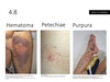
Accumulation of blood within a tissue.
Hematoma(TOPNOTCH)Robbins Basic Pathology, 8th ed. p.86 *SEE SLIDE 4.8

1-2mm hemorrhages into skin, mucous membranes, or serosal surfaces.
Petechiae(TOPNOTCH)Robbins Basic Pathology, 8th ed. p.86 *SEE SLIDE 4.8

3-5mm hemorrhages which can occur with trauma, vascular inflammation, or increased vascular fragility.
Purpura(TOPNOTCH)Robbins Basic Pathology, 8th ed. p.86 *SEE SLIDE 4.8
1-2cm subcutaneous hematomas/bruises.
Ecchymoses (TOPNOTCH)Robbins Basic Pathology, 8th ed. p.86 *SEE SLIDE 4.8


