Week 6 - the digestive system Flashcards

1
Q

What is the gastrointestinal system composed of and what are the main functional processes that occur in the gastrointestinal tract?

A
  • Gastrointestinal system is composed of hollow organs from the mouth to the anus
  • Functional processes occuring here include:
    • Digestion
    • Motility
    • Secretion
    • Cirulation
    • Regulation
  • The regulation of these processes are critical for maintaining gastrointestinal health
2
Q

How is the gastrointestinal tract divided?

A
  • Gastrointestinal tract is divided into the upper and lower tracts
    • Upper GI tract is composed of esophagus, stomach and duodenum
    • Lower GI tract is composed of the small and large intestine
3
Q

What mechanisms are involve in making nutrients available to every cell in the body?

A
  • Ingestion
  • Propulsion
  • Digestion
  • Secretion
  • Absorption
  • Elimination
  • Regulation
4
Q

With regards mechanisms of the digestive system ,what does ingestion refer to?

A
  • Ingestion is the process of taking food into the mouth
5
Q

With regards mechanisms of the digestive system ,what does propulsion mean?

A
  • The moving of food along the GI tract including peristalsis and segmentation (the main process being peristalsis - contractions and relaxations of smooth muscle that lines the walls)
6
Q

With regards mechanisms of the digestive system ,what does digestion​mean?

A
  • Digestion is a range of processes that break down ingested food into simpler nutrients
    • Includes chewing and churning of the stomach and small intestine
7
Q

With regards mechanisms of the digestive system ,what does secretion​mean?

A
  • Secreion is the release of digestive enzymes
8
Q

With regards mechanisms of the digestive system, what does absorption ​mean?

A
  • Absorption is the movement of digested material, via passive diffusion or active transport, the the GI mucosa and into the internal environment
9
Q

With regards mechanisms of the digestive system, what does elimination ​mean?

A
  • Elimination is the exretion of waste material
10
Q

With regards mechanisms of the digestive system, what does regulation ​mean?

A
  • The GI tract is controlled by both intrinsic and extrinsic neuronal and endocrine signals
11
Q

Outline the digestive process

A
  1. Food ingested, teeth break up food and salivary amylase digests starch and moistens food
  2. Food is swallowed (pushed down from throat into esophagus), muscles in esophagus wall contract and push food along (peristalsis)
  3. Stomach has elastic walls which stretch, stomach muscles contract to churn food, stomach lining is folded and contains gastric pits each containing a gland producing gastric juice (acidic and contains pepsin)
  4. Once food a liquid its released into the first part of small intestine and mixed with pancreatic juice (containing amylase, pepsin &lipase)
  5. Inner surface of small intestine lined with villae to increase S.A. for absorption of molecules into the blood stream, intestine wall also folded
  6. Fibre is not absorbed and passes out of body through anus
12
Q

Structure of the mouth: what is the roof of the mouth formed of?

A
  • Roof of the mouth is made of hard and soft palates
    • Palatine and maxillary bones form the hard palate
      • Maxilla is continuous with the rest of the skull
    • Soft palate is formed mainly of muscle and connective tissue
13
Q

Structure of the mouth: what is the function of the uvula and soft palate?

A
  • The uvula and soft palate prevent ingested food from entering the nasal cavities above the mouth
14
Q

Structure of the mouth, how is the floor of the mouth and the tongue formed?

A
  • The arch of the mandible supports the sling of muscles that forms the floor of the mouth and of the tongue
15
Q

Structure of the mouth, describe the tongue

A
  • The tongue consists of skeletal muscle covered with a mucous membrane
16
Q

Structure of the mouth: what attaches the tongue to the floor of the mouth?

A
  • The frenulum is a thin membrane that attaches the tongue to the floor of the mouth
17
Q

Structure of the mouth: describe the cheeks

A
  • The cheek muscles in the sides of the mouth are mainly buccinators and supporting tissue
18
Q

Structure of the mouth: what is the mouth and gums lined with?

A
  • Entire mouth and gums are lined with non-cornified stratified squamous epithelium, which changes to cornified stratified squamous epithelium (skin) at the border of the lips
19
Q

Structure of the mouth: describe the teeth

A
  • Teeth are living structures with vascular and nerve supply to the pulp (centre of each tooth)
  • Dentine is a bony layer that surrounds the pulp and a calcified layer called enamel surrounds the dentine
20
Q

Describe the process of digestion in the mouth

A
  • Food ingested into GI tract through mouth
  • Mouth, cheek and teeth cut, break, grind and moisten what can be chewed and prepare a round smooth bolus that can be swallowed and passed onto the rest of the digestive system
  • Lips, cheek and tongue keep food moving and position food effectively for chewing
21
Q

How can a typical tooth be divided?

A
  • It can be broken down into three main parts:
  1. Crown
  2. Neck
  3. Root
22
Q

Describe the specialisation of teeth for specific tasks

A
  • Incisors : flat sharp edges that are used to cut through food
  • Canines : sharp and pointed ends for gripping and piercing food
  • Premolars and molars : flattened surfaces that are responsible for grinding food
23
Q

Outline some common disorders of the mouth

A
  • Herpes simplex : infection of the mouth causing cold sores that erupt on lips
  • Dental caries : tooth loss with advancing age, caused by bacterial infection in periodontal membrane and gums which is assisted by sugar-rich food residues left in mouth
  • Plaque : hard impenetrable layer arises when bacteria grow between enamel and gums, bacterial metabolic waste products (e.g. organic acids) cause damage to enamel. Infections can penetrate the pulp causing an abscess and chronic infection destroys pulp
24
Q

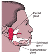
What are the salivsry glands and what are the three main pairs?

A
  • Salivary glands and a group of enxocrine glands that secrete saliva
  • Three main pairs of salivary glands are:
  1. Parotid
  2. Submandibular
  3. Sublingual glands
  • All three secrete saliva into the mouth via secretory ducts
25
Q

Outline the parotid glands

A
  • Largest of salivary glands, situated on side of face, in front of ears at the angle of the jaw
  • Facial nerves travel through the parotid gland
26
Q

Outline the submandibular glands

A
  • Situated below mandibular angle and are irregular in shape
  • Duct of submandibular gland opens onto the mouth at the side of the base of the tongue
27
Q

Outline the sublingual glands

A
  • Smallest of salivry glands
  • Situated in front of the submandibular glands beneath the mucous membrane covering the floor of the mouth
28
Q

Describe the structure of the salivary glands

A
  • They are lobulated with partitions between the lobules
  • The functional unit is the acinus, which is spherical in shape and consists of a single layer of secretory epithelial cells around the central lumen
  • Secretory cells of pyrimidal shaped and the majority are seromucoid - secreting a thick mucoid fluid that contains proteins
29
Q

How much saliva is produced in the mouth each day?

A
  • 1-2 litres and almost all is swallowed and reabsorbed
30
Q

What are the functions of saliva in the mouth?

A
  • Lubricate the mouth and teeth, provide antibacterial properties and digestive enzymes, maintain the chemical balance of tooth enamel
31
Q

Why is the lubrication of food by saliva important?

A
  • It is paramount for chewing and forming a bolus of food that can be easily swallowed
32
Q

How is the secretion of saliva controlled?

A
  • Secretion of saliva is under automonic control
  • Food in mouth stimulates nerve fibres that stimulate salivary nuclei in the mid-brain
  • Salivation also stimulated by smell and sight and anticipation of food through nerve impulses from the cortex stimulating the brainstem salivary nuclei
33
Q

Outline some common disorders of the salivary glands

A
  • Mumps virus : attack on salivary glands, pancreas, ovaries and testicles
    • Parotid inflammation causes the swollen cheeks appearance of mumps
  • Anticholingeric drugs : cause decreased saliva production and dry mouth (xerostomia), Less comon causes include autoimmune damage to salivary glands
    • Xerostomia is a serious condition since chewing and swallowing depends on adequate saliva
34
Q

Describe the structure of the esophagus

A
  • Muscular tube which starts at the pharynx and ends in the stomach
  • Transverse to the neck and throat and is situated close to the trachea
  • Epiglottis can flap over the larynx covering it during swallowing and funneling of food towards the esophagus
35
Q

How are the walls of the esophagus formed?

A
  • They are formed from outside to inside by:
  • Longitudinal muscle layer
  • Circular muscle layer
  • Submucosal layer
  • Muscularis mucosae
  • Mucosa and epithlium
36
Q

Describe the innervation of the esophagus

A
  • Vagus nerve runs alongisde the esophagus and innervates the esophageal muscle directly through intrinsic nerves
37
Q

Describe vascular supply to the esophagus

A
  • Venous drainage ofthe esophagus forms a submucosal venous plexus that drains directly into the systemic circulation, avoiding the hepatic portal vein and liver
38
Q

What is the function of the esophagus?

A
  • Esophagus transports food, liquid and saliva from the mouth to the stomach by peristalsis
39
Q

What is peristalsis and what is it controlled by?

A
  • Rhthymic wave of contraction behind the bolus of food with relaxation infront of the bolus
    • Coordinated movement propels the bolus forwards
  • Involuntary as a result of intrisic neuromuscular reflexes in the intestinal wall
  • External stimuli modulate frequency and strength of peristaltic activity
40
Q

What is the significance of vomiting and peristalsis?

A
  • In vomiting, peristaltic waves travel in the reverse direction, propelling food away from the stomach and towards the mouth
41
Q

Outline some disorders of the esophagus

A
  • Dysphagia : is difficulty in swallowing and odnyophagia is painful swallowing
    • Obstruction to flow down esophagus causes dysphagia and in severe caases can stop swallowing of saliva so that patients drool
      • Pneumonia can be causes by chronic obstruction leading to aspiration of food into larynx
  • Gastroesophageal reflux disease : backward flow of stomach acid up the esophagus causing heartburn and acid indigestion
    • Severe & frequent episode can cause asthma attacks, severe chest pains, bleeding, narrowing and irritation of the esophagus
42
Q

Describe the structure of the stomach

A
  • “J” shaped and consists of curvatures
  • When empty its lining lies in folds called rugae
  • Thick mucous layer protects against trauma from HCl and proteolytic enzymes
  • Gastric pits narrow invaginations of the epithelium connected to gastric glands situated at the beginning of the stomach
43
Q

What are gastric glands?

A
  • Consists of specialised cells for the production of HCl, pepsin and mucous-producing goblet cells
44
Q

What are the five regions that the stomach is composed of?

A
  • The cardia
  • Fundus
  • Body (corpus)
  • Antrum
  • the pylorus forms a sphincter separating the stomach from the duodenum
45
Q

Describe the formation of the walls of the stomach

A
  • From outside-in the stomach wall is formed of:
  1. Serosa
  2. Longitudinal muscle layer
  3. Circular muscle layer
  4. Oblique muscle layer
  5. Submucosa
  6. Muscularis musosae
  7. Mucosa
46
Q

Where does food enter the stomach?

A
  • Enters the stomach by passing through the muscular lower esophageal sphincter (a.k.a cardiac sphincter) which prevents food from reentering the esophagus when the stomach contracts
47
Q

What is the function of the stomach?

A
  • Stomach is adapted for churning, storage and digestion of food and plays a role in neuro-endocrine coordination of intestinal function
48
Q

How does the stomach ensure that food is mixed properly and that no large food particles enter the duodenum?

A
  • Churning action of the gastric muscle against a closed pyloric sphincter ensures that food is mixed thoroughly
  • The pylorus only opens to permit chyme, a semiliquid material, to enter the duodenum and therefore preventing the passage of large food particles
49
Q

What is the gastric slow wave?

A
  • Rhthymic electrical activity in the stomach produces peristaltic waves known as the gastric slow wave
50
Q

How is gastric secretion stimulated?

A
  • Anticipation of food stimulates gastric secretion (cephalic phase) and by food reaching the stomach (gastric phase)
51
Q

What is the signficance of HCl in the stomach and how is its secretion stimulated?

A
  • HCl activates plasminogen to produce pepsin which stimulates the digestion of proteins
  • Secretion of HCl is stimulated by a variety of substances including acetylcholine
52
Q

Outline some common diseases of the stomach

A
  • Peptic ulcers and gastritis are associted with: infection of Helicobacter pylori bacterium, use of non-steroidal anti-inflammatory drugs (NSAIDs) and gastric carcinoma
  • Majority of patients with peptic ulcers are as a results of H. pylori
  • During peptic ulcers the epithelial cells of stomach or duodenum are damage and ulcerate causing inflammation to extend into the underlying mucosa and submucosa
  • Further damage to adjacent tissues and blood vessels is caused by the penetration of gastric acid and digestibe enzymes
53
Q

What is the small intestine composed of ?

A
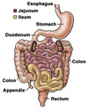
  • Small intestine is composed of duodenum, jejunum and ileum
54
Q

Small intestine, where is the duodenum located?

A
  • The duodenum extends from the pylorus and the first part is called the bulb
  • Second part receives bile and pacreatic enzymes via the ampulla of Vater and is situated adjactent to the pancreas
55
Q

What are the walls of the duodenum made of?

A
  • Walls of the duodenum are composed of:
    • Serosa
    • Circular muscle layer
    • Submucosa
    • Muscularis mucosae
    • Mucosal layer
56
Q

Describe the lining of the duodenum

A
  • Intestinal lining is composed of columnar cells called enterocytes
    • On the luminal face microvillae increase SA for digestion & absorption
    • Glycoproteins, enzymes and mucins cover the surface of microvillae forming a brush border
    • Enterocytes synthesise digestive enzymes and secrete them onto the brush border
57
Q

Describe how the duodenum is responsible for digestion

A
  • It receive chyme from the stomach and mixes it with alkaline bile and pancreatic enzymes, these neutralise the stomach acid
  • Pancreatic enzymes secreted into the duodenum support rapid and effective digestion
  • Disaccharidases and peptidases catalyse the final stages of digestion on the brush border of enterocytes
  • Biles salts emulsify fatty foods and permit digestive enzymes to operate effectively
58
Q

How is the duodenum responsible for absorption?

A
  • Transport proteins on the brush border actively absorb sugars, amino acids and electrolytes into the enterocytes
  • Cholesterol and fatty acids enter via diffusion
  • In the duodenum cholecystokinin and secretin are secreted from entero-endocrine cells in response to food which stimulates the contaction of the gallbladder, pancreatic secretion and inhibition of gastric motility
    • Secretion of cholecystokinin hormone is an example of hormonal regulation of the GI mobility
59
Q

Give an example of hormonal regulation of hormonal mobility

A
  • The secretion of cholecystokinin
60
Q

Describe the structure of the jejunum and the ileum

A
  • Jejunum begins at the junction with the duodenum
  • ileum makes up most of distal small intestine ending in the caecum
  • Jejunum and ileum are attached to he abdominal wall by mesentery that permits free movement and rotation
  • Structure of jejenum and ileum is similar to duodenum
    • Jejunal villae are long, broad and leaf-shaped whilst ileal villae are shorter
61
Q

What are the functions of the jejunum and the ileum?

A
  • They are the main absorptive surfaces of the GI tract (most chemical digestion occurs in the duodenum)
  • Jejunal epithelial cells have enzymatic pathways that process and absorb dietary folic acid
  • Ileal epithelial cells are specialised for the absoption of vitamin B12
  • All fats are digested, bile salts are released and resaborbed in the terminal ileum via transport proteins
    • Bile salts are then recycled in the liver
62
Q

Outline some common disorders of the small intestine

A
  • Tropical sprue : is linked with bacterial infection of the small intesine causing malabsorption as a result of damage to the small intestinal mucosa
  • Crohn’s disease : can affect any part of the intestine but mainly the terminal ileum causing mucosal ulceration and inflammation.
    • Crohn’s disease of terminal ileum is assocated with gene mutations
63
Q

Describe the structure of the liver

A
  • Situated in upper quadrant of abdomen & dividied into 9 functional segments based on biliary drainage and vascular supply
  • The main hepatic vein joins the inferior vena cava that traverses the liver
  • Enclosed in tough fibrous capsule and is covered by peritoneum
  • Gallbadder is situated below the liver and is connected to the bile duct the the cystic duct
  • Hepatocytes form three dimensional cords and plates in the liver
64
Q

What are the functions of the liver?

A
  • The liver is the largest organ in the body and performs vital metabolic functions:
    • Regulating homeostasis
    • Storing nutrients, e.g. glycogen fats and vitamins
    • Producing and secreting plasma proteins and lipoproteins
    • Synthesing bile salts for lipid digestion
    • Detoxifying and secreting bilirubin
    • Clearing toxins
65
Q

Describe the formation of bile and what it is used for

A
  • Bile is formed by hepatocytes and is modified by the biliary epithelium
  • It is an exocrine secretion required for digestion and it is an excretion product for the removal of toxins and metabolic waste
66
Q

Outline cirrhosis of the liver

A
  • Cirrhosis and scarring is caused by long-lasting damage to the liver
    • Several types of liver injury produce cirrhosis and the precise cause is required to determine the treatment
67
Q

Outline chronic liver disease

A
  • Main effects of chronic liver disease due to a reduction in hepatocytes and disruption of the normal architecture eventually causing an increae in pressure in the portal veins and alters the blood flow to the liver
  • Reduction in hepatic function causes accumulation and bilirubin and causes jaundice
  • Since liver is the main regulator of metabolism chronic liver disease causes metabolic dysregulation, weight loss and wasting
68
Q

Outline gallstones as a disorder of the liver and gallbladder

A
  • Common and asymptomatic
  • Form when constituents such as cholesterol or bile pigments are partially soluble, which reach high concentrations and cystallize around a bacterial cell
  • Formation of gallstones is likely to occur if the gallbladder does not empty regularly and chemically imbalance or cholesterol laden bile reamins in the gallbladder for long periods
69
Q

Outline the structure and function of the pancreas

A
  • Lies transversely on the posterior wall and is covered by peritoneum and the main pancreatic duct stretches the length of the gland
  • Critical for intestinal digestion
  • Exocrine gland that secretes the majority of enzymes into the intestine
  • Endocrine function is to produce insulin and glucagon in the α and β cells of the pancreatic islets of Langerhans
70
Q

Why is pancreatic juice important?

A
  • Pancreatic juice contains enzymes that digest the three major types of food
  • Also contains sodium bicarbonate to neutralise HCl in the gastric juice that enters the intestines
71
Q

What happens to the enzymes produced by the pancreas?

A
  • The digestive enzymes produced by the pacreas are stored as pro-enzymes (inactive precursors) to avoid autodigestion
72
Q

Outline some common disorders of the pancreas

A
  • Acute pancreatitis : a serious illness and the most common cause is excess alcohol ingestion. Inflamed pancreas releases enzymes into the systemic circulation affecting the whole body
  • Chronic pancreatitis : may follow many repeat sessions of acute pancreatitis, main symptoms are abdominal pain and malabsorption as a result of failure of exocrine pancreas
73
Q

Describe the caecum and appendix

A
  • Caecum is the most proximal part of the large intesine into which the ileum opens
  • Appendix protrudes from the caecum
  • Both caecum and appendix have no special function in humans, in other species they are highly developed containing bacteria that metabolise plant carbohydrates which cannot be digested by mammalian enzymes
  • Appendix hsa lymphatic tissue that may contribute to immune regulation
74
Q

Describe a condition of the appendix

A
  • Appendicitis is a condition that arises from obstruction of the appendiceal lumen that causes inflammation and infection
75
Q

Describe the structure of the large intestine

A
  • Colon comprises most of the large intesine, colon is divied into four parts:
  1. ascending - begins at top of caecum
  2. transverse
  3. descending
  4. sigmoid - joins rectum
  • Colon wall is same as organisation of the intestinal tract except the external longitudinal muscle is discontinuous
76
Q

What is the function of the colon?

A
  • Main function of colon is to reabsorb water from liquid intestinal contents which remains after digestion and reabsorption in the jejenum and ileum
  • This process converts faecal liquid into a semisolid mass hat is then excreted
  • Muscular action of the colon mixes and compact the faecal matter and directs it towards the rectum
77
Q

What are the functions of the rectum and anus?

How is defecation stimulated?

A
  • Rectum serves as a reservoir for faeces and the anus is a sphincter controlling defecation
  • Defecation is stimulated by distension of the rectum causing increased pressure which stimulates intrisic nerves to increase peristalsis
78
Q

Identify and describe the layers of the digestive wall

A
  • There are 4 typical layers tat comprise the wall of the digestive tracts:
  1. Mucosa
  2. Submucosa
  3. Muscularis
  4. Serosa
79
Q

What are macronutrients and micronutrients and what is their function?

A
  • Carbohydrates, lipids and proteins form the main part of the diet and are referred to as macronutrients
    • Macronutrients are the source for dietary energy and structural materials needed for body tissues
    • Robust mechanisms efficiently extract and absorb macronutrients from diet
  • Micronutrients such as vitamins are required in small quantities
80
Q

What is required for the absorption of digested food?

A
  • Small intestine is the main absorptive surface and absorption of digested food requires a large surface area
81
Q

Describe the digestion of carbohydrates

A
  • Carbohydrates are ingested as sugars and starch (a polysaccharide)
  • Sucrase, maltase and lactase are produced by enterocytes which convert disaccharides into monosaccharides
  • Specialised transporters such as sodium-glucose co-transporter are present on surface of enterocytes and transport molecules into the cytoplasm of the cell
  • Aborbed monosaccharides leave the enterocytes, by facilitated diffusion, which then enter circulation via capillary network in the villus
82
Q

Describe the digestion of proteins

A
  • Digestion of proteins begins in the stomach by proteases (e.g. pepsin) which is secreated by the pancreas
  • Endopeptidases and exopeptidases cleave the peptide chain into smaller fragments
  • Peptidases on the brush border complete digestion of peptides permitting the absorption of single amino acids
  • From the cytoplasm of enterocytes, amino acids enter the circulaton via selective channels
83
Q

Describe the digestion of lipids

A
  • In contrast to proteins and carbohydrates which are hydrophilic and are easily accessible to enzymes, lipids require a partition into a hydrophibic environment
  • Most fats are undigested until after emulsification by bile in the duodenum
  • After emulsification pancreatic lipases digest the fat molecules into fatty acids and glycerol
  • The digested lipds are absorbed across the cell membrane into the enterocyte cytoplasm where they are re-esterified
  • Lipids are mainly digested in the small intestine
84
Q

Outline a common digestive disorder

A
  • Inability to digest and absorb macronutrients leads to wasting of muscle and fat
  • Consequently, essential tissues cannot be maintained and patients can die from multigrain failure
    • Such changes are also apparent during starvation